

")


Diagnostic challenge: innovative approach in use of magnetic resonance imaging in aortic aneurysm
- Authors: Kobelev E.1, Pak N.T.1, Bobrikova E.E.1, Ussov W.Y.2, Kliver E.E.1, Sirota D.A.1, Chernyavskiy A.M.1, Bergen T.A.1
-
Affiliations:
- E. Meshalkin National Medical Research Center
- Tomsk National Research Medical Center
- Issue: Vol 3, No 3 (2022)
- Pages: 332-339
- Section: Case reports
- URL: https://journals.rcsi.science/DD/article/view/108404
- DOI: https://doi.org/10.17816/DD108404
- ID: 108404
Cite item

Abstract
Here we report a case of technological innovation: the use of magnetic resonance imaging to determine surgical strategy.
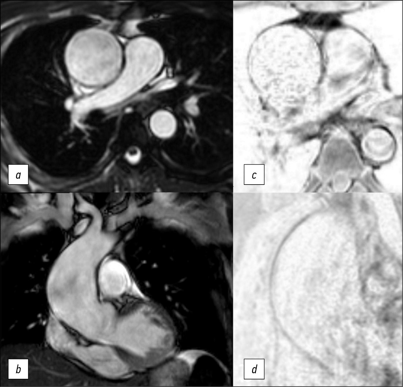
Here is a 47-year-old man who underwent an magnetic resonance imaging and subsequent surgical treatment of the aortic aneurysm. Unlike echocardiography, magnetic resonance imaging enabled us to view the entire thoracic aorta. Unlike computer tomography, magnetic resonance imaging enabled us to detect changes in the aortic wall accurately. Thus, in this case, the use of magnetic resonance imaging allowed us to determine the distal resection edge. The patient`s postoperative course was unremarkable. Use of electrocardiogram-synchronized magnetic resonance imaging of thoracic aorta allows detecting structural changes of the aortic wall and its mechanical properties. It is significant that magnetic resonance imaging results of the aortic wall correlate with histologic examination.
The extent of changes in the aortic wall must be determined to accurately plan surgical treatment of patients with aortic aneurism.
Magnetic resonance imaging of the aortic wall is promising for further study in multicenter research.
Keywords
Full Text
##article.viewOnOriginalSite##About the authors
Evgenii Kobelev
E. Meshalkin National Medical Research Center
Email: kobelev_e@meshalkin.ru
ORCID iD: 0000-0002-5901-2271
SPIN-code: 7828-9713
Russian Federation, Novosibirsk
Natalya T. Pak
E. Meshalkin National Medical Research Center
Email: n_pak@meshalkin.ru
ORCID iD: 0000-0002-7842-9881
SPIN-code: 1896-8447
MD, Cand. Sci. (Med.)
Russian Federation, NovosibirskEvgeniya E. Bobrikova
E. Meshalkin National Medical Research Center
Email: bobrikova_e@meshalkin.ru
ORCID iD: 0000-0001-5985-4076
SPIN-code: 6315-9772
Russian Federation, Novosibirsk
Wladimir Y. Ussov
Tomsk National Research Medical Center
Email: ussov1962@yandex.ru
ORCID iD: 0000-0001-7978-5514
SPIN-code: 1299-2074
MD, Dr. Sci. (Med.), Professor
Russian Federation, TomskEvgeniy E. Kliver
E. Meshalkin National Medical Research Center
Email: ee_kliver@meshalkin.ru
ORCID iD: 0000-0002-3915-3616
SPIN-code: 1511-3814
MD, Dr. Sci. (Med.)
Russian Federation, NovosibirskDmitriy A. Sirota
E. Meshalkin National Medical Research Center
Email: d_sirota@meshalkin.ru
ORCID iD: 0000-0002-9940-3541
SPIN-code: 4706-7549
MD, Cand. Sci. (Med.)
Russian Federation, NovosibirskAleksandr M. Chernyavskiy
E. Meshalkin National Medical Research Center
Email: a_cherniavsky@meshalkin.ru
ORCID iD: 0000-0001-9818-8678
SPIN-code: 5286-6950
MD, Dr. Sci. (Med.), Professor, Corresponding member of the Russian Academy of Sciences
Russian Federation, NovosibirskTatyanа A. Bergen
E. Meshalkin National Medical Research Center
Author for correspondence.
Email: bergen_t@meshalkin.ru
ORCID iD: 0000-0003-1530-1327
SPIN-code: 5467-7347
MD, Dr. Sci. (Med.)
Russian Federation, NovosibirskReferences
- Erbel R, Aboyans V, Boileau C. et al. 2014 ESC Guidelines on the diagnosis and treatment of aortic diseases: document covering acute and chronic aortic diseases of the thoracic and abdominal aorta of the adult. The task force for the diagnosis and treatment of aortic diseases of the European society of cardiology (ESC). Eur Heart J. 2014;35(41):2873–926. doi: 10.1093/eurheartj/ehu281
- Hiratzka LF, Bakris GL, Beckman JA, et al. Guidelines for the diagnosis and management of patients with thoracic aortic disease. A report of the American College of Cardiology Foundation/American Heart Association Task Force on Practice Guidelines, American Association for Thoracic Surgery, American College of Radiology, American Stroke Association, Society of Cardiovascular Anesthesiologists, Society for Cardiovascular Angiography and Interventions, Society of Interventional Radiology, Society of Thoracic Surgeons, and Society for Vascular Medicine. J Am Coll Cardiol. 2010;55(14):e27–e129.
- Galizia MS, Bolen MA, Flamm S. MRI of the thoracic aorta. Applied Radiology. 2015;44(8):22–26. doi: 10.37549/ar2207
- Saliba E, Sia Y. The ascending aortic aneurysm: When to intervene? IJC Heart Vasculature. 2015;6:91–100. doi: 10.1016/j.ijcha.2015.01.009
Supplementary files


