

")


诊断能力:在主动脉瘤中使用磁共振成像的创新方法
- 作者: Kobelev E.1, Pak N.T.1, Bobrikova E.E.1, Ussov W.Y.2, Kliver E.E.1, Sirota D.A.1, Chernyavskiy A.M.1, Bergen T.A.1
-
隶属关系:
- E. Meshalkin National Medical Research Center
- Tomsk National Research Medical Center
- 期: 卷 3, 编号 3 (2022)
- 页面: 332-339
- 栏目: 临床病例及临床病例的系列
- URL: https://journals.rcsi.science/DD/article/view/108404
- DOI: https://doi.org/10.17816/DD108404
- ID: 108404
如何引用文章

详细
本文介绍了磁共振成像在确定手术策略方面的创新应用。
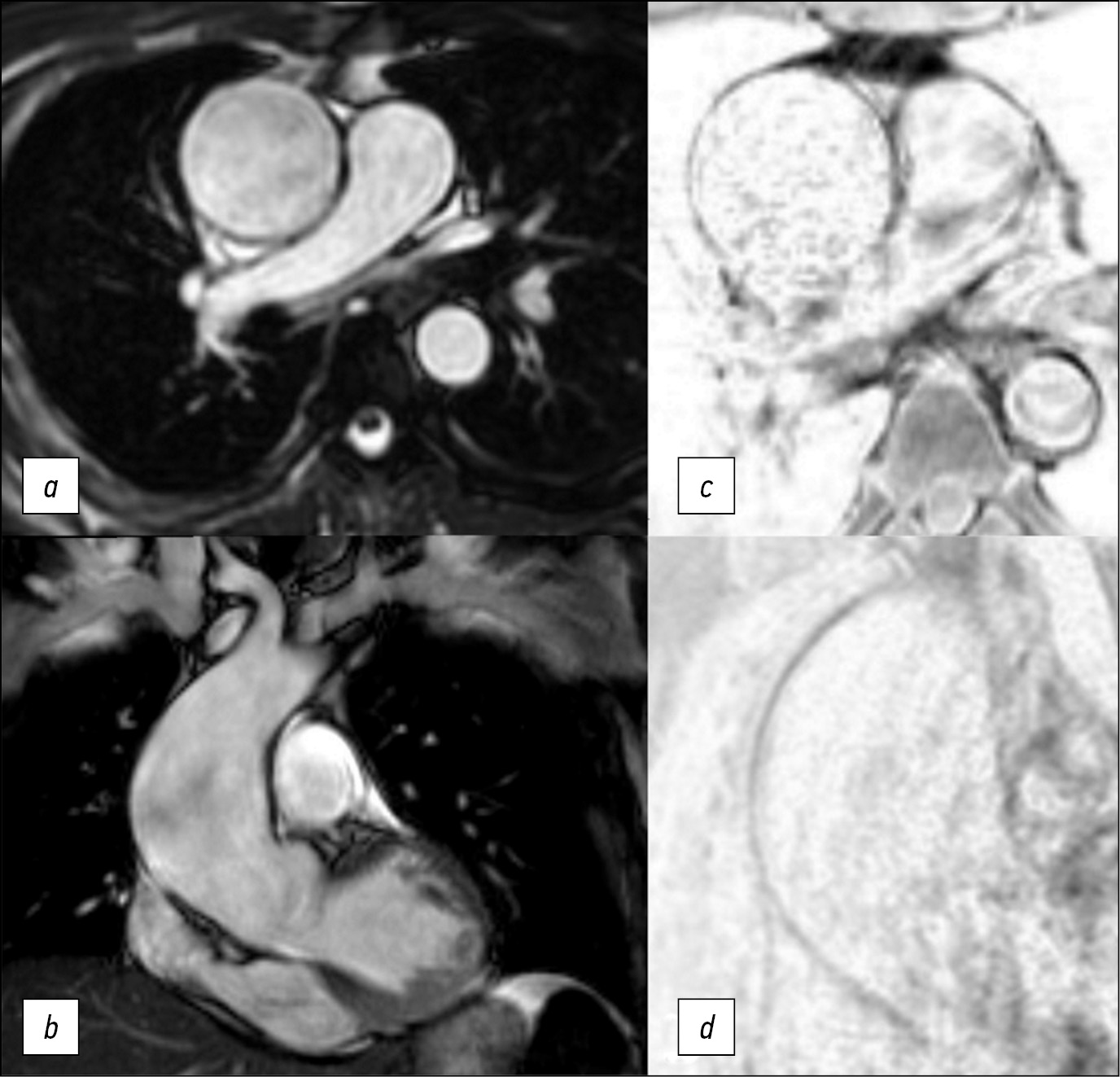
作者描述了一名47岁患者的病例,该患者接受了磁共振成像,随后对主动脉瘤进行了手术治疗。与超声心动图不同,此类诊断方法可以看到整个胸主动脉,与计算机断层扫描不同,它有助于识别主动脉壁的变化。使用磁共振成像,我们能够确定切除的远心端。术后期间一切顺利。根据与心电图同步的数据,我们评估了主动脉壁的结构变化及其力学性能。值得注意的是,磁共振成像的结果与组织学检查的结果相关。
为了对主动脉瘤患者进行有效的手术治疗,必须确定血管壁的变化程度。
主动脉壁的磁共振成像是一个很有前途的诊断方向,需要在多中心研究中进一步研究。
作者简介
Evgenii Kobelev
E. Meshalkin National Medical Research Center
Email: kobelev_e@meshalkin.ru
ORCID iD: 0000-0002-5901-2271
SPIN 代码: 7828-9713
俄罗斯联邦, Novosibirsk
Natalya T. Pak
E. Meshalkin National Medical Research Center
Email: n_pak@meshalkin.ru
ORCID iD: 0000-0002-7842-9881
SPIN 代码: 1896-8447
MD, Cand. Sci. (Med.)
俄罗斯联邦, NovosibirskEvgeniya E. Bobrikova
E. Meshalkin National Medical Research Center
Email: bobrikova_e@meshalkin.ru
ORCID iD: 0000-0001-5985-4076
SPIN 代码: 6315-9772
俄罗斯联邦, Novosibirsk
Wladimir Y. Ussov
Tomsk National Research Medical Center
Email: ussov1962@yandex.ru
ORCID iD: 0000-0001-7978-5514
SPIN 代码: 1299-2074
MD, Dr. Sci. (Med.), Professor
俄罗斯联邦, TomskEvgeniy E. Kliver
E. Meshalkin National Medical Research Center
Email: ee_kliver@meshalkin.ru
ORCID iD: 0000-0002-3915-3616
SPIN 代码: 1511-3814
MD, Dr. Sci. (Med.)
俄罗斯联邦, NovosibirskDmitriy A. Sirota
E. Meshalkin National Medical Research Center
Email: d_sirota@meshalkin.ru
ORCID iD: 0000-0002-9940-3541
SPIN 代码: 4706-7549
MD, Cand. Sci. (Med.)
俄罗斯联邦, NovosibirskAleksandr M. Chernyavskiy
E. Meshalkin National Medical Research Center
Email: a_cherniavsky@meshalkin.ru
ORCID iD: 0000-0001-9818-8678
SPIN 代码: 5286-6950
MD, Dr. Sci. (Med.), Professor, Corresponding member of the Russian Academy of Sciences
俄罗斯联邦, NovosibirskTatyanа A. Bergen
E. Meshalkin National Medical Research Center
编辑信件的主要联系方式.
Email: bergen_t@meshalkin.ru
ORCID iD: 0000-0003-1530-1327
SPIN 代码: 5467-7347
MD, Dr. Sci. (Med.)
俄罗斯联邦, Novosibirsk参考
- Erbel R, Aboyans V, Boileau C. et al. 2014 ESC Guidelines on the diagnosis and treatment of aortic diseases: document covering acute and chronic aortic diseases of the thoracic and abdominal aorta of the adult. The task force for the diagnosis and treatment of aortic diseases of the European society of cardiology (ESC). Eur Heart J. 2014;35(41):2873–926. doi: 10.1093/eurheartj/ehu281
- Hiratzka LF, Bakris GL, Beckman JA, et al. Guidelines for the diagnosis and management of patients with thoracic aortic disease. A report of the American College of Cardiology Foundation/American Heart Association Task Force on Practice Guidelines, American Association for Thoracic Surgery, American College of Radiology, American Stroke Association, Society of Cardiovascular Anesthesiologists, Society for Cardiovascular Angiography and Interventions, Society of Interventional Radiology, Society of Thoracic Surgeons, and Society for Vascular Medicine. J Am Coll Cardiol. 2010;55(14):e27–e129.
- Galizia MS, Bolen MA, Flamm S. MRI of the thoracic aorta. Applied Radiology. 2015;44(8):22–26. doi: 10.37549/ar2207
- Saliba E, Sia Y. The ascending aortic aneurysm: When to intervene? IJC Heart Vasculature. 2015;6:91–100. doi: 10.1016/j.ijcha.2015.01.009
补充文件


