

")


Оценка диаметра лёгочной артерии при различной степени тяжести течения COVID-19 (по данным бесконтрастной компьютерной томографии лёгких)
- Авторы: Алиев А.Ф.1,2, Кудрявцев Н.Д.1,3, Петряйкин А.В.3, Артюкова З.Р.3, Шкода А.С.4, Морозов С.П.3
-
Учреждения:
- Городская клиническая больница № 67 имени Л.А. Ворохобова Департамента здравоохранения города Москвы
- Московский городской научно-практический центр борьбы с туберкулезом Департамента здравоохранения г. Москвы
- Научно-практический клинический центр диагностики и телемедицинских технологий Департамента здравоохранения г. Москвы
- Городская клиническая больница №67 им.Л.А.Ворохобова Департамента здравоохранения города Москвы
- Выпуск: Том 2, № 3 (2021)
- Страницы: 249-260
- Раздел: Оригинальные исследования
- URL: https://journals.rcsi.science/DD/article/view/76726
- DOI: https://doi.org/10.17816/DD76726
- ID: 76726
Цитировать

Аннотация
Обоснование. Компьютерная томография является методом выбора при оценке объёма поражения лёгких при вирусных пневмониях, в том числе ассоциированных с COVID-19. Помимо оценки объёма поражения лёгких, компьютерная томография позволяет определить размеры магистральных сосудов грудной клетки. Это позволило проанализировать связь между тяжестью течения COVID-19 и наличием изменения диаметров лёгочной артерии и восходящей аорты. Расширение лёгочной артерии является признаком лёгочной гипертензии. Изучение данных закономерностей может иметь клиническое значение в отношении определения тактики лечения и прогноза течения заболевания COVID-19.
Цель ― оценить зависимость между диаметром лёгочной артерии и степенью тяжести течения COVID-19 у пациентов различного возраста.
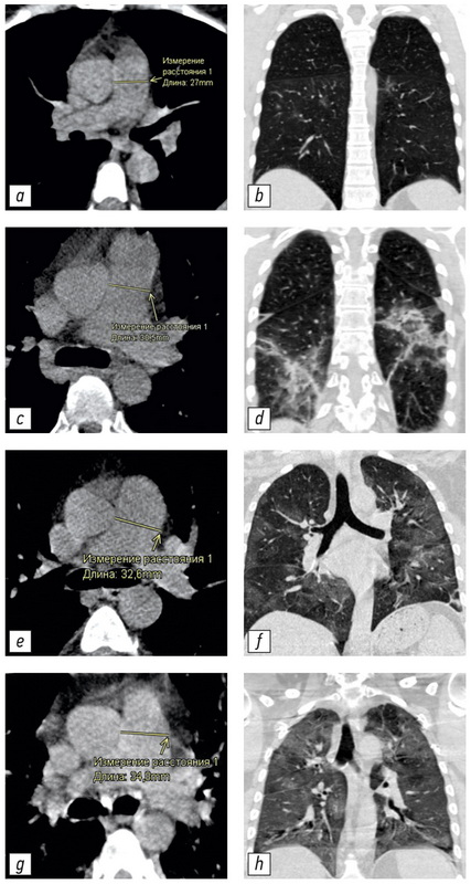
Материалы и методы. Одноцентровое одномоментное сплошное неконтролируемое исследование выполнено в группе пациентов (n=511, 267 мужчин, медиана 59 лет, IQR 49,0–65,0, размах от 31 до 84 лет), проходивших лечение во временном госпитале для лечения пациентов с COVID-19. При госпитализации все пациенты прошли компьютерное томографическое исследование органов грудной клетки с помощью мобильной системы Airo TruCT (Stryker, США). Степень поражения лёгочной ткани оценивалась по шкале КТ 1–4. Измерение диаметра лёгочной артерии и восходящей аорты проводилось стандартными инструментами рабочей станции врача-рентгенолога перпендикулярно длинной оси сосуда.
Результаты. Получены следующие статистически значимые закономерности: расширение лёгочной артерии и увеличение отношения лёгочной артерии/восходящей аорты было связано с увеличением степени поражения лёгких при COVID-19 (критерий Краскела–Уоллиса, p <0,001; медианный тест, p <0,001); диаметр восходящего отдела аорты достоверно увеличивается с возрастом пациента (критерий Краскела–Уоллиса, p <0,001; медианный тест, p <0,001). Показаны недостоверная связь между увеличением диаметра лёгочной артерии и возрастом пациента (критерий Краскела–Уоллиса, p=0,094; медианный тест, p=0,311) и недостоверная связь между изменением диаметра восходящей аорты и степенью поражения лёгких (критерий Краскела–Уоллиса, p=0,061; медианный тест, p=0,165). Во всех возрастных группах с тяжёлым течением заболевания и большим объёмом поражения лёгких (КТ-3 и КТ-4) показано достоверно большее количество пациентов с признаками лёгочной гипертензии (расширенная от 29 мм и более лёгочная артерия).
Заключение. Дилатация лёгочной артерии и увеличение отношения диаметров лёгочной артерии/восходящей аорты достоверно связано с увеличением объёма поражения лёгких при COVID-19 во всех возрастных группах.
Ключевые слова
Полный текст
Открыть статью на сайте журналаОб авторах
Александр Физулиевич Алиев
Городская клиническая больница № 67 имени Л.А. Ворохобова Департамента здравоохранения города Москвы; Московский городской научно-практический центр борьбы с туберкулезом Департамента здравоохранения г. Москвы
Email: alijealex83@gmail.com
ORCID iD: 0000-0003-3282-0567
SPIN-код: 7891-9314
кандидат медицинских наук, врач-рентгенолог высшей категории кабинета компьютерной томографии Московского научно-практического центра борьбы с туберкулезом
Россия, 123423, Москва, ул. Саляма Адиля, д. 2/44; 107014, Москва, ул. Стромынка, 10, стр. 1Никита Дмитриевич Кудрявцев
Городская клиническая больница № 67 имени Л.А. Ворохобова Департамента здравоохранения города Москвы; Научно-практический клинический центр диагностики и телемедицинских технологий Департамента здравоохранения г. Москвы
Email: n.kudryavtsev@npcmr.ru
ORCID iD: 0000-0003-4203-0630
SPIN-код: 1125-8637
врач-рентгенолог, младший научный сотрудник
Россия, 123423, Москва, ул. Саляма Адиля, д. 2/44; 127051, Москва, ул. Петровка, 24 с1Алексей Владимирович Петряйкин
Научно-практический клинический центр диагностики и телемедицинских технологий Департамента здравоохранения г. Москвы
Email: alexeypetraikin@gmail.com
ORCID iD: 0000-0003-1694-4682
SPIN-код: 6193-1656
кандидат медицинских наук, врач-рентгенологов, доцент, ведущий научный сотрудник
Россия, 127051, Москва, ул. Петровка, 24 с1Злата Романовна Артюкова
Научно-практический клинический центр диагностики и телемедицинских технологий Департамента здравоохранения г. Москвы
Email: z.artyukova@npcmr.ru
ORCID iD: 0000-0003-2960-9787
SPIN-код: 7550-2441
врач-рентгенолог, младший научный сотрудник
Россия, 127051, Москва, ул. Петровка, 24 с1Андрей Сергеевич Шкода
Городская клиническая больница №67 им.Л.А.Ворохобова Департамента здравоохранения города Москвы
Email: gkb67@zdrav.mos.ru
ORCID iD: 0000-0002-9783-1796
доктор медицинских наук, врач высшей квалификационной категории по специальности «Организация здравоохранения и общественное здоровье», главный врач
Россия, 123423, Москва, ул. Саляма Адиля, 2/44Сергей Павлович Морозов
Научно-практический клинический центр диагностики и телемедицинских технологий Департамента здравоохранения г. Москвы
Автор, ответственный за переписку.
Email: morozov@npcmr.ru
ORCID iD: 0000-0001-6545-6170
SPIN-код: 8542-1720
доктор медицинских наук, профессор, директор
Россия, 127051, Москва, ул. Петровка, 24 с1Список литературы
- Морозов С.П., Проценко Д.Н., Сметанина С.В., и др. Лучевая диагностика коронавирусной болезни (COVID-19): организация, методология, интерпретация результатов: препринт № ЦДТ-2020-II. Версия 2 от 17.04.2020. Серия «Лучшие практики лучевой и инструментальной диагностики». Вып. 65. Москва: ГБУЗ «НПКЦ ДиТ ДЗМ», 2020. 78 с.
- Профилактика, диагностика и лечение новой коронавирусной инфекции (COVID-19): Временные методические рекомендации. Версия 10 (08.02.2020). 2020. 261 с.
- Фомин В.В., Терновой С.К., Серова Н.С. Рекомендации по лучевой диагностике у пациентов с COVID-19 (опыт Cеченовского Университета)//REJR. 2020. T. 10, № 2. С. 8–13. doi: 10.21569/2222-7415-2020-10-2-8-13
- Henkel M., Weikert T., Marston K., et al. Lethal COVID-19: radiological-pathological correlation of the lungs//Radiol Cardiothorac Imaging. 2020. Vol. 2, N 6. P. e200406. doi: 10.1148/ryct.2020200406
- Sun Z., Zhang N., Li Y., et al. A systematic review of chest imaging findings in COVID-19//Quant Imaging Med Surg. 2020. Vol. 10, N 5. P. 1058–1079. doi: 10.21037/qims-20-564
- Salehi S., Abedi A., Balakrishnan S., et al. Coronavirus disease 2019 (COVID-19): A systematic review of imaging findings in 919 patients//AJR Am J Roentgenol. 2020. Vol. 215, N 1. P. 87–93. doi: 10.2214/AJR.20.23034
- Qanadli S.D., Beigelman-Aubry C., Rotzinger D.C. Vascular changes detected with thoracic CT in coronavirus disease (COVID-19) might be significant determinants for accurate diagnosis and optimal patient management//AJR Am J Roentgenol. 2020. Vol. 215, N 1. P. 15. doi: 10.2214/AJR.20.23185
- Li X., Ma X. Acute respiratory failure in COVID-19: Is it "typical" ARDS?//Crit Care. 2020. Vol. 24, N 1. P. 198. doi: 10.1186/s13054-020-02911-9
- Spagnolo P., Cozzi A., Foà R.A., et al. CT-derived pulmonary vascular metrics and clinical outcome in COVID-19 patients//Quant Imaging Med Surg. 2020. Vol. 10, N 6. P. 1325–1333. doi: 10.21037/qims-20-546
- Lv H., Chen T., Pan Y., et al. Pulmonary vascular enlargement on thoracic CT for diagnosis and differential diagnosis of COVID-19: a systematic review and meta-analysis//Ann Transl Med. 2020. Vol. 8, N 14. P. 878–878. doi: 10.21037/atm-20-4955
- Chang Y.C., Yu C.J., Chang S.C., et al. Pulmonary sequelae in convalescent patients after severe acute respiratory syndrome: Evaluation with thin-section CT//Radiology. 2005. Vol. 236, N 3. P. 1067–1075. doi: 10.1148/radiol.2363040958
- Prokop M., van Everdingen W., van Rees Vellinga T., et al. CO-RADS: A categorical CT assessment scheme for patients suspected of having COVID-19-definition and evaluation//Radiology. 2020. Vol. 296, N 2. P. E97–E104. doi: 10.1148/radiol.2020201473
- Corson N., Armato S.G., Labby Z.E., et al. CT-based pulmonary artery measurements for the assessment of pulmonary hypertension//Acad Radiol. 2014. Vol. 21, N 4. P. 523–530. doi: 10.1016/j.acra.2013.12.015
- Truong Q.A., Massaro J.M., Rogers I.S., et al. Reference values for normal pulmonary artery dimensions by noncontrast cardiac computed tomography the Framingham heart study//Circ Cardiovasc Imaging. 2012. Vol. 5, N 1. P. 147–154. doi: 10.1161/CIRCIMAGING.111.968610
- Collins J.A., Munoz J.V., Patel T.R., et al. The anatomy of the aging aorta//Clin Anat. 2014. Vol. 27, N 3. P. 463–466. doi: 10.1002/ca.22384
- Compton G.L., Florence J., MacDonald C., et al. Main pulmonary artery-to-ascending aorta diameter ratio in healthy children on MDCT//AJR Am J Roentgenol. 2015. Vol. 205, N 6. P. 1322–1325. doi: 10.2214/AJR.15.14301
- Galiè N., Humbert M., Vachiery J.L., et al. 2015 ESC/ERS guidelines for the diagnosis and treatment of pulmonary hypertension: the joint task force for the diagnosis and treatment of pulmonary hypertension of the European society of cardiology (ESC) and the European Respiratory Society (ERS): Endorsed by: Association for European Paediatric and Congenital Cardiology (AEPC), International Society for Heart and Lung Transplantation (ISHLT)//European Heart Journal. 2016. Vol. 37, N 1. P. 67–119. doi: 10.1093/eurheartj/ehv317
- Parasuraman S., Walker S., Loudon B.L., et al. Assessment of pulmonary artery pressure by echocardiography — A comprehensive review//Int J Cardiol Heart Vasc. 2016. Vol. 12. P. 45–51. doi: 10.1016/j.ijcha.2016.05.011
- Чучалин А.Г., Авдеев С.Н., Айсанов З.Р., и др. Диагностика и лечение идиопатического легочного фиброза. Федеральные клинические рекомендации//Пульмонология. 2016. Т. 26, № 4. С. 399–419. doi: 10.18093/0869-0189-2016-26-4-399-419
- Черняев А.Л., Самсонова М.В. Патологическая анатомия лёгких. Атлас. 2-е изд., испр. и доп. Серия монографий Российского респираторного общества / под ред. А.Г. Чучалина. Москва: Атмосфера, 2011. 111 с.
- Dolhnikoff M., Duarte-Neto A.N., de Almeida Monteiro R.A., et al. Pathological evidence of pulmonary thrombotic phenomena in severe COVID-19//J Thromb Haemost. 2020. Vol. 18, N 6. P. 1517–1519. doi: 10.1111/jth.14844
Дополнительные файлы


