

")


Changing of pulmonary artery diameter in accordance with severity of COVID-19 (assessment based on non-contrast computer tomography)
- Авторлар: Aliev A.F.1,2, Kudryavtsev N.D.1,3, Petraikin A.V.3, Artyukova Z.R.3, Shkoda A.S.4, Morozov S.P.3
-
Мекемелер:
- L.A. Vorokhobov Municipal Clinical Hospital No 67 of the Moscow Health Department
- The Moscow Research and Clinical Center for Tuberculosis Control of the Moscow Health Department
- Moscow Center for Diagnostics and Telemedicine
- Vorokhobov City Clinical Hospital No. 67
- Шығарылым: Том 2, № 3 (2021)
- Беттер: 249-260
- Бөлім: Original Study Articles
- URL: https://journals.rcsi.science/DD/article/view/76726
- DOI: https://doi.org/10.17816/DD76726
- ID: 76726
Дәйексөз келтіру

Аннотация
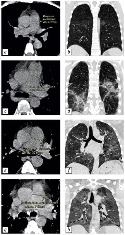
BACKGROUND: Computed tomography is the method of choice for assessing the volume of lung damage in viral pneumonia, including those associated with COVID-19. In addition, computed tomography can determine the main vessels size of the thorax. This allowed us to analyze the relationship between the severity of COVID-19 and the changes in the diameters of the pulmonary artery (PA) and ascending aorta (Ao). Dilation of the PA is a sign of pulmonary hypertension. The study of these patterns may be of clinical significance in determining the treatment tactics and prognosis of the course of COVID-19 disease.
AIM: To evaluate the relationship between PA diameter and the severity of the COVID-19 course in patients of different ages.
MATERIALS AND METHODS: This study is a single-centered, cross-section, continuous, uncontrolled study performed on a group of patients (n=511, 267 men, median 59 years, IQR 49.0–65.0, ages 31–84 years) who were treated in a temporary hospital to treat patient with COVID-19. During hospitalization all patients were examined by CT scan of the chest. All studies were carried out using a mobile CT scan system Airo TruCT (Stryker, USA). The degree of damage to the lung tissue was assessed using the CT volume scale 1–4. Measurement of the LA and Ao diameters was carried out using standard instruments of the radiologist’s CT workstation perpendicular to the long axis of the vessel.
RESULTS: The following statistically significant regularities were obtained: the detection of a dilated pulmonary artery (PA) and an increased PA/Ao ratio correlated to an increase in the degree of lung damage in COVID-19 (Kruskal-Wallis test, K-W p <0.001; median test, MT p <0.001), the diameter of the ascending aorta (Ao) significantly increases with the patient’s age (K-W p <0.001; MT p <0.001). An insignificant correlation between an increase in the diameter of the pulmonary artery (PA) and the patient’s age (K-W p=0.094; MT p=0.311) and an insignificant correlation between detection of a change in aortic (Ao) diameter and the degree of lung damage (K-W p=0.061; MT p=0.165) were shown. In groups with a severe course of the disease and a large volume of lung lesions (CT-3 and CT-4), a significantly greater number of patients with signs of pulmonary hypertension (detection of the dilated pulmonary artery: 29 mm and more) was shown for all age groups.
CONCLUSION: The study showed that PA dilatation and increased PA/Ao diameter ratio were significantly associated with increased pulmonary lesion volume in COVID-19 in all age groups.
Негізгі сөздер
Толық мәтін
##article.viewOnOriginalSite##Авторлар туралы
Alexander Aliev
L.A. Vorokhobov Municipal Clinical Hospital No 67 of the Moscow Health Department; The Moscow Research and Clinical Center for Tuberculosis Control of the Moscow Health Department
Email: alijealex83@gmail.com
ORCID iD: 0000-0003-3282-0567
SPIN-код: 7891-9314
MD, Cand. Sci. (Med.)
Ресей, 2/44, Salyama Adilya street, 123423 Moscow; 10 c1, Stromynka Street, 107014 MoscowNikita Kudryavtsev
L.A. Vorokhobov Municipal Clinical Hospital No 67 of the Moscow Health Department; Moscow Center for Diagnostics and Telemedicine
Email: n.kudryavtsev@npcmr.ru
ORCID iD: 0000-0003-4203-0630
SPIN-код: 1125-8637
diagnostic radiologist, junior researcher
Ресей, 2/44, Salyama Adilya street, 123423 Moscow; 24 с1, Petrovka street, 127051 MoscowAlexey Petraikin
Moscow Center for Diagnostics and Telemedicine
Email: alexeypetraikin@gmail.com
ORCID iD: 0000-0003-1694-4682
SPIN-код: 6193-1656
MD, Cand. Sci. (Med.), Associate Professor, leading researcher, diagnostic radiologist
Ресей, 24 с1, Petrovka street, 127051 MoscowZlata Artyukova
Moscow Center for Diagnostics and Telemedicine
Email: z.artyukova@npcmr.ru
ORCID iD: 0000-0003-2960-9787
SPIN-код: 7550-2441
MD, junior researcher, radiologist
Ресей, 24 с1, Petrovka street, 127051 MoscowAndrey Shkoda
Vorokhobov City Clinical Hospital No. 67
Email: gkb67@zdrav.mos.ru
ORCID iD: 0000-0002-9783-1796
MD, Dr. Sci. (Med.), Professor, doctor of the highest qualification category in the specialty, chief physician
Ресей, 2/44, Salama Adil street, 123423 MoscowSergey Morozov
Moscow Center for Diagnostics and Telemedicine
Хат алмасуға жауапты Автор.
Email: morozov@npcmr.ru
ORCID iD: 0000-0001-6545-6170
SPIN-код: 8542-1720
MD, Dr. Sci. (Med.), Professor
Ресей, 24 с1, Petrovka street, 127051 MoscowӘдебиет тізімі
- Morozov SP, Protsenko DN, Smetanina SV, et al. Radiation diagnostics of coronavirus disease (COVID-19): organization, methodology, interpretation of results: preprint No. CDT-2020-II. Version 2 from 17.04.2020. The series "Best practices of radiation and instrumental diagnostics". Issue 65. Moscow: Scientific and Practical Clinical Center for Diagnostics and Telemedicine Technologies of the Department of Healthcare of the City of Moscow; 2020. 78 p. (In Russ).
- Prevention, diagnosis and treatment of a new coronavirus infection (COVID-19): Temporary methodological recommendations. Version 10 (08.02.2020). 2020. 261 p. (In Russ).
- Fomin VV, Ternovoy SK, Serova NS. Radiological guidelines in patients with COVID-19 (Sechenov University experience). REJR. 2020;10(2):8–13. (In Russ). doi: 21569/2222-7415-2020-10-2-8-13
- Henkel M, Weikert T, Marston K, et al. Lethal COVID-19: radiological-pathological correlation of the lungs. Radiol Cardiothorac Imaging. 2020;2(6):e200406. doi: 10.1148/ryct.2020200406
- Sun Z, Zhang N, Li Y, et al. A systematic review of chest imaging findings in COVID-19. Quant Imaging Med Surg. 2020;10(5): 1058–1079. doi: 10.21037/qims-20-564
- Salehi S, Abedi A, Balakrishnan S, et al. Coronavirus disease 2019 (COVID-19): A systematic review of imaging findings in 919 patients. AJR Am J Roentgenol. 2020;215(1):87–93. doi: 10.2214/AJR.20.23034
- Qanadli SD, Beigelman-Aubry C, Rotzinger DC. Vascular changes detected with thoracic CT in coronavirus disease (COVID-19) might be significant determinants for accurate diagnosis and optimal patient management. AJR Am J Roentgenol. 2020;215(1):15. doi: 10.2214/AJR.20.23185
- Li X, Ma X. Acute respiratory failure in COVID-19: Is it "typical" ARDS? Crit Care. 2020;24(1):198. doi: 10.1186/s13054-020-02911-9
- Spagnolo P, Cozzi A, Foà RA, et al. CT-derived pulmonary vascular metrics and clinical outcome in COVID-19 patients. Quant Imaging Med Surg. 2020;10(6):1325–1333. doi: 10.21037/qims-20-546
- Lv H, Chen T, Pan Y, et al. Pulmonary vascular enlargement on thoracic CT for diagnosis and differential diagnosis of COVID-19: a systematic review and meta-analysis. Ann Transl Med. 2020; 8(14):878–878. doi: 10.21037/atm-20-4955
- Chang YC, Yu CJ, Chang SC, et al. Pulmonary sequelae in convalescent patients after severe acute respiratory syndrome: Evaluation with thin-section CT. Radiology. 2005;236(3):1067–1075. doi: 10.1148/radiol.2363040958
- Prokop M, van Everdingen W, van Rees Vellinga T, et al. CO-RADS: A categorical CT assessment scheme for patients suspected of having COVID-19-definition and evaluation. Radiology. 2020;296(2):E97–E104. doi: 10.1148/radiol.2020201473
- Corson N, Armato SG, Labby ZE, et al. CT-based pulmonary artery measurements for the assessment of pulmonary hypertension. Acad Radiol. 2014;21(4):523–530. doi: 10.1016/j.acra.2013.12.015
- Truong QA, Massaro JM, Rogers IS, et al. Reference values for normal pulmonary artery dimensions by noncontrast cardiac computed tomography the framingham heart study. Circ Cardiovasc Imaging. 2012;5(1):147–154. doi: 10.1161/CIRCIMAGING.111.968610
- Collins JA, Munoz JV, Patel TR, et al. The anatomy of the aging aorta. Clin Anat. 2014;27(3):463–466. doi: 10.1002/ca.22384
- Compton GL, Florence J, MacDonald C, et al. Main pulmonary artery-to-ascending aorta diameter ratio in healthy children on MDCT. AJR Am J Roentgenol. 2015;205(6):1322–1325. doi: 10.2214/AJR.15.14301
- Galiè N, Humbert M, Vachiery JL, et al. 2015 ESC/ERS guidelines for the diagnosis and treatment of pulmonary hypertension: the joint task force for the diagnosis and treatment of pulmonary hypertension of the European society of cardiology (ESC) and the European Respiratory Society (ERS): Endorsed by: Association for European Paediatric and Congenital Cardiology (AEPC), International Society for Heart and Lung Transplantation (ISHLT). European Heart Journal. 2016;37(1):67–119. doi: 10.1093/eurheartj/ehv317
- Parasuraman S, Walker S, Loudon BL, et al. Assessment of pulmonary artery pressure by echocardiography — A comprehensive review. Int J Cardiol Heart Vasc. 2016;12:45–51. doi: 10.1016/j.ijcha.2016.05.011
- Chuchalin AG, Avdeev SN, Aysanov ZR, et al. Diagnosis and treatment of idiopathic pulmonary fibrosis federal clinical guidelines. Pulmonology. 2016;26(4):399–419. (In Russ). doi: 10.18093/0869-0189-2016-26-4-399-419
- Chernyaev AL, Samsonova MV. Pathological anatomy of the lungs. Atlas. 2nd ed., revised and updated. A series of monographs of the Russian Respiratory Society. Ed. by A.G. Chuchalin. Moscow: Atmosfera; 2011. 111 p. (In Russ).
- Dolhnikoff M, Duarte-Neto AN, de Almeida Monteiro RA, et al. Pathological evidence of pulmonary thrombotic phenomena in severe COVID-19. J Thromb Haemost. 2020;18(6):1517–1519. doi: 10.1111/jth.14844
Қосымша файлдар

