

")


Surface-based morphometry of the cerebral cortex in cognitive impairments of varying severity in patients with age-related cerebral small vessel disease
- Authors: Kremneva E.I.1, Dobrynina L.A.1, Shamtieva K.V.1, Trubitsyna V.V.1, Gadzhieva Z.S.1, Makarova A.G.1, Tsypushtanova M.M.1, Krotenkova M.V.1
-
Affiliations:
- Research Center of Neurology
- Issue: Vol 5, No 3 (2024)
- Pages: 436-449
- Section: Original Study Articles
- URL: https://journals.rcsi.science/DD/article/view/310029
- DOI: https://doi.org/10.17816/DD631162
- ID: 310029
Cite item
Full Text

Abstract
BACKGROUND: Analysis of structural magnetic resonance images is essential to assessing the main substrate of cognitive impairment in sporadic age-related cerebral small vessel disease, accounting for up to 45% of all dementia cases. Variations in the results of magnetic resonance morphometry applied in cerebral small vessel disease require extensive studies and clinical correlation.
AIM: To assess cerebral atrophy features in cognitive impairment in patients with cerebral small vessel disease by surface-based morphometry.
MATERIALS AND METHODS: A prospective study was conducted to assess patients with cerebral small vessel disease and cognitive impairments of varying severity levels (subjective, moderate, and dementia) and sex- and age-matched groups of volunteers. The assessment included the analysis of signs of cerebral small vessel disease based on the results of magnetic resonance imaging with the computation of general cerebral small vessel disease index and processing T1 multiplanar reconstruction images by surface-based morphometry to quantify general and regional brain parameters, including the thickness of the cerebral cortex.
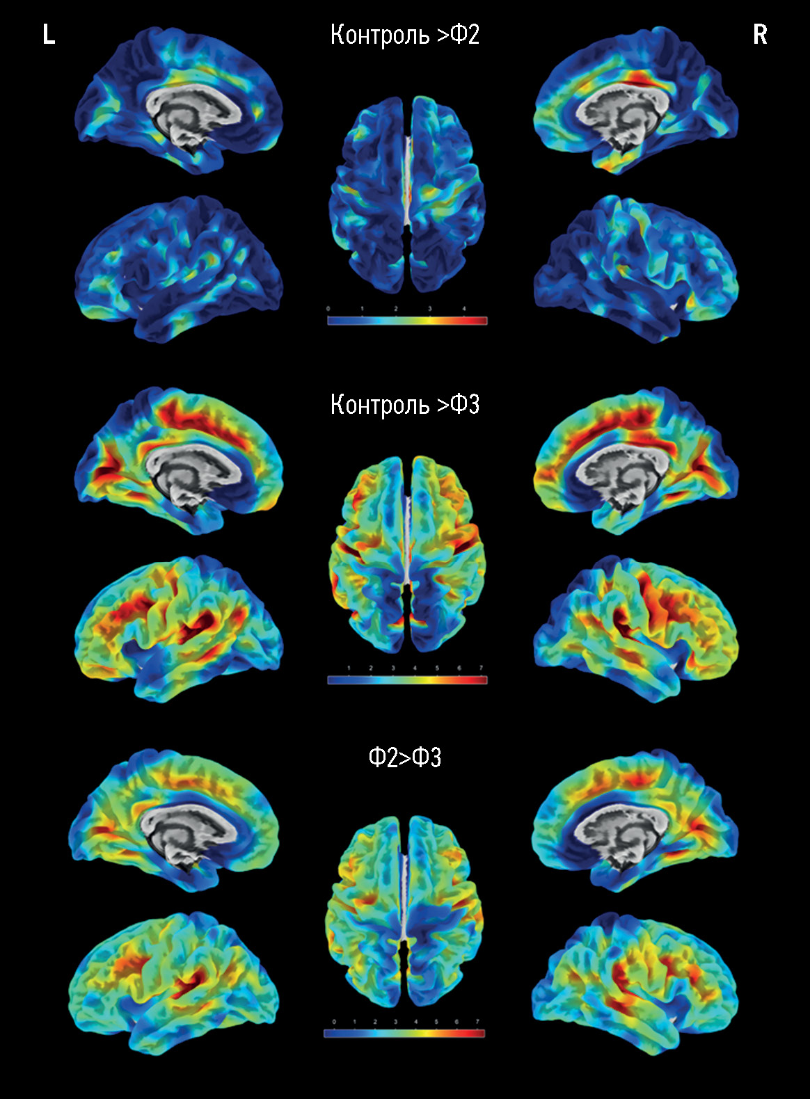
RESULTS: The main group consisted of 173 patients with cerebral small vessel disease, whereas the control group included 47 healthy volunteers. As the severity of brain structural changes and cognitive impairments increased, a significant (p <0.05) decrease in the cortical thickness of certain regions following a similar pattern was reported, particularly in the cingulate gyri, mainly their posterior sections; medial and middle sections of the frontal lobes, various areas of the insular cortex, and temporoparietal areas, particularly the supramarginal gyri. The brain volumes (overall, gray matter, and white matter volumes) in cerebral small vessel disease were significantly different only in controls but not between patients with cognitive impairment of different severity levels. The hyperintense white matter volume was significantly different between patients with dementia and moderate cognitive impairment, dementia, and subjective cognitive impairment (p <0.0001).
CONCLUSIONS: The results confirm secondary/mixed atrophy in cerebral small vessel disease. The clarification of the severity level of cognitive impairment in cerebral small vessel disease based on atrophy data is limited by the wide variety of regions with significant cortical thinning. Thus, the quantification of the cortex can only be a supplementary method in predicting cerebral small vessel disease progression.
Full Text
##article.viewOnOriginalSite##About the authors
Elena I. Kremneva
Research Center of Neurology
Author for correspondence.
Email: kremneva@neurology.ru
ORCID iD: 0000-0001-9396-6063
SPIN-code: 8799-8092
MD, Dr. Sci. (Medicine)
Russian Federation, MoscowLarisa A. Dobrynina
Research Center of Neurology
Email: dobrla@mail.ru
ORCID iD: 0000-0001-9929-2725
SPIN-code: 2824-8750
MD, Dr. Sci. (Medicine), Assistant Professor
Russian Federation, MoscowKamila V. Shamtieva
Research Center of Neurology
Email: kamila.shamt@gmail.com
ORCID iD: 0000-0002-6995-1352
SPIN-code: 5645-8768
MD, Cand. Sci. (Medicine)
Russian Federation, MoscowVictoria V. Trubitsyna
Research Center of Neurology
Email: pobeda-1994@mail.ru
ORCID iD: 0000-0001-7898-6541
Russian Federation, Moscow
Zukhra S. Gadzhieva
Research Center of Neurology
Email: zuhradoc@mail.ru
ORCID iD: 0000-0001-7498-4063
SPIN-code: 7015-5970
MD, Cand. Sci. (Medicine)
Russian Federation, MoscowAngelina G. Makarova
Research Center of Neurology
Email: angelinagm@mail.ru
ORCID iD: 0000-0001-8862-654X
MD, Cand. Sci. (Medicine)
Russian Federation, MoscowMaria M. Tsypushtanova
Research Center of Neurology
Email: tzipushtanova@mail.ru
ORCID iD: 0000-0002-4231-3895
MD, Cand. Sci. (Medicine)
Russian Federation, MoscowMarina V. Krotenkova
Research Center of Neurology
Email: krotenkova_mrt@mail.ru
ORCID iD: 0000-0003-3820-4554
SPIN-code: 9663-8828
MD, Dr. Sci. (Medicine), Assistant Professor
Russian Federation, MoscowReferences
- Pantoni L, Gorelick PB. Cerebral small vessel disease. Cambridge University Press; 2014.
- Gorelick PB, Scuteri A, Black SE, et al. Vascular contributions to cognitive impairment and dementia: A statement for healthcare professionals from the American Heart Association/American Stroke Association. Stroke. 2011;42(9):2672–2713. EDN: PIHATP doi: 10.1161/STR.0b013e3182299496
- Duering M, Biessels GJ, Brodtmann A, et al. Neuroimaging standards for research into small vessel disease-advances since 2013. Lancet Neurol. 2023;22(7):602–618. EDN: AEXCGU doi: 10.1016/S1474-4422(23)00131-X
- Jagust WJ, Zheng L, Harvey DJ, et al. Neuropathological basis of magnetic resonance images in aging and dementia. Ann Neurol. 2008;63(1):72–80. doi: 10.1002/ana.21296
- Godin O, Maillard P, Crivello F, et al. Association of white-matter lesions with brain atrophy markers: The three-city Dijon MRI study. Cerebrovascular Dis. 2009;28(2):177–184. doi: 10.1159/000226117
- Peres R, De Guio F, Chabriat H, et al. Alterations of the cerebral cortex in sporadic small vessel disease: A systematic review of in vivo MRI data. J Cerebral Blood Flow Metabolism. 2016;36(4):681–695. doi: 10.1177/0271678X15625352
- Tuladhar AM, van Norden AG, de Laat KF, et al. White matter integrity in small vessel disease is related to cognition. NeuroImage Clin. 2015;7:518–524. doi: 10.1016/j.nicl.2015.02.003
- Bethlehem RA, Seidlitz J, White SR, et al. Brain charts for the human lifespan. Nature. 2022;604(7906):525–533. EDN: BEAJBV doi: 10.1038/s41586-022-04554-y
- Ashburner J, Friston KJ. Voxel-based morphometry: the methods. Neuroimage. 2000;11(6):805–821. doi: 10.1006/nimg.2000.0582
- Dale AM, Fischl B, Sereno MI. Cortical surface-based analysis. I. Segmentation and surface reconstruction. Neuroimage. 1999;9(2):179–194. doi: 10.1006/nimg.1998.0395
- Goto M, Abe O, Hagiwara A, et al. Advantages of using both voxel-and surface-based morphometry in cortical morphology analysis: A review of various applications. Magnetic Res Med Sci. 2022;21(1):41–57. EDN: VWVUEU doi: 10.2463/mrms.rev.2021-0096
- Damulina AI, Konovalov RN, Kadykov AS. Significance of voxel-based morphometry in studying mild cognitive impairments. Ann Clin Experimental Neurol. 2015;9(3):42–48. EDN: VKPNWD
- Ozzoude M, Ramirez J, Raamana PR, et al. Cortical thickness estimation in individuals with cerebral small vessel disease, focal atrophy, and chronic stroke lesions. Front Neurosci. 2020;14:598868. EDN: QRNNGX doi: 10.3389/fnins.2020.598868
- Mo Y, Huang L, Qin R, et al. Decreased cortical thickness and normal regional homogeneity underlying cognitive impairment in cerebral small vessel disease. Adv Neuro. 2022;1(1):48. doi: 10.36922/an.v1i1.48
- Bookstein FL. “Voxel-based morphometry” should not be used with imperfectly registered images. Neuroimage. 2001;14(6):1454–1462. doi: 10.1006/nimg.2001.0770
- Staals J, Booth T, Morris Z, et al. Total MRI load of cerebral small vessel disease and cognitive ability in older people. Neurobiol Aging. 2015;36(10):2806–2811. doi: 10.1016/j.neurobiolaging.2015.06.024
- Gaser C, Dahnke R, Kurth K, et al. CAT-a computational anatomy toolbox for the analysis of structural MRI data. BioRxiv. 2022. doi: 10.1101/2022.06.11.495736
- Desikan RS, Ségonne F, Fischl B, et al. An automated labeling system for subdividing the human cerebral cortex on MRI scans into gyral based regions of interest. Neuroimage. 2006;31(3): 968–980. doi: 10.1016/j.neuroimage.2006.01.021
- Smith EE, Beaudin AE. New insights into cerebral small vessel disease and vascular cognitive impairment from MRI. Curr Opinion Neurol. 2018;31(1):36–43. doi: 10.1097/WCO.0000000000000513
- Dobrynina LA, Gadzhieva ZSh, Kremneva EI, et al. Survival, cognitive functions, and brain MRI in patients with cSVD: 5-year observation. Ann Clin Exp Neurol. 2022;16(4):18–28. EDN: EZEIVD doi: 10.54101/ACEN.2022.4.3
- Kremneva EI, Maximov II, Dobrynina LA, Krotenkova MV. The assessment of cerebral white matter microstructure in cerebral small vessel disease based on the diffusion-weighted magnetic resonance imaging. Ann Clin Exp Neurol. 2020;14(1):33–43. EDN: RXKAYI doi: 10.25692/ACEN.2020.1.4
- Gasquoine PG. Localization of function in anterior cingulate cortex: From psychosurgery to functional neuroimaging. Neurosci Biobehavioral Rev. 2013;37(3):340–348. doi: 10.1016/j.neubiorev.2013.01.002
- Dobrynina LA, Gadzhieva ZS, Morozova SN, et al. Executive functions: FMRI of healthy volunteers during stroop test and the serial count test. S.S Korsakov J Neurol Psychiatry. 2018;118(11):64–71. EDN: YRLKTZ doi: 10.17116/jnevro201811811164
- Sergeeva AN, Seliverstova EV, Dobrynina LA, et al. Pulsed arterial spin labeling (pasl) in receiving perfusion and functional data: Technique abilities. Russ Electronic J Radiol. 2019;9(1):148–159. EDN: ZJQHTK doi: 10.21569/2222-7415-2019-9-1-148-159
- Liu R, Wu W, Ye Q, et al. Distinctive and pervasive alterations of functional brain networks in cerebral small vessel disease with and without cognitive impairment. Dementia Geriatric Cognitive Disorders. 2019;47(1-2):55–67. doi: 10.1159/000496455
- Aribisala BS, Hernandez MC, Royle NA, et al. Brain atrophy associations with white matter lesions in the ageing brain: The Lothian birth cohort 1936. Eur Radiol. 2013;23(4):1084–1092. EDN: UYIACD doi: 10.1007/s00330-012-2677-x
- LADIS Study Group. 2001–2011: A decade of the LADIS (Leukoaraiosis and DISability) Study: What have we learned about white matter changes and small-vessel disease? Cerebrovascular Dis. 2011;32(6):577–588. doi: 10.1159/000334498
- Lawrence AJ, Patel B, Morris RG, et al. Mechanisms of cognitive impairment in cerebral small vessel disease: Multimodal MRI results from the St George’s cognition and neuroimaging in stroke (SCANS) study. PloS One. 2013;8(4):e61014. doi: 10.1371/journal.pone.0061014
- Kremneva EI. Age-dependent cerebral microangiopathy: MRI equivalents of cognitive disorders, severity and mechanisms of progression [dissertation]: 3.1.24; 3.1.25. Place of protection: Scientific Centre of Neurology. Moscow; 2023. (In Russ.) Available from: https://neurology.ru/upload/medialibrary/e7e/kas58vctp2as9tqb8pobkp30jztwpbrt/Kremneva-Elena-Igorevna-_-dissertatsiya.pdf. Accessed: 21.04.2024.
- Struyfs H, Sima DM, Wittens M. Automated MRI volumetry as a diagnostic tool for Alzheimer’s disease: Validation of icobrain dm. Neuroimage Clin. 2020;26:102243. doi: 10.1016/j.nicl.2020.102243
- Wonderlick JS, Ziegler DA, Hosseini-Varnamkhasti P, et al. Reliability of MRI-derived cortical and subcortical morphometric measures: Effects of pulse sequence, voxel geometry, and parallel imaging. Neuroimage. 2009;44(4):1324–1333. doi: 10.1016/j.neuroimage.2008.10.037
Supplementary files


