

")


类风湿性关节炎患者肩关节磁共振成像上的米粒体症状
- 作者: Ageeva S.F.1, Filatova D.A.1, Mershina E.A.1, Sinitsyn V.Е.1
-
隶属关系:
- Lomonosov Moscow State University
- 期: 卷 4, 编号 4 (2023)
- 页面: 616-624
- 栏目: 临床病例及临床病例的系列
- URL: https://journals.rcsi.science/DD/article/view/262986
- DOI: https://doi.org/10.17816/DD508786
- ID: 262986
如何引用文章

详细
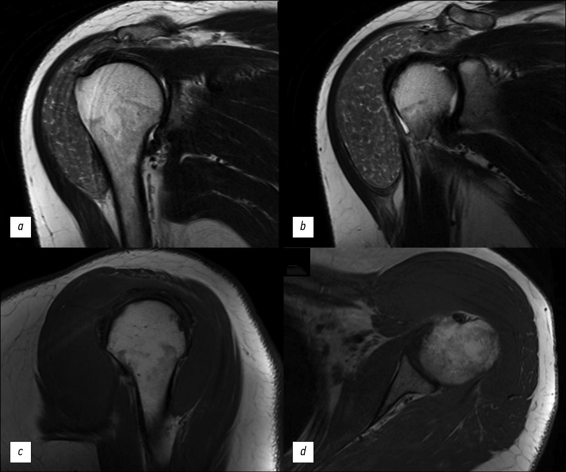
类风湿性关节炎患者肩关节磁共振成像(MRI)上的米粒体症状是一种罕见但特殊的发现。其特征是关节滑液、滑膜囊或滑膜鞘中出现多个圆形小结构。这些结构彼此相似,就像米粒一样。米粒体的成因至今不明。据推测,米粒体是因类风湿性关节炎或其他炎症性关节疾病患者滑膜微梗塞而形成的。在临床上,米粒体的存在可能会使患者感到疼痛。然而,情况并非总是如此。在放射诊断方法中,磁共振成像在检测米粒体方面发挥着主导作用。
本文介绍一例在肩关节磁共振成像中发现该症状的临床病例。该症状是在一名有长期类风湿性关节炎病史的患者身上发现的。患者因左肩部无痛性增大而就医。对左肩关节进行了电子计算机断层扫描(CT)和磁共振成像检查。医生通过这些检查发现了,米粒体症状是基础疾病的一种表现形式。这些检查有助于确定进一步的治疗策略。
作者简介
Sofia F. Ageeva
Lomonosov Moscow State University
编辑信件的主要联系方式.
Email: son.ageeva13@gmail.com
ORCID iD: 0000-0003-4726-0806
SPIN 代码: 9695-3717
俄罗斯联邦, Moscow
Daria A. Filatova
Lomonosov Moscow State University
Email: dariafilatova.msu@mail.ru
ORCID iD: 0000-0002-0894-1994
SPIN 代码: 2665-5973
俄罗斯联邦, Moscow
Elena A. Mershina
Lomonosov Moscow State University
Email: elena_mershina@mail.ru
ORCID iD: 0000-0002-1266-4926
SPIN 代码: 6897-9641
MD, Cand. Sci. (Med.), Аssistant professor
俄罗斯联邦, MoscowValentin Е. Sinitsyn
Lomonosov Moscow State University
Email: vsini@mail.ru
ORCID iD: 0000-0002-5649-2193
SPIN 代码: 8449-6590
MD, Dr. Sci. (Med.), Professor
俄罗斯联邦, Moscow参考
- Nasonov EL, Karateev DE, Balabanova RM. Rheumatoid arthritis. In: Nasonov EL, Nasonova VA, editors. Rheumatology. National manual. Moscow : GEOTAR-Media. 2008. P. 290–331 (In Russ)
- Rheumatoid arthritis. Clinical Guidelines. ID 250. Approved by the Scientific and Practical Council of the Ministry of Health of the Russian Federation. 2021. Available from: https://cr.minzdrav.gov.ru/schema/250 (In Russ)
- Bullock J, Rizvi SA, Saleh AM, et al. Rheumatoid Arthritis: A Brief Overview of the Treatment. Medical Principles and Practice. 2018;27(6):501–507. doi: 10.1159/000493390
- who.int [Internet]. World Health Organization [cited 6 September 2023]. Available from: https://www.who.int
- Kay J, Upchurch KS. ACR/EULAR 2010 rheumatoid arthritis classification criteria. Rheumatology. 2012;51 Suppl. 6:vi5–vi9. doi: 10.1093/rheumatology/kes279
- Narvaez JA, Narváez J, De Lama E, et al. MR Imaging of Early Rheumatoid Arthritis. RadioGraphics. 2010;30(1):143–163. doi: 10.1148/rg.301095089
- Edison MN, Caram A, Flores M, et al. Rice Body Formation Within a Peri-Articular Shoulder Mass. Cureus. 2016;8(8):e718. doi: 10.7759/cureus.718
- Forse CL, Mucha BL, Santos MLZ., et al. Rice body formation without rheumatic disease or tuberculosis infection: a case report and literature review. Clinical Rheumatology. 2012;31(12):1753–1756. doi: 10.1007/s10067-012-2063-8
- Narváez JA, Narváez J, Roca Y, et al. MR imaging assessment of clinical problems in rheumatoid arthritis. European Radiology. 2002;12(7):1819–1828. doi: 10.1007/s00330-001-1207-z
- Griffith JF, Peh WCG, Evans NS, et al. Multiple rice body formation in chronic subacromial/subdeltoid bursitis: MR appearances. Clinical Radiology. 1996;51(7):511–514. doi: 10.1016/s0009-9260(96)80193-0
- Kataria RK, Chaiamnuay S, Jacobson LD, et al. Subacromial bursitis with rice bodies as the presenting manifestation of rheumatoid arthritis. The Journal of rheumatology. 2003;30(6):1354–1355.
- Popert AJ, Scott DL, Wainwright AC, et al. Frequency of occurrence, mode of development, and significance or rice bodies in rheumatoid joints. Annals of the Rheumatic Diseases. 1982;41(2):109–117. doi: 10.1136/ard.41.2.109
- Reid HS, McNally E, Carr A. Soft tissue mass around the shoulder. Annals of the Rheumatic Diseases. 1998;57(1):6–8. doi: 10.1136/ard.57.1.6
补充文件


