

")


“Rice bodies” symptoms on magnetic resonance imaging of the shoulder in a patient with rheumatoid arthritis
- Авторлар: Ageeva S.F.1, Filatova D.A.1, Mershina E.A.1, Sinitsyn V.E.1
-
Мекемелер:
- Lomonosov Moscow State University
- Шығарылым: Том 4, № 4 (2023)
- Беттер: 616-624
- Бөлім: Case reports
- URL: https://journals.rcsi.science/DD/article/view/262986
- DOI: https://doi.org/10.17816/DD508786
- ID: 262986
Дәйексөз келтіру

Аннотация
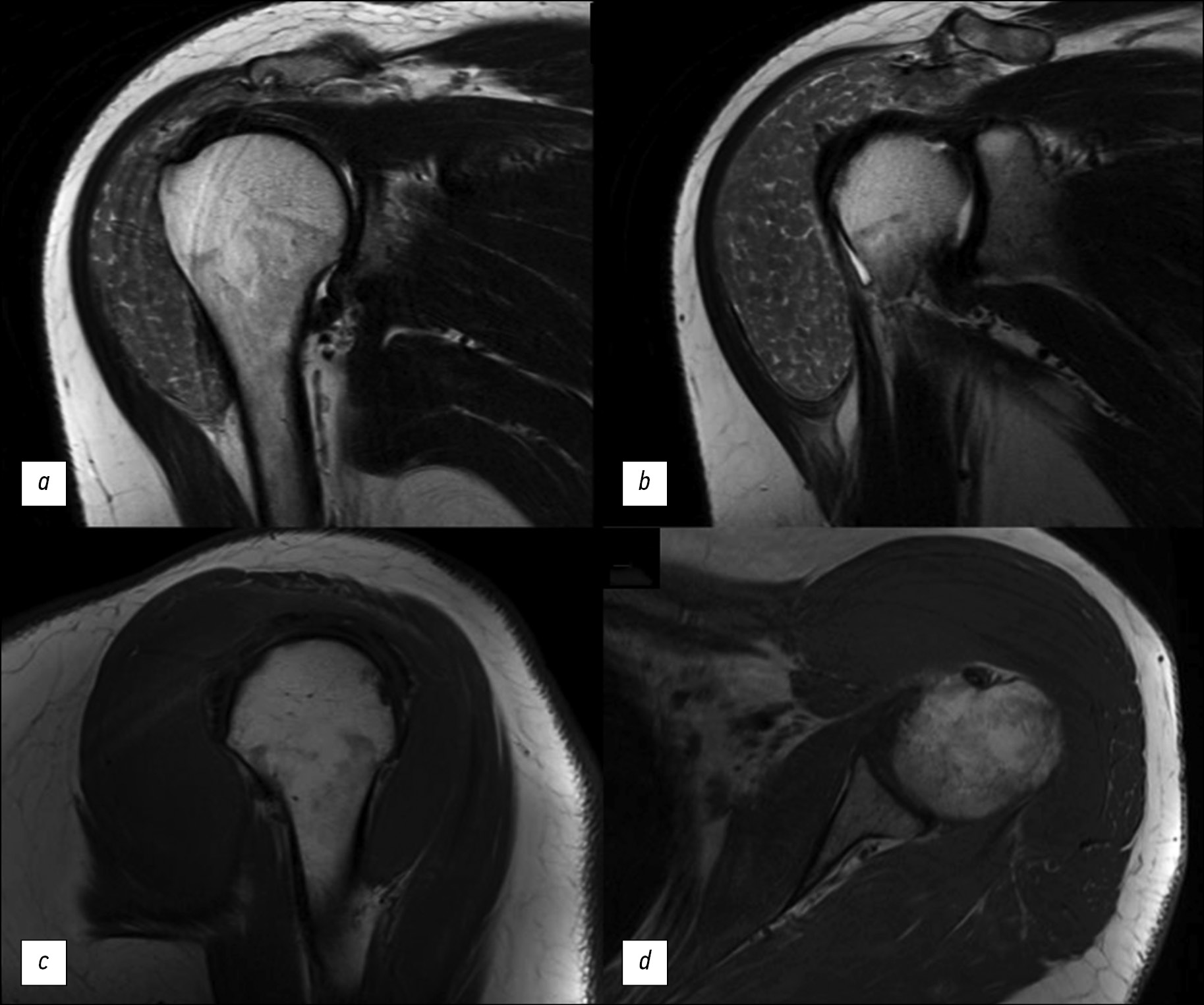
The “rice bodies” symptom on magnetic resonance imaging of the shoulder joint in patients with rheumatoid arthritis is a rare but specific finding characterized by the presence of multiple small, round, rice-grain-like structures in the synovial fluid of the joint, synovial pouches, or sheaths. The etiology of the “rice bodies” is still not fully understood. They are suggested as the result of microinfarcts of the synovial membrane in patients with rheumatoid arthritis or other inflammatory joint diseases. Clinically, the “rice bodies” symptom may cause pain, but not in every case. Among radiological diagnostic methods, magnetic resonance imaging is the leading method for the detection of rice bodies. This article presents a clinical case of “rice bodies” symptoms diagnosed by magnetic resonance imaging in a patient with a long history of rheumatoid arthritis who presented with a painless enlargement in the left shoulder. Computed tomography and magnetic resonance imaging of the left shoulder could detect “rice bodies” as a manifestation of an underlying disease and determine further treatment techniques.
Толық мәтін
##article.viewOnOriginalSite##Авторлар туралы
Sofia Ageeva
Lomonosov Moscow State University
Хат алмасуға жауапты Автор.
Email: son.ageeva13@gmail.com
ORCID iD: 0000-0003-4726-0806
SPIN-код: 9695-3717
Ресей, Moscow
Daria Filatova
Lomonosov Moscow State University
Email: dariafilatova.msu@mail.ru
ORCID iD: 0000-0002-0894-1994
SPIN-код: 2665-5973
Ресей, Moscow
Elena Mershina
Lomonosov Moscow State University
Email: elena_mershina@mail.ru
ORCID iD: 0000-0002-1266-4926
SPIN-код: 6897-9641
MD, Cand. Sci. (Med.), Аssistant professor
Ресей, MoscowValentin Sinitsyn
Lomonosov Moscow State University
Email: vsini@mail.ru
ORCID iD: 0000-0002-5649-2193
SPIN-код: 8449-6590
MD, Dr. Sci. (Med.), Professor
Ресей, MoscowӘдебиет тізімі
- Nasonov EL, Karateev DE, Balabanova RM. Rheumatoid arthritis. In: Nasonov EL, Nasonova VA, editors. Rheumatology. National manual. Moscow : GEOTAR-Media. 2008. P. 290–331 (In Russ)
- Rheumatoid arthritis. Clinical Guidelines. ID 250. Approved by the Scientific and Practical Council of the Ministry of Health of the Russian Federation. 2021. Available from: https://cr.minzdrav.gov.ru/schema/250 (In Russ)
- Bullock J, Rizvi SA, Saleh AM, et al. Rheumatoid Arthritis: A Brief Overview of the Treatment. Medical Principles and Practice. 2018;27(6):501–507. doi: 10.1159/000493390
- who.int [Internet]. World Health Organization [cited 6 September 2023]. Available from: https://www.who.int
- Kay J, Upchurch KS. ACR/EULAR 2010 rheumatoid arthritis classification criteria. Rheumatology. 2012;51 Suppl. 6:vi5–vi9. doi: 10.1093/rheumatology/kes279
- Narvaez JA, Narváez J, De Lama E, et al. MR Imaging of Early Rheumatoid Arthritis. RadioGraphics. 2010;30(1):143–163. doi: 10.1148/rg.301095089
- Edison MN, Caram A, Flores M, et al. Rice Body Formation Within a Peri-Articular Shoulder Mass. Cureus. 2016;8(8):e718. doi: 10.7759/cureus.718
- Forse CL, Mucha BL, Santos MLZ., et al. Rice body formation without rheumatic disease or tuberculosis infection: a case report and literature review. Clinical Rheumatology. 2012;31(12):1753–1756. doi: 10.1007/s10067-012-2063-8
- Narváez JA, Narváez J, Roca Y, et al. MR imaging assessment of clinical problems in rheumatoid arthritis. European Radiology. 2002;12(7):1819–1828. doi: 10.1007/s00330-001-1207-z
- Griffith JF, Peh WCG, Evans NS, et al. Multiple rice body formation in chronic subacromial/subdeltoid bursitis: MR appearances. Clinical Radiology. 1996;51(7):511–514. doi: 10.1016/s0009-9260(96)80193-0
- Kataria RK, Chaiamnuay S, Jacobson LD, et al. Subacromial bursitis with rice bodies as the presenting manifestation of rheumatoid arthritis. The Journal of rheumatology. 2003;30(6):1354–1355.
- Popert AJ, Scott DL, Wainwright AC, et al. Frequency of occurrence, mode of development, and significance or rice bodies in rheumatoid joints. Annals of the Rheumatic Diseases. 1982;41(2):109–117. doi: 10.1136/ard.41.2.109
- Reid HS, McNally E, Carr A. Soft tissue mass around the shoulder. Annals of the Rheumatic Diseases. 1998;57(1):6–8. doi: 10.1136/ard.57.1.6
Қосымша файлдар


