

")


COVID-19患者的肺部动态磁共振成像: 一系列临床病例
- 作者: Vasilev Y.A.1, Grik E.A.2, Panina O.Y.1,3,4, Khoruzhaya A.N.1, Semenov D.S.1, Bazhin A.V.1, Vasileva Y.N.4
-
隶属关系:
- Moscow Center for Diagnostics and Telemedicine
- Lincoln Medical Center
- City Clinical Oncological Hospital 1 of the Department of healthcare of the city of Moscow
- Moscow State University of Medicine and Dentistry named after A.I. Evdokimov
- 期: 卷 4, 编号 1 (2023)
- 页面: 71-79
- 栏目: 临床病例及临床病例的系列
- URL: https://journals.rcsi.science/DD/article/view/146877
- DOI: https://doi.org/10.17816/DD114723
- ID: 146877
如何引用文章

详细
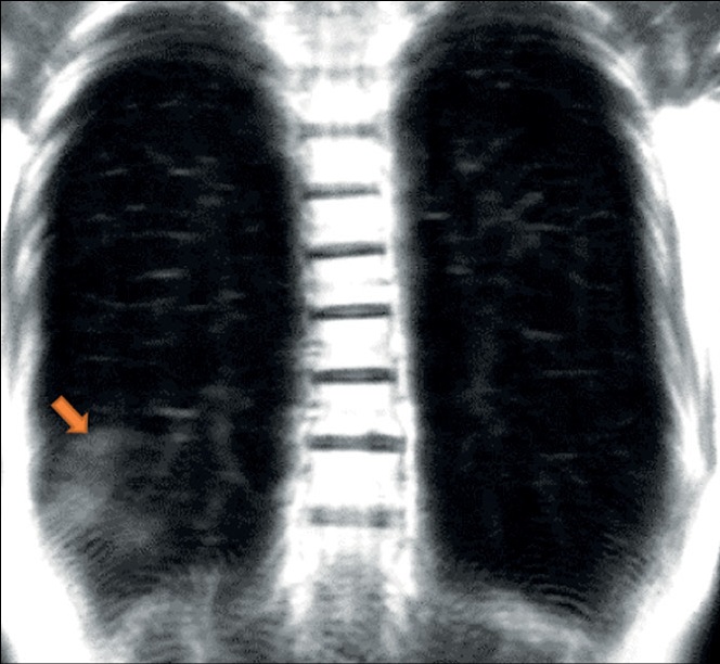
一场新冠状病毒感染(COVID-19)的广泛流行导致了对其诊断特征的积极性研究。与COVID-19相关的急性病毒性肺炎是已经在计算机断层扫描(CT)、辐射成像和静态磁共振成像(MRI)的研究中详细描述的。然而,文献中有很少关于通过动态MRI观察到的描述性图片的数据。鉴于综合诊断方法,放射科医生知道如何通过MRI正确识别和解释COVID-19是很重要的。在这一系列临床病例中,我们展示了动态MRI工作方法的威力,使我们能够看到“cloudy sky”的迹象,并将其与COVID-19患者的固结区分开来,使我们能够推测区分早期或轻微的变化和渐进的临床过程。因此,MRI作为一种无辐射的工具,在进行CT扫描的机会有限和需要动态形态功能成像的情况下是非常有用的。
作者简介
Yuriy A. Vasilev
Moscow Center for Diagnostics and Telemedicine
Email: y.vasilev@npcmr.ru
ORCID iD: 0000-0002-0208-5218
SPIN 代码: 4458-5608
MD, Cand. Sci. (Med.)
俄罗斯联邦, MoscowEvgeniia A. Grik
Lincoln Medical Center
Email: evgeniyagrik@gmail.com
ORCID iD: 0000-0002-7908-3982
SPIN 代码: 5558-7307
美国, The Bronx, NY
Olga Yu. Panina
Moscow Center for Diagnostics and Telemedicine; City Clinical Oncological Hospital 1 of the Department of healthcare of the city of Moscow; Moscow State University of Medicine and Dentistry named after A.I. Evdokimov
Email: o.panina@npcmr.ru
ORCID iD: 0000-0002-8684-775X
SPIN 代码: 5504-8136
俄罗斯联邦, Moscow; Moscow; Moscow
Anna N. Khoruzhaya
Moscow Center for Diagnostics and Telemedicine
编辑信件的主要联系方式.
Email: a.khoruzhaya@npcmr.ru
ORCID iD: 0000-0003-4857-5404
SPIN 代码: 7948-6427
俄罗斯联邦, Moscow
Dmitriy S. Semenov
Moscow Center for Diagnostics and Telemedicine
Email: d.semenov@npcmr.ru
ORCID iD: 0000-0002-4293-2514
SPIN 代码: 2278-7290
俄罗斯联邦, Moscow
Alexander V. Bazhin
Moscow Center for Diagnostics and Telemedicine
Email: a.bazhin@npcmr.ru
ORCID iD: 0000-0003-3198-1334
SPIN 代码: 6122-5786
俄罗斯联邦, Moscow
Yulia N. Vasileva
Moscow State University of Medicine and Dentistry named after A.I. Evdokimov
Email: drugya@yandex.ru
ORCID iD: 0000-0002-1066-3989
SPIN 代码: 9777-2067
MD, Cand. Sci. (Med.)
俄罗斯联邦, Moscow参考
- Shi H, Han X, Jiang N, et al. Radiological findings from 81 patients with COVID-19 pneumonia in Wuhan, China: A descriptive study. Lancet Infectious Dis. 2020;20(4):425–434. doi: 10.1016/S1473-3099(20)30086-4
- Salehi S, Abedi A, Balakrishnan S, Gholamrezanezhad A. Coronavirus Disease 2019 (COVID-19): A systematic review of imaging findings in 919 patients. Am J Roentgenol. 2020;215(1):87–93. doi: 10.2214/AJR.20.23034
- ACR Recommendations for the use of chest radiography and computed tomography (CT) for suspected COVID-19 infection [cited March 11, 2020]. Available from: https://www.acr.org/Advocacy-and-Economics/ACR-Position-Statements/Recommendations-for-Chest-Radiography-and-CT-for-Suspected-COVID19-Infection. Accessed: 15.01.2023.
- Temporary guidelines “Prevention, diagnosis and treatment of new coronavirus infection (COVID-19)”. Version 16 (08/18/2022). Moscow; 2022. 249 p. (In Russ).
- Vasilev YA, Sergunova KA, Bazhin AV, et al. Chest MRI of patients with COVID-19. Magn Reson Imaging. 2021;(79):13–19. doi: 10.1016/j.mri.2021.03.005
- Vasilev YuA, Sergunova KA, BazhinАV, et al. Chest MRI of a pregnant woman with COVID-19 pneumonia. Digital Diagnostics. 2020;1(1):61−68. doi: 10.17816/DD46800
- Vasiliev YuA, Panina OYu, Kudryavtsev ND, et al. Magnetic resonance imaging of the lungs: methodological recommendations. Issue 92. Moscow; 2022. 102 р. (Series «The best practices of radiation and instrumental diagnostics»). (In Russ).
- Dong D, Tang Z, Wang S, et al. The role of imaging in the detection and management of COVID-19: A review. IEEE Rev Biomed Eng. 2021;(14):16–29. doi: 10.1109/RBME.2020.2990959
- Fields BK, Demirjian NL, Dadgar H, Gholamrezanezhad A. Imaging of COVID-19: CT, MRI, and PET. Semin Nucl Med. 2021;51(4):312–320. doi: 10.1053/j.semnuclmed.2020.11.003
- Langenbach MC, Hokamp GN, Persigehl T, Bratke G. MRI appearance of COVID-19 infection. Diagn Interv Radiol. 2020;26(4):377–378. doi: 10.5152/dir.2020.20152
- Fonseca EK, Chate RC, Neto RS, et al. Findings of COVID-19 on magnetic resonance imaging. Radiology Cardiothoracic Imaging. 2020;2(2):e200193. doi: 10.1148/ryct.2020200193
- Torkian P, Rajebi H, Zamani T, et al. Magnetic resonance imaging features of coronavirus disease 2019 (COVID-19) pneumonia: The first preliminary case series. Clin Imaging. 2021;(69):261–265. doi: 10.1016/j.clinimag.2020.09.002
- Ates OF, Taydas O, Dheir H. Thorax magnetic resonance imaging findings in patients with coronavirus disease (COVID-19). Acad Radiol. 2020;27(10):1373–1378. doi: 10.1016/j.acra.2020.08.009
- Ussov WY, Nudnov NV, Ignatenko G, et al. Primary and prospective imaging of the chest using magnetic resonance imaging in patients with viral lung damage in COVID-19. Med Visualizat. 2020;24(4):11–26. (In Russ). doi: 10.24835/1607-0763-2020-4-11-26
补充文件


