

")


Dynamic MRI in a COVID-19 patient: a case series
- Авторлар: Vasilev Y.A.1, Grik E.A.2, Panina O.Y.1,3,4, Khoruzhaya A.N.1, Semenov D.S.1, Bazhin A.V.1, Vasileva Y.N.4
-
Мекемелер:
- Moscow Center for Diagnostics and Telemedicine
- Lincoln Medical Center
- City Clinical Oncological Hospital 1 of the Department of healthcare of the city of Moscow
- Moscow State University of Medicine and Dentistry named after A.I. Evdokimov
- Шығарылым: Том 4, № 1 (2023)
- Беттер: 71-79
- Бөлім: Case reports
- URL: https://journals.rcsi.science/DD/article/view/146877
- DOI: https://doi.org/10.17816/DD114723
- ID: 146877
Дәйексөз келтіру

Аннотация
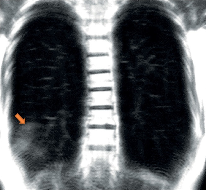
Extensive spread of the coronavirus disease (COVID-19) prompted an investigation of its diagnostic features. Acute viral pneumonia associated with COVID-19 has been described in detail using CT, radiography, and MRI. There is no data in the literature on the descriptive picture observed with dynamic MRI. Considering a comprehensive diagnostic approach, radiologists should know how to correctly recognize and interpret COVID-19 on MRI. This case series demonstrated the ability of dynamic MRI to detect the cloudy sky sign and distinguish it from consolidation in COVID-19 patients, thus presumably distinguishing between early or mild changes and a progressive clinical course. These changes in dynamic lung images on MRI can be recorded depending on the phase of the respiratory cycle. Thus, MRI, as a radiation-free tool that can be used to examine a patient with acute viral pneumonia COVID-19, can be useful in cases where access to computed tomography is limited and dynamic morphofunctional imaging is required.
Негізгі сөздер
Толық мәтін
##article.viewOnOriginalSite##Авторлар туралы
Yuriy Vasilev
Moscow Center for Diagnostics and Telemedicine
Email: y.vasilev@npcmr.ru
ORCID iD: 0000-0002-0208-5218
SPIN-код: 4458-5608
MD, Cand. Sci. (Med.)
Ресей, MoscowEvgeniia Grik
Lincoln Medical Center
Email: evgeniyagrik@gmail.com
ORCID iD: 0000-0002-7908-3982
SPIN-код: 5558-7307
АҚШ, The Bronx, NY
Olga Panina
Moscow Center for Diagnostics and Telemedicine; City Clinical Oncological Hospital 1 of the Department of healthcare of the city of Moscow; Moscow State University of Medicine and Dentistry named after A.I. Evdokimov
Email: o.panina@npcmr.ru
ORCID iD: 0000-0002-8684-775X
SPIN-код: 5504-8136
Ресей, Moscow; Moscow; Moscow
Anna Khoruzhaya
Moscow Center for Diagnostics and Telemedicine
Хат алмасуға жауапты Автор.
Email: a.khoruzhaya@npcmr.ru
ORCID iD: 0000-0003-4857-5404
SPIN-код: 7948-6427
Ресей, Moscow
Dmitriy Semenov
Moscow Center for Diagnostics and Telemedicine
Email: d.semenov@npcmr.ru
ORCID iD: 0000-0002-4293-2514
SPIN-код: 2278-7290
Ресей, Moscow
Alexander Bazhin
Moscow Center for Diagnostics and Telemedicine
Email: a.bazhin@npcmr.ru
ORCID iD: 0000-0003-3198-1334
SPIN-код: 6122-5786
Ресей, Moscow
Yulia Vasileva
Moscow State University of Medicine and Dentistry named after A.I. Evdokimov
Email: drugya@yandex.ru
ORCID iD: 0000-0002-1066-3989
SPIN-код: 9777-2067
MD, Cand. Sci. (Med.)
Ресей, MoscowӘдебиет тізімі
- Shi H, Han X, Jiang N, et al. Radiological findings from 81 patients with COVID-19 pneumonia in Wuhan, China: A descriptive study. Lancet Infectious Dis. 2020;20(4):425–434. doi: 10.1016/S1473-3099(20)30086-4
- Salehi S, Abedi A, Balakrishnan S, Gholamrezanezhad A. Coronavirus Disease 2019 (COVID-19): A systematic review of imaging findings in 919 patients. Am J Roentgenol. 2020;215(1):87–93. doi: 10.2214/AJR.20.23034
- ACR Recommendations for the use of chest radiography and computed tomography (CT) for suspected COVID-19 infection [cited March 11, 2020]. Available from: https://www.acr.org/Advocacy-and-Economics/ACR-Position-Statements/Recommendations-for-Chest-Radiography-and-CT-for-Suspected-COVID19-Infection. Accessed: 15.01.2023.
- Temporary guidelines “Prevention, diagnosis and treatment of new coronavirus infection (COVID-19)”. Version 16 (08/18/2022). Moscow; 2022. 249 p. (In Russ).
- Vasilev YA, Sergunova KA, Bazhin AV, et al. Chest MRI of patients with COVID-19. Magn Reson Imaging. 2021;(79):13–19. doi: 10.1016/j.mri.2021.03.005
- Vasilev YuA, Sergunova KA, BazhinАV, et al. Chest MRI of a pregnant woman with COVID-19 pneumonia. Digital Diagnostics. 2020;1(1):61−68. doi: 10.17816/DD46800
- Vasiliev YuA, Panina OYu, Kudryavtsev ND, et al. Magnetic resonance imaging of the lungs: methodological recommendations. Issue 92. Moscow; 2022. 102 р. (Series «The best practices of radiation and instrumental diagnostics»). (In Russ).
- Dong D, Tang Z, Wang S, et al. The role of imaging in the detection and management of COVID-19: A review. IEEE Rev Biomed Eng. 2021;(14):16–29. doi: 10.1109/RBME.2020.2990959
- Fields BK, Demirjian NL, Dadgar H, Gholamrezanezhad A. Imaging of COVID-19: CT, MRI, and PET. Semin Nucl Med. 2021;51(4):312–320. doi: 10.1053/j.semnuclmed.2020.11.003
- Langenbach MC, Hokamp GN, Persigehl T, Bratke G. MRI appearance of COVID-19 infection. Diagn Interv Radiol. 2020;26(4):377–378. doi: 10.5152/dir.2020.20152
- Fonseca EK, Chate RC, Neto RS, et al. Findings of COVID-19 on magnetic resonance imaging. Radiology Cardiothoracic Imaging. 2020;2(2):e200193. doi: 10.1148/ryct.2020200193
- Torkian P, Rajebi H, Zamani T, et al. Magnetic resonance imaging features of coronavirus disease 2019 (COVID-19) pneumonia: The first preliminary case series. Clin Imaging. 2021;(69):261–265. doi: 10.1016/j.clinimag.2020.09.002
- Ates OF, Taydas O, Dheir H. Thorax magnetic resonance imaging findings in patients with coronavirus disease (COVID-19). Acad Radiol. 2020;27(10):1373–1378. doi: 10.1016/j.acra.2020.08.009
- Ussov WY, Nudnov NV, Ignatenko G, et al. Primary and prospective imaging of the chest using magnetic resonance imaging in patients with viral lung damage in COVID-19. Med Visualizat. 2020;24(4):11–26. (In Russ). doi: 10.24835/1607-0763-2020-4-11-26
Қосымша файлдар


