

")


Миксома створки митрального клапана
- Авторы: Вишнякова М.В.1, Абраменко А.С.1, Вишнякова М.В.1, Шумаков Д.В.1
-
Учреждения:
- Московский областной научно-исследовательский клинический институт имени М.Ф. Владимирского
- Выпуск: Том 3, № 1 (2022)
- Страницы: 64-70
- Раздел: Клинические случаи и серии клинических случаев
- URL: https://journals.rcsi.science/DD/article/view/100281
- DOI: https://doi.org/10.17816/DD100281
- ID: 100281
Цитировать

Аннотация
Первичные опухоли сердца являются крайне редким заболеванием, распространённость их в популяции, по разным данным, составляет 0,0017–0,03%.
В большинстве случаев опухоли сердца имеют доброкачественный характер, более половины подобных образований представлены миксомами сердца. Миксома, поражающая створки клапанов сердца, является редчайшей патологией. Впервые подобный вариант изменений был описан в 1934 году. Наиболее часто миксомы сердца локализуются на уровне межпредсердной перегородки в непосредственной близости от овальной ямки. Одним из типичных признаков миксом является узкая ножка и неровная поверхность, что обусловливает риск эмболии. Эхокардиографическое исследование и магнитно-резонансная томография на настоящий момент являются методами выбора при подозрении на объёмное образование сердца. При подобной нетипичной локализации опухоли обязательна дифференциальная диагностика с вегетациями на клапанах сердца и папиллярной фиброэластомой.
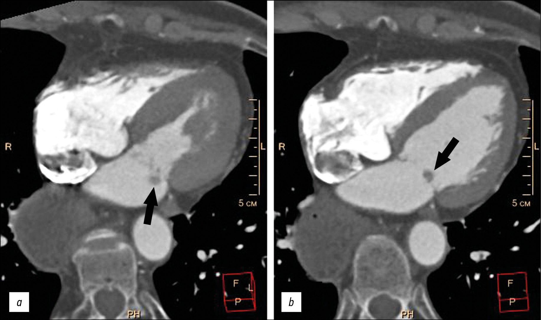
Представлен случай пожилой пациентки с жалобами на одышку, колющие боли в левой половине грудной клетки, аритмии, в анамнезе которой имелась аспирационная пневмония, экстирпация пищевода с эзофагогастропластикой желудка. При обследовании у пациентки выявлены пароксизмальная форма фибрилляции предсердий (вне пароксизма), хроническая сердечная недостаточность, артериальная гипертензия. Клинические данные больной были нехарактерны для инфекционного эндокардита с вегетациями на клапанах. Благодаря эхокардиографическому исследованию и мультиспиральной компьютерной томографии с болюсным контрастным усилением на атриальной поверхности задней створки митрального клапана обнаружено дополнительное объёмное образование размерами 5–9 мм, округ-лой формы, с чёткими неровными контурами, смещаемое вместе со створкой клапана в полость левого желудочка в систолу предсердий. Оптимальная визуализация образования получена в режиме Fiesta-CINE в модифицированных двух- и четырёхкамерных проекциях. Пациентке выполнено удаление образования с шовной пластикой митрального клапана в условиях искусственного кровообращения. При гистологическом исследовании образования получена характерная морфологическая картина миксомы. Послеоперационный период протекал без осложнений.
Ключевые слова
Полный текст
Открыть статью на сайте журналаОб авторах
Марина Валентиновна Вишнякова
Московский областной научно-исследовательский клинический институт имени М.Ф. Владимирского
Автор, ответственный за переписку.
Email: cherridra@mail.ru
ORCID iD: 0000-0003-3838-636X
SPIN-код: 1137-2991
доктор медицинских наук
Россия, 129110, Москва, ул. Щепкина, д. 61/2Александр Сергеевич Абраменко
Московский областной научно-исследовательский клинический институт имени М.Ф. Владимирского
Email: a.s.abramenko@gmail.com
ORCID iD: 0000-0002-6286-2162
SPIN-код: 9743-3001
Научный сотрудник отделения лучевой диагностики
Россия, 129110, Москва, ул. Щепкина, д. 61/2Мария Валентиновна Вишнякова
Московский областной научно-исследовательский клинический институт имени М.Ф. Владимирского
Email: cherridra@list.ru
ORCID iD: 0000-0002-2649-4198
SPIN-код: 7748-1831
доктор медицинских наук
Россия, 129110, Москва, ул. Щепкина, д. 61/2Дмитрий Валерьевич Шумаков
Московский областной научно-исследовательский клинический институт имени М.Ф. Владимирского
Email: cherridra@mail.ru
ORCID iD: 0000-0003-4204-8865
SPIN-код: 2545-2978
доктор медицинских наук, чл.-корр. РАН
Россия, 129110, Москва, ул. Щепкина, д. 61/2Список литературы
- Centofanti P., Di Rosa E., Deorsola L., et al. Primary cardiac tumors: early and late results of surgical treatment in 91 patients // Ann Thorac Surg. 1999. Vol. 68, N 4. Р. 1236–1241. doi: 10.1016/s0003-4975(99)00700-6
- Hoey E.T., Shahid M., Ganeshan A., et al. MRI assessment of cardiac tumours: part 1, multiparametric imaging protocols and spectrum of appearances of histologically benign lesions // Quant Imaging Med Surg. 2014. Vol. 4, N 6. Р. 478–488. doi: 10.3978/j.issn.2223-4292.2014.11.23
- Желтовский Ю.В., Батеха В.И., Подкаменный В.А., и др. Диагностика и лечение миксом сердца // Acta Biomedica Sci. 2017. Т. 2, № 6. С. 21–26.
- Lam K.Y., Dickens P., Chan A.C. Tumors of the heart. A 20-year experience with a review of 12,485 consecutive autopsies // Arch Pathol Lab Med. 1993. Vol. 117, N 10. Р. 1027–1031.
- Li X., Chen Y., Liu J., et al. Cardiac magnetic resonance imaging of primary cardiac tumors // Quant Imaging Med Surg. 2020. Vol. 10, N 1. Р. 294–313. doi: 10.21037/qims.2019.11.13
- Aggeli C., Dimitroglou Y., Raftopoulos L., et al. Cardiac masses: the role of cardiovascular imaging in the differential diagnosis // Diagnostics. 2020. Vol. 10, N 12. Р. 1088. doi: 10.3390/diagnostics10121088
- Kallstrom E., Kallus E., Erbe K., et al. Differentiation of left atrial myxomas by multimodality imaging // J Diagnostic Med Sonography. 2020. Vol. 36, N 1. Р. 52–63. doi: 10.1177/8756479319872153
- Jaleski T.C. Myxoma of the heart valves: report of a case // Am J Pathol. 1934. Vol. 10, N 3. Р. 399–406.
- Wold L.E., Lie J.T. Cardiac myxomas: a clinicopathologic profile // Am J Pathol. 1980. Vol. 101, N 1. Р. 219–240.
- Yoon J.H., Kim J.H., Sung Y.J., et al. Cardiac myxoma originating from the anterior mitral valve leaflet // J Cardiovasc Ultrasound. 2011. Vol. 19, N 4. Р. 228–231. doi: 10.4250/jcu.2011.19.4.228
- Rajiah P., Moore A., Saboo S., et al. Multimodality imaging of complications of cardiac valve surgeries // RadioGraphics. 2019. Vol. 39, N 4. Р. 932–956. doi: 10.1148/rg.2019180177
- El Ouazzani J., Jandou I., Christophe Thuaire I. Thrombus or vegetation? Importance of cardiac MRI as a diagnostic tool based on case report and literature review // Ann Med Surg (Lond). 2020. Vol. 60. Р. 690–694. doi: 10.1016/j.amsu.2020.12.007
- Anand S., Sydow N., Janardhanan R. Papillary fibroelastoma diagnosed through multimodality cardiac imaging: a rare tumour in an uncommon location with review of literature // BMJ Case Rep. 2017. Vol. 2017. Р. bcr2017219327. doi: 10.1136/bcr-2017-219327
Дополнительные файлы


