

")


Cardiac myxoma originating from mitral valve leaflet
- Авторлар: Vishniakova M.V.1, Abramenko A.S.1, Vishniakova M.V.1, Shumakov D.V.1
-
Мекемелер:
- Moscow Regional Research and Clinical Institute
- Шығарылым: Том 3, № 1 (2022)
- Беттер: 64-70
- Бөлім: Case reports
- URL: https://journals.rcsi.science/DD/article/view/100281
- DOI: https://doi.org/10.17816/DD100281
- ID: 100281
Дәйексөз келтіру

Аннотация
Primary heart tumors are an extremely rare disease, with a prevalence of 0.0017%–0.03% in the population according to various data.
Heart tumors are benign in most cases, and more than half of such formations are represented by cardiac myxomas. Myxoma is the most common primary cardiac tumor; however, its number is extremely small among the general population. Myxoma that affects the cardiac valves is a rare pathology. For the first time, such variance of changes was described in 1934. Most often, cardiac myxomas are localized at the atrial septum level near the oval fossa. One of the typical signs of myxoma is a narrow leg and an uneven surface, which causes the risk of embolism. Echocardiographic examination and magnetic resonance imaging are currently the methods of choice when suspecting the presence of volumetric heart formation. With such atypical tumor localization, conducting a mandatory differential diagnosis with heart valve vegetations and papillary fibroelastoma is necessary.
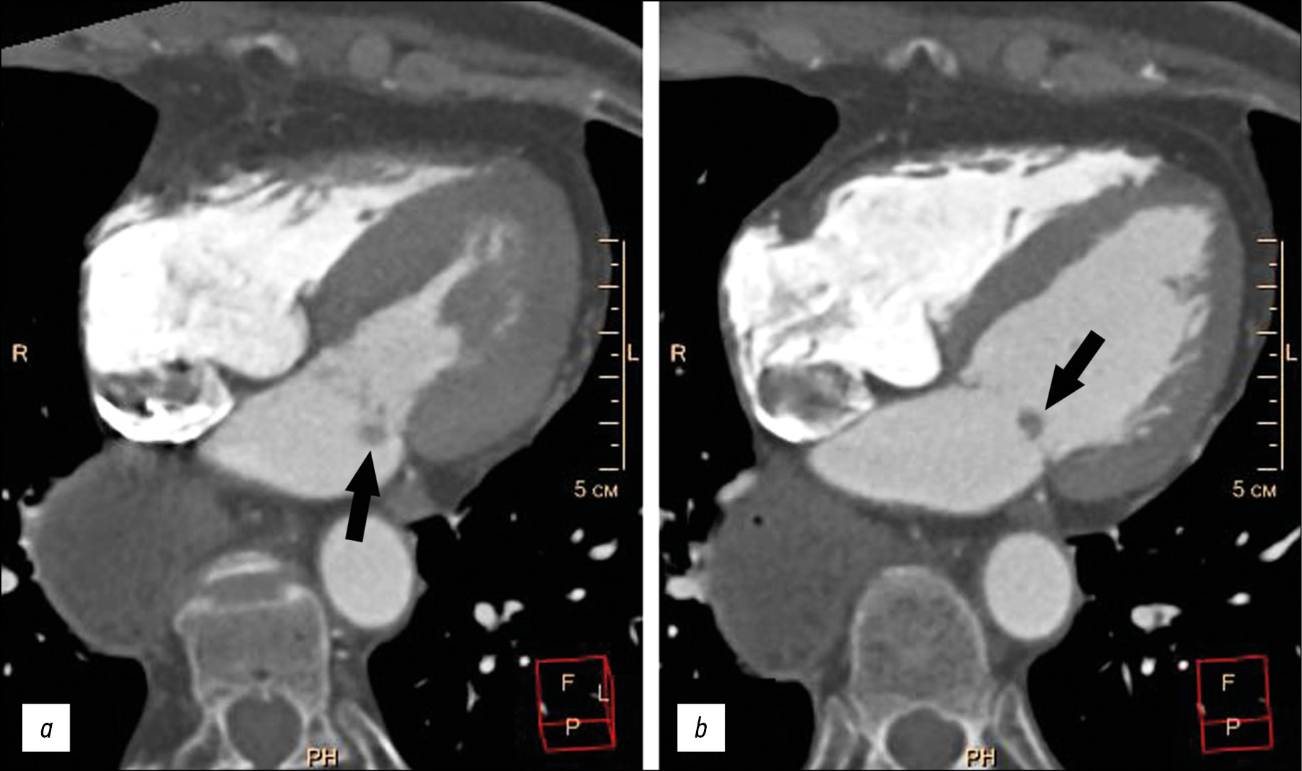
Herein, presented an elderly patient with complaints of shortness of breath, stabbing pains in the left half of the chest, and arrhythmias with a history of aspiration pneumonia and esophageal extirpation with stomach esophagogastroplasty. During the examination, the patient revealed a paroxysmal form of atrial fibrillation (outside of paroxysm), chronic heart failure, and arterial hypertension. The clinical data of the patient were not characteristic enough for the possibility of infectious endocarditis with valvular vegetations. The echocardiographic examination and multispiral computed tomography with bolus contrast enhancement on the atrial surface of the posterior flap of the mitral valve revealed an additional volume formation of 5–9 mm in size, rounded shape, with clear uneven contours, together with the valve flap into the left ventricular cavity into the atrial systole. The formation was optimally visualized using the Fiesta-CINE mode in modified two- and four-chamber projections. The formation was removed with suture plasty of the mitral valve in artificial blood circulation conditions. The histological examination of the formation revealed a morphological characteristic of myxoma. The postoperative period proceeded without complications.
Негізгі сөздер
Толық мәтін
##article.viewOnOriginalSite##Авторлар туралы
Marina Vishniakova
Moscow Regional Research and Clinical Institute
Хат алмасуға жауапты Автор.
Email: cherridra@mail.ru
ORCID iD: 0000-0003-3838-636X
SPIN-код: 1137-2991
MD, Dr. Sci. (Med)
Ресей, 61/2, Shepkina street, Moscow,129110Alexander Abramenko
Moscow Regional Research and Clinical Institute
Email: a.s.abramenko@gmail.com
ORCID iD: 0000-0002-6286-2162
SPIN-код: 9743-3001
Научный сотрудник отделения лучевой диагностики
Ресей, 61/2, Shepkina street, Moscow,129110Mariya Vishniakova
Moscow Regional Research and Clinical Institute
Email: cherridra@list.ru
ORCID iD: 0000-0002-2649-4198
SPIN-код: 7748-1831
MD, Dr. Sci. (Med)
Ресей, 61/2, Shepkina street, Moscow,129110Dmitry Shumakov
Moscow Regional Research and Clinical Institute
Email: cherridra@mail.ru
ORCID iD: 0000-0003-4204-8865
SPIN-код: 2545-2978
MD, Dr. Sci. (Med), Corresponding Member of the Russian Academy of Sciences
Ресей, 61/2, Shepkina street, Moscow,129110Әдебиет тізімі
- Centofanti P, Di Rosa E, Deorsola L, et al. Primary cardiac tumors: early and late results of surgical treatment in 91 patients. Ann Thorac Surg. 1999;68(4):1236–1241. doi: 10.1016/s0003-4975(99)00700-6
- Hoey ET, Shahid M, Ganeshan A, et al. MRI assessment of cardiac tumours: part 1, multiparametric imaging protocols and spectrum of appearances of histologically benign lesions. Quant Imaging Med Surg. 2014;4(6):478–488. doi: 10.3978/j.issn.2223-4292.2014.11.23
- Zheltovsky YV, Batekh VI, Podkamenny VA, et al. Diagnosis and treatment with a heart mix. Acta Biomedica Sci. 2017;2(6):21–26. (In Russ).
- Lam KY, Dickens P, Chan AC. Tumors of the heart. A 20-year experience with a review of 12,485 consecutive autopsies. Arch Pathol Lab Med. 1993;117(10):1027–1031.
- Li X, Chen Y, Liu J, et al. Cardiac magnetic resonance imaging of primary cardiac tumors. Quant Imaging Med Surg. 2020;10(1):294–313. doi: 10.21037/qims.2019.11.13
- Aggeli C, Dimitroglou Y, Raftopoulos L, et al. Cardiac masses: the role of cardiovascular imaging in the differential diagnosis. Diagnostics. 2020;10(12):1088. doi: 10.3390/diagnostics10121088
- Kallstrom E, Kallus E, Erbe K, et al. Differentiation of left atrial myxomas by multimodality imaging. J Diagnostic Med Sonography. 2020;36(1):52–63. doi: 10.1177/8756479319872153
- Jaleski TC. Myxoma of the heart valves: report of a case. Am J Pathol. 1934;10(3):399–406.
- Wold LE, Lie JT. Cardiac myxomas: a clinicopathologic profile. Am J Pathol. 1980;101(1):219–240.
- Yoon JH, Kim JH, Sung YJ, et al. Cardiac myxoma originating from the anterior mitral valve leaflet. J Cardiovasc Ultrasound. 2011;19(4):228–231. doi: 10.4250/jcu.2011.19.4.228
- Rajiah P, Moore A, Saboo S, et al. Multimodality imaging of complications of cardiac valve surgeries. Radio Graphics. 2019;39(4):932–956. doi: 10.1148/rg.2019180177
- El Ouazzani J, Jandou I, Christophe Thuaire I. Thrombus or vegetation? Importance of cardiac MRI as a diagnostic tool based on case report and literature review. Ann Med Surg (Lond). 2020;60:690–694. doi: 10.1016/j.amsu.2020.12.007
- Anand S, Sydow N, Janardhanan R. Papillary fibroelastoma diagnosed through multimodality cardiac imaging: a rare tumour in an uncommon location with review of literature. BMJ Case Rep. 2017;2017:bcr2017219327. doi: 10.1136/bcr-2017-219327
Қосымша файлдар

