

")


二尖瓣粘液瘤
- 作者: Vishniakova M.V.1, Abramenko A.S.1, Vishniakova M.V.1, Shumakov D.V.1
-
隶属关系:
- Moscow Regional Research and Clinical Institute
- 期: 卷 3, 编号 1 (2022)
- 页面: 64-70
- 栏目: 临床病例及临床病例的系列
- URL: https://journals.rcsi.science/DD/article/view/100281
- DOI: https://doi.org/10.17816/DD100281
- ID: 100281
如何引用文章

详细
原发性心脏肿瘤是一种极为罕见的疾病,根据各种来源,其在人群中的患病率为0.0017–0.03%。
在大多数情况下,心脏肿瘤为良性,其中一半以上为心脏粘液瘤。影响瓣叶的粘液瘤是一种非常罕见的病理现象。1934年首次描述了这种类型的变化。大多数情况下,心脏粘液瘤位于卵圆窝附近的房间隔水平。粘液瘤的典型症状之一是狭窄的腿和不平整的表面,这会导致栓塞的风险。超声心动图和磁共振成像目前是疑似心脏肿块的选择法。在此类非典型的肿瘤定位的情况下,必须与心脏瓣膜上的赘生物和乳头状弹力纤维瘤进行鉴别诊断。
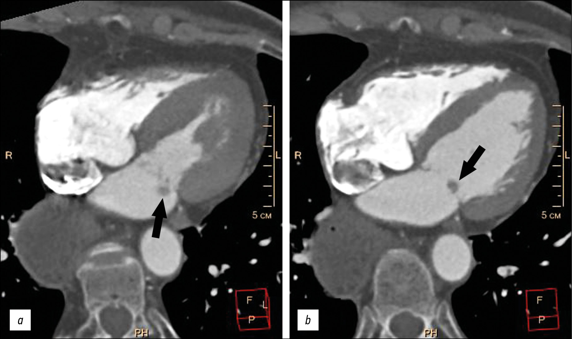
介绍了一名老年患者的病例,主诉喘息,左胸刺痛,心律失常,此患的病史包括吸引性肺炎,食管胃吻合术,食道摘除术。患者检查发现阵发性心房颤动(阵发外)、慢性心力衰竭、动脉高血压。患者的临床资料不具感染性心内膜炎特征,瓣膜上有赘生物。超声心动图和多螺旋计算机断层扫描在二尖瓣后叶的心房表面进行快速对比增强,显示额外的体积形成 5-9 毫米,圆形,轮廓明显不均匀,与瓣叶一起移位进入心房收缩期左心室腔。在Fiesta-CINE模式下,在修改后的两室和四室投影中获得了肿块的最佳可视化。患者在体外循环下接受了肿块的切除,并进行了二尖瓣成形术。肿块的组织学检查揭示了粘液瘤的特征性形态情况。术后期间一切顺利。
作者简介
Marina V. Vishniakova
Moscow Regional Research and Clinical Institute
编辑信件的主要联系方式.
Email: cherridra@mail.ru
ORCID iD: 0000-0003-3838-636X
SPIN 代码: 1137-2991
MD, Dr. Sci. (Med)
俄罗斯联邦, 61/2, Shepkina street, Moscow,129110Alexander S. Abramenko
Moscow Regional Research and Clinical Institute
Email: a.s.abramenko@gmail.com
ORCID iD: 0000-0002-6286-2162
SPIN 代码: 9743-3001
Научный сотрудник отделения лучевой диагностики
俄罗斯联邦, 61/2, Shepkina street, Moscow,129110Mariya V. Vishniakova
Moscow Regional Research and Clinical Institute
Email: cherridra@list.ru
ORCID iD: 0000-0002-2649-4198
SPIN 代码: 7748-1831
MD, Dr. Sci. (Med)
俄罗斯联邦, 61/2, Shepkina street, Moscow,129110Dmitry V. Shumakov
Moscow Regional Research and Clinical Institute
Email: cherridra@mail.ru
ORCID iD: 0000-0003-4204-8865
SPIN 代码: 2545-2978
MD, Dr. Sci. (Med), Corresponding Member of the Russian Academy of Sciences
俄罗斯联邦, 61/2, Shepkina street, Moscow,129110参考
- Centofanti P, Di Rosa E, Deorsola L, et al. Primary cardiac tumors: early and late results of surgical treatment in 91 patients. Ann Thorac Surg. 1999;68(4):1236–1241. doi: 10.1016/s0003-4975(99)00700-6
- Hoey ET, Shahid M, Ganeshan A, et al. MRI assessment of cardiac tumours: part 1, multiparametric imaging protocols and spectrum of appearances of histologically benign lesions. Quant Imaging Med Surg. 2014;4(6):478–488. doi: 10.3978/j.issn.2223-4292.2014.11.23
- Zheltovsky YV, Batekh VI, Podkamenny VA, et al. Diagnosis and treatment with a heart mix. Acta Biomedica Sci. 2017;2(6):21–26. (In Russ).
- Lam KY, Dickens P, Chan AC. Tumors of the heart. A 20-year experience with a review of 12,485 consecutive autopsies. Arch Pathol Lab Med. 1993;117(10):1027–1031.
- Li X, Chen Y, Liu J, et al. Cardiac magnetic resonance imaging of primary cardiac tumors. Quant Imaging Med Surg. 2020;10(1):294–313. doi: 10.21037/qims.2019.11.13
- Aggeli C, Dimitroglou Y, Raftopoulos L, et al. Cardiac masses: the role of cardiovascular imaging in the differential diagnosis. Diagnostics. 2020;10(12):1088. doi: 10.3390/diagnostics10121088
- Kallstrom E, Kallus E, Erbe K, et al. Differentiation of left atrial myxomas by multimodality imaging. J Diagnostic Med Sonography. 2020;36(1):52–63. doi: 10.1177/8756479319872153
- Jaleski TC. Myxoma of the heart valves: report of a case. Am J Pathol. 1934;10(3):399–406.
- Wold LE, Lie JT. Cardiac myxomas: a clinicopathologic profile. Am J Pathol. 1980;101(1):219–240.
- Yoon JH, Kim JH, Sung YJ, et al. Cardiac myxoma originating from the anterior mitral valve leaflet. J Cardiovasc Ultrasound. 2011;19(4):228–231. doi: 10.4250/jcu.2011.19.4.228
- Rajiah P, Moore A, Saboo S, et al. Multimodality imaging of complications of cardiac valve surgeries. Radio Graphics. 2019;39(4):932–956. doi: 10.1148/rg.2019180177
- El Ouazzani J, Jandou I, Christophe Thuaire I. Thrombus or vegetation? Importance of cardiac MRI as a diagnostic tool based on case report and literature review. Ann Med Surg (Lond). 2020;60:690–694. doi: 10.1016/j.amsu.2020.12.007
- Anand S, Sydow N, Janardhanan R. Papillary fibroelastoma diagnosed through multimodality cardiac imaging: a rare tumour in an uncommon location with review of literature. BMJ Case Rep. 2017;2017:bcr2017219327. doi: 10.1136/bcr-2017-219327
补充文件


