

")


Basic pulse sequences in the diagnosis of abdominal pathology
- Авторлар: Syrkashev E.M.1,2, Kadyrberdieva F.Z.2, Abuladze L.R.1, Semenov D.S.1, Privalova E.G.1
-
Мекемелер:
- Moscow Center for Diagnostics and Telemedicine
- National Medical Research Center for Obstetrics, Gynecology and Perinatology
- Шығарылым: Том 4, № 1 (2023)
- Беттер: 39-50
- Бөлім: Reviews
- URL: https://journals.rcsi.science/DD/article/view/146874
- DOI: https://doi.org/10.17816/DD123543
- ID: 146874
Дәйексөз келтіру

Аннотация
Magnetic resonance imaging is used for diagnosing abdominal and retroperitoneal space pathology, which allows visualizing focal or diffuse lesions in the parenchymal and hollow viscera with high diagnostic accuracy and reproducibility. Magnetic resonance imaging has advantages over computed tomography in the sensitivity and specificity of determining pathological changes in parenchymal organs, bile ducts and ducts of the pancreas, peritoneum, and retroperitoneal space.
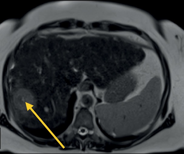
The multiparametric protocol provides information about the mutual topography of organs and their structure and the functional state of tissues. This allows to move from structural to functional evaluation. In most cases, the standard abdominal protocol includes T1-weighted images, T2-weighted images, diffusion-weighted images, and magnetic resonance cholangiopancreatography. Depending on the objectives and patient’s condition, this protocol can be significantly reduced or supplemented.
Existing technical developments and achievements make it possible to simplify the scanning process and reduce the time for obtaining images while increasing the reproducibility of techniques in different healthcare institutions.
Негізгі сөздер
Толық мәтін
##article.viewOnOriginalSite##Авторлар туралы
Egor Syrkashev
Moscow Center for Diagnostics and Telemedicine; National Medical Research Center for Obstetrics, Gynecology and Perinatology
Хат алмасуға жауапты Автор.
Email: egorsrkshv@mail.ru
ORCID iD: 0000-0003-4043-907X
SPIN-код: 1901-5364
MD, Cand Sci (Med.)
Ресей, Moscow; MoscowFaina Kadyrberdieva
National Medical Research Center for Obstetrics, Gynecology and Perinatology
Email: k.faina1992@mail.ru
ORCID iD: 0009-0004-7787-3413
MD, Cand Sci (Med.)
Ресей, MoscowLiya Abuladze
Moscow Center for Diagnostics and Telemedicine
Email: AbuladzeLR@zdrav.mos.ru
ORCID iD: 0000-0001-6745-1672
SPIN-код: 8640-9989
MD
Ресей, MoscowDmitriy Semenov
Moscow Center for Diagnostics and Telemedicine
Email: semenovds4@zdrav.mos.ru
ORCID iD: 0000-0002-4293-2514
SPIN-код: 2278-7290
Ресей, Moscow
Ekaterina Privalova
Moscow Center for Diagnostics and Telemedicine
Email: e-privalova@mail.ru
ORCID iD: 0000-0002-9851-9390
SPIN-код: 6546-5135
MD, Dr.Sci. (Med.)
Ресей, MoscowӘдебиет тізімі
- Hussain SM, Sorrell MF. Liver MRI: Correlation with other imaging modalities and histopathology, second edition. Springer; 2015. doi: 10.1007/978-3-319-06004-0
- Runge VM, Clanton JA, Partain CL, James AE. Respiratory gating in magnetic resonance imaging at 0.5 Tesla. Radiology. 1984;151(2):521–523. doi: 10.1148/radiology.151.2.6709928
- Bailes DR, Gilderdale DJ, Bydder GM, et al. Respiratory ordered phase encoding (ROPE): A method for reducing respiratory motion artefacts in MR imaging. J Comput Assist Tomogr. 1985;9(4):835–838.
- American College of Radiology [online]. ACR-SAR-SPR practice parameter for the performance of magnetic resonance (MR) Enterography. Available from: https://www.acr.org/-/media/ACR/Files/Practice-Parameters/MR-Enterog.pdf. Accessed: 17.01.2023.
- Semelka RC, Kelekis NL, Thomasson D, et al. HASTE MR imaging: Description of technique and preliminary results in the abdomen. J Mag Reson Imaging. 1996;6(4):698–699. doi: 10.1002/jmri.1880060420
- Rofsky NM, Lee VS, Laub G, et al. Abdominal MR imaging with a volumetric interpolated breath-hold examination. Radiology. 1999;212(3):876–884. doi: 10.1148/radiology.212.3.r99se34876
- Syrkashev EM, Solopova A, Kulabukhova EA. Magnetic resonance imaging in the diagnosis of peritoneal carcinomatosis indisseminated ovarian cancer. Obstetrics Gynecology. 2020;(9):38–47. doi: 10.18565/aig.2020.9.38-47
- Vanderveen KA, Hussain HK. Magnetic resonance imaging of cholangiocarcinoma. Cancer Imaging. 2004;4(2):104–115. doi: 10.1102/1470-7330.2004.0018
- Kilcoyne A, Kaplan JL, Gee MS. Inflammatory bowel disease imaging: Current practice and future directions. World J Gastroenterol. 2016;22(3):917–932. doi: 10.3748/wjg.v22.i3.917
- Koh DM, Collins DJ. Diffusion-weighted MRI in the body: Applications and challenges in oncology. AJR Am J Roentgenol. 2007;188(6):1622–1635. doi: 10.2214/AJR.06.1403
- Abuladze LR, Semenov DS, Panina OY, Vasilev YA. Optimized biparametric magnetic resonance imaging protocol for prostate cancer detection. Digit Diagnostics. 2022;3(3):166–177. doi: 10.17816/dd108484
- Thoeny HC, De Keyzer F. Extracranial applications of diffusion-weighted magnetic resonance imaging. Eur Radiol. 2007;17(6):1385–1393. doi: 10.1007/s00330-006-0547-0
- Charles-Edwards EM, De Souza NM. Diffusion-weighted magnetic resonance imaging and its application to cancer. Cancer Imaging. 2006;6(1):135–143. doi: 10.1102/1470-7330.2006.0021
- Kele PG, van der Jagt EJ. Diffusion weighted imaging in the liver. World J Gastroenterol. 2010;16(13):1567–1576. doi: 10.3748/wjg.v16.i13.1567
- Monticciolo L, Podberesky DJ, Pollack MS, et al. ACR-SAR-SPR practice parameter for the performance of magnetic resonance imaging (MRI) of the abdomen (Excluding the Liver). Semantic Scholar; 2015. Available from: https://www.semanticscholar.org/paper/ACR-%E2%80%93-SAR-%E2%80%93-SPR-PRACTICE-PARAMETER-FOR-THE-OF-(-MRI-Monticciolo-Podberesky/7dc9771a1b5aaec215c99fd74ab5e659738cf4fd. Accessed: 17.01.2023.
- Taron J, Martirosian P, Erb M, et al. Simultaneous multislice diffusion-weighted MRI of the liver: Analysis of different breathing schemes in comparison to standard sequences. J Magn Reson Imaging. 2016;44(4):865–879. doi: 10.1002/jmri.25204
Қосымша файлдар


