

")


Possibilities of Dixon sequences in magnetic resonance imaging for fat fraction quantification: a phantom study
- Authors: Panina O.Y.1,2, Gromov A.I.3,4, Ahkmad E.S.1, Semenov D.S.1, Kivasev S.A.5, Petraikin A.V.1, Nechaev V.A.2
-
Affiliations:
- Research and Practical Clinical Center for Diagnostics and Telemedicine Technologies
- Moscow City Hospital named after S.S. Yudin
- Russian University of Medicine
- National Medical Research Radiological Center
- Central Clinical Hospital “RZD-Medicine”
- Issue: Vol 6, No 2 (2025)
- Pages: 191-202
- Section: Original Study Articles
- URL: https://journals.rcsi.science/DD/article/view/310209
- DOI: https://doi.org/10.17816/DD633802
- EDN: https://elibrary.ru/WDZWBY
- ID: 310209
Cite item
Full Text

Abstract
BACKGROUND: The accuracy of quantitative parameters obtained using magnetic resonance imaging is of scientific and practical interest. Monitoring of scan parameters and standardization of commonly used approaches to assess fat fraction remain challenging in radiology.
AIM: This study aimed to evaluate the possibility of fat fraction quantification using standard Dixon pulse sequences through phantom modeling.
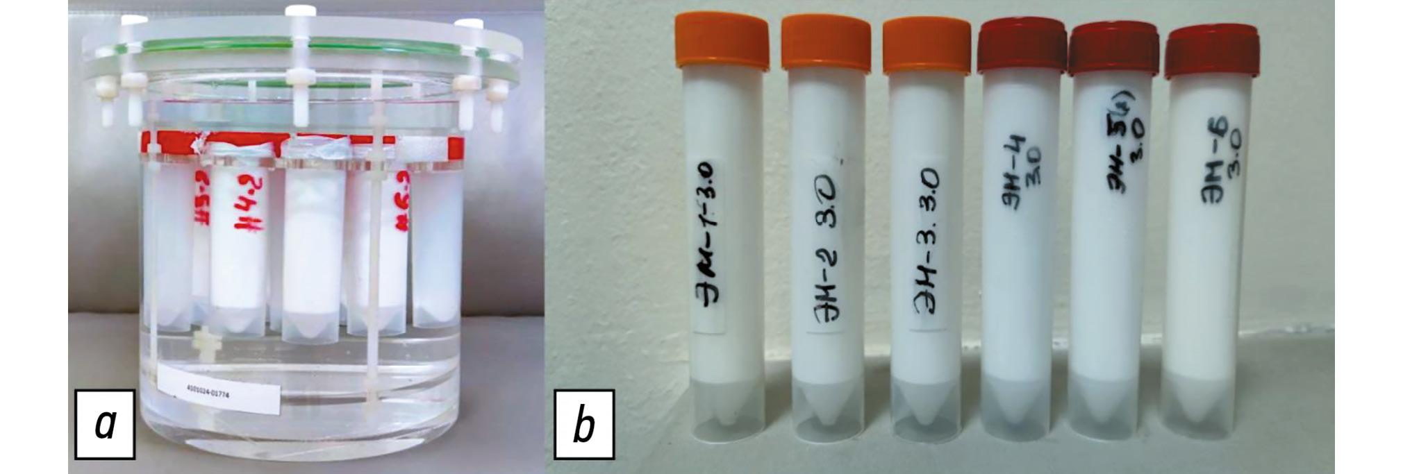
METHODS: This multicenter, cross-sectional, nonblinded experimental study used direct oil-in-water emulsions to model substances with varying fat concentrations. Test tubes containing these emulsions were placed in a cylindrical phantom. The emulsions were prepared with mixtures of vegetable oils, with fat fraction values of 10%–60%. Several tests were conducted using scanners from different manufacturers and with varying magnetic field strengths: Optima MR450w, 1.5 T; MAGNETOM Skyra, 3 T; Ingenia, 1.5 T; and Ingenia Achieva dStream, 3.0 T at different medical centers. Fat fraction was obtained using standard formulas based on signal intensity measurements. A regression analysis was conducted to assess the linear relationship between the measured and predefined fat fraction concentrations and an F-test to evaluate variability.
RESULTS: Phantom modeling was employed to determine the performance of Dixon pulse sequences across different magnetic resonance imaging scanners for quantitative fat fraction estimation using relevant formulas. In assessing the accuracy of fat fraction quantification, a weak linear correlation was found between the obtained values and predefined fat fraction concentrations. Additionally, significant deviations >5% were observed for certain scanners. Reproducibility analysis demonstrated variability in fat fraction concentration across different scanner models and within the same model.
CONCLUSION: Obtained results confirm that fat fraction quantification using Dixon pulse sequences and relevant formulas should be performed only after preliminary phantom scanning. The use of a phantom ensures adequate quality control and calibration of the magnetic resonance imaging scanner, making accurate quantitative fat measurement more reliable and widely accessible.
Full Text
##article.viewOnOriginalSite##About the authors
Olga Yu. Panina
Research and Practical Clinical Center for Diagnostics and Telemedicine Technologies; Moscow City Hospital named after S.S. Yudin
Author for correspondence.
Email: olgayurpanina@gmail.com
ORCID iD: 0000-0002-8684-775X
SPIN-code: 5504-8136
MD
Russian Federation, 24 Petrovka st, bldg 1, Moscow, 127051; MoscowAlexander I. Gromov
Russian University of Medicine; National Medical Research Radiological Center
Email: gai8@mail.ru
ORCID iD: 0000-0002-9014-9022
SPIN-code: 6842-8684
MD, Dr. Sci. (Medicine), Professor
Russian Federation, Moscow; MoscowEkaterina S. Ahkmad
Research and Practical Clinical Center for Diagnostics and Telemedicine Technologies
Email: akhmades@zdrav.mos.ru
ORCID iD: 0000-0002-8235-9361
SPIN-code: 5891-4384
Russian Federation, 24 Petrovka st, bldg 1, Moscow, 127051
Dmitry S. Semenov
Research and Practical Clinical Center for Diagnostics and Telemedicine Technologies
Email: semenovds4@zdrav.mos.ru
ORCID iD: 0000-0002-4293-2514
SPIN-code: 2278-7290
Cand. Sci. (Engineering)
Russian Federation, 24 Petrovka st, bldg 1, Moscow, 127051Stanislav A. Kivasev
Central Clinical Hospital “RZD-Medicine”
Email: Kivasev@yahoo.com
ORCID iD: 0000-0003-1160-5905
SPIN-code: 9883-3406
MD
Russian Federation, MoscowAlexey V. Petraikin
Research and Practical Clinical Center for Diagnostics and Telemedicine Technologies
Email: PetryajkinAV@zdrav.mos.ru
ORCID iD: 0000-0003-1694-4682
SPIN-code: 6193-1656
MD, Dr. Sci. (Medicine)
Russian Federation, 24 Petrovka st, bldg 1, Moscow, 127051Valentin A. Nechaev
Moscow City Hospital named after S.S. Yudin
Email: NechaevVA1@zdrav.mos.ru
ORCID iD: 0000-0002-6716-5593
SPIN-code: 2527-0130
MD, Cand. Sci. (Medicine)
Russian Federation, MoscowReferences
- Outwater EK, Blasbalg R, Siegelman ES, Vala M. Detection of lipid in abdominal tissues with opposed-phase gradient-echo images at 1.5 T: techniques and diagnostic importance. RadioGraphics. 1998;18(6):1465–1480. doi: 10.1148/radiographics.18.6.9821195
- Panina OYu, Gromov AI, Akhmad ES, et al. Accuracy of fat fraction estimation using DIXON: experimental phantom study. Medical Visualization. 2022;26(4):147–158. doi: 10.24835/1607-0763-1160 EDN: JDIWXI
- Bray TJP, Chouhan MD, Punwani S, et al. Fat fraction mapping using magnetic resonance imaging: insight into pathophysiology. The British Journal of Radiology. 2017;91(1089):20170344. doi: 10.1259/bjr.20170344
- Bhat V, Velandai S, Belliappa V, et al. Quantification of liver fat with mDIXON magnetic resonance imaging, comparison with the computed tomography and the biopsy. Journal of Clinical and Diagnostic Research. 2017;11(7):TC06–TC10. doi: 10.7860/JCDR/2017/26317.10234
- Bainbridge A, Bray TJP, Sengupta R, Hall-Craggs MA. Practical approaches to bone marrow fat fraction quantification across magnetic resonance imaging platforms. Journal of Magnetic Resonance Imaging. 2020;52(1):298–306. doi: 10.1002/jmri.27039 EDN: WCMNIG
- Gulani V, Seiberlich N. Quantitative MRI: rationale and challenges. Advances in Magnetic Resonance Technology and Applications. 2020;1:xxxvii–li. doi: 10.1016/B978-0-12-817057-1.00001-9
- Vasilev YuA, Semenov DS, Akhmad ES, et al. A method for assessing the effect of metal artifact reduction algorithms on quantitative characteristics of CT images. Biomedical Engineering. 2020;54(4):285–288. doi: 10.1007/s10527-020-10023-5 EDN: YEHJTT
- Morozov S, Sergunova K, Petraikin A, et al. Diffusion processes modeling in magnetic resonance imaging. Insights into Imaging. 2020;11(1):60. doi: 10.1186/s13244-020-00863-w EDN: QEFDVK
- Sergunova KA. Using siloxane-based inverse emulsions to control the measured diffusion coefficient in magnetic resonance imaging. Biomedical Engineering. 2019;(5):22–25. (In Russ.) EDN: HUPRTQ
- Petraikin AV, Ivanova DV, Akhmad ES, et al. Phantom modeling for selection of optimum reconstruction filters in the quantitative computer tomography. Meditsinskaya fizika. 2020;(2):34–44. EDN: TLLBVQ
- Vasilev YuA, Cherkasskaya MV, Akhmad ES, et al. Phantom modelling in magnetic resonance imaging: an overview of materials for simulating tissue relaxation time (review). Polymer materials and technologies. 2023;9(4):6–20. doi: 10.32864/polymmattech-2023-9-4-6-20 EDN: TCSKRR
- van Vucht N, Santiago R, Lottmann B, et al. The Dixon technique for MRI of the bone marrow. Skeletal Radiology. 2019;48(12):1861–1874. doi: 10.1007/s00256-019-03271-4
- Gromov AI, Gorinov AV, Galljamov EA. Mesenteric chillous lymphangioma. Visualization features on opposed-phase MR images. Medical Visualization. 2019;23(4):86–92. doi: 10.24835/1607-0763-2019-4-86-92 EDN: UCRGCC
- Zhao Y, Huang M, Ding J, et al. Prediction of abnormal bone density and osteoporosis from lumbar spine MR using modified dixon quant in 257 subjects with quantitative computed tomography as reference. Journal of Magnetic Resonance Imaging. 2018;49(2):390–399. doi: 10.1002/jmri.26233
- Maeder Y, Dunet V, Richard R, et al. Bone marrow metastases: T2-weighted Dixon Spin-Echo Fat Images Can Replace T1-weighted Spin-Echo Images. Radiology. 2018;286(3):948–959. doi: 10.1148/radiol.2017170325
- Chow LTC, Ng AWH, Wong SKC. Focal nodular and diffuse haematopoietic marrow hyperplasia in patients with underlying malignancies: a radiological mimic of malignancy in need of recognition. Clinical Radiology. 2017;72(3):265.e7–265.e23. doi: 10.1016/j.crad.2016.10.015
- Omoumi P. Update on Advances in Musculoskeletal Magnetic Resonance Imaging. Seminars in Musculoskeletal Radiology. 2015;19(04):319–320. doi: 10.1055/s-0035-1565876
- Pezeshk P, Alian A, Chhabra A. Role of chemical shift and Dixon based techniques in musculoskeletal MR imaging. European Journal of Radiology. 2017;94:93–100. doi: 10.1016/j.ejrad.2017.06.011
- Fukuzawa K, Hayashi T, Takahashi J, et al. Evaluation of six-point modified dixon and magnetic resonance spectroscopy for fat quantification: a fat–water–iron phantom study. Radiological Physics and Technology. 2017;10(3):349–358. doi: 10.1007/s12194-017-0410-9
Supplementary files


