

")


Using a mobile computer tomography scanner in a field hospital setting to manage patients with COVID-19
- Authors: Kudryavtsev N.D.1, Petraikin A.V.1, Ahkmad E.S.1, Kiselev F.A.1, Burashov V.V.1, Mukhortova A.N.1, Soldatov I.V.1, Shkoda A.S.2
-
Affiliations:
- Moscow Center for Diagnostics and Telemedicine
- City Clinical Hospital No. 67 named after L.A. Vorokhobov
- Issue: Vol 4, No 3 (2023)
- Pages: 427-438
- Section: Correspondence
- URL: https://journals.rcsi.science/DD/article/view/254080
- DOI: https://doi.org/10.17816/DD321670
- ID: 254080
Cite item

Abstract
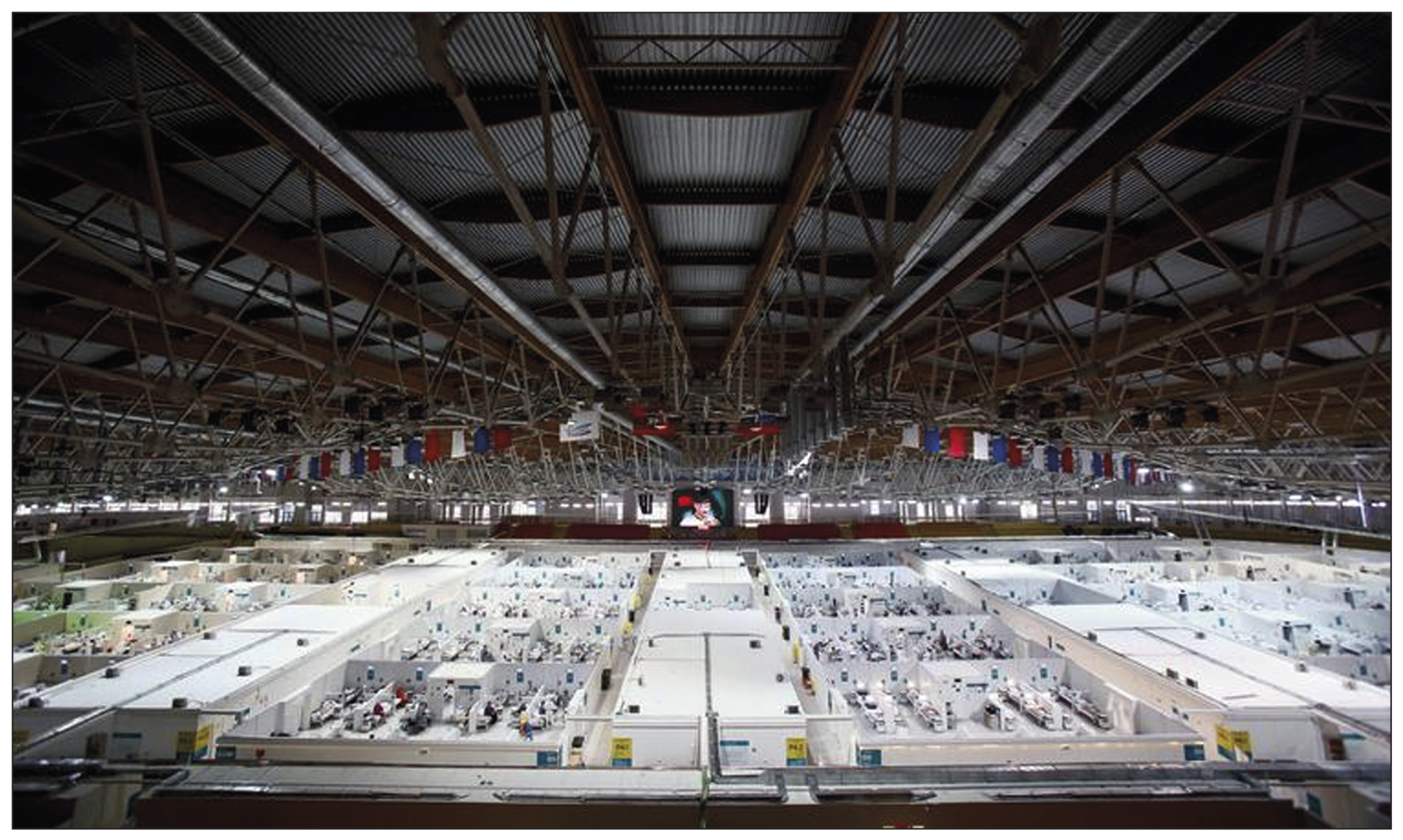
The global outbreak of COVID-19 has posed unprecedented challenges to healthcare systems worldwide. Healthcare administrators had to make quick and effective decisions to ensure high quality of medical care standards in new conditions. The need to form a reserve bed fund during the pandemic was due to the high load on city hospitals in Moscow. Due to this fact, temporary reserved hospitals for COVID-19 patients were organized in non-core facilities, such as ice arenas, shopping malls, and exhibition pavilions. This urgency prompted a search for solutions that could provide the necessary level of diagnosis and treatment appropriate to specialized medical facility. Given the technical and time constraints associated with the installation of a fixed computer tomographic scanner, the deployment of mobile computer tomographic scanners emerged as a viable option.
The study aims to share insights gained from using a mobile computer tomographic scanner within a temporary backup hospital setting to treating patients with COVID-19 coronavirus infection. The paper discusses the features, advantages, and disadvantages of mobile computer tomography. It also presents hardware and control room layouts, along with the placement options for the computer tomography device. The research includes the results of dosimetry studies and provides a clinical assessment of the applicability of this type of diagnostic devices.
Full Text
##article.viewOnOriginalSite##About the authors
Nikita D. Kudryavtsev
Moscow Center for Diagnostics and Telemedicine
Author for correspondence.
Email: n.kudryavtsev@npcmr.ru
ORCID iD: 0000-0003-4203-0630
SPIN-code: 1125-8637
Russian Federation, Moscow
Alexey V. Petraikin
Moscow Center for Diagnostics and Telemedicine
Email: PetryajkinAV@zdrav.mos.ru
ORCID iD: 0000-0003-1694-4682
SPIN-code: 6193-1656
MD, Dr. Sci. (Med.), Associate Professor
Ekaterina S. Ahkmad
Moscow Center for Diagnostics and Telemedicine
Email: e.ahkmad@npcmr.ru
ORCID iD: 0000-0002-8235-9361
SPIN-code: 5891-4384
Russian Federation, Moscow
Fyodor A. Kiselev
Moscow Center for Diagnostics and Telemedicine
Email: KiselevFA@zdrav.mos.ru
ORCID iD: 0009-0006-6472-8940
Russian Federation, Moscow
Vyacheslav V. Burashov
Moscow Center for Diagnostics and Telemedicine
Email: BurashovVV@zdrav.mos.ru
ORCID iD: 0000-0001-9250-0667
SPIN-code: 4308-0912
Russian Federation, Moscow
Anna N. Mukhortova
Moscow Center for Diagnostics and Telemedicine
Email: a.mukhortova@npcmr.ru
ORCID iD: 0000-0001-9814-3533
SPIN-code: 9051-1130
Russian Federation, Moscow
Iliya V. Soldatov
Moscow Center for Diagnostics and Telemedicine
Email: i.soldatov@npcmr.ru
ORCID iD: 0000-0002-4867-0746
SPIN-code: 4065-6048
Russian Federation, Moscow
Andrey S. Shkoda
City Clinical Hospital No. 67 named after L.A. Vorokhobov
Email: a.shkoda@67gkb.ru
ORCID iD: 0000-0002-9783-1796
SPIN-code: 4520-2141
MD, Dr. Sci. (Med), Professor
Russian Federation, MoscowReferences
- Morozov SP, Kuzmina ES, Ledikhova NV, et al. Mobilizing the academic and practical potential of diagnostic radiology during the COVID-19 pandemic in Moscow. Digital Diagnostics. 2020;1(1):5–12. (In Russ). doi: 10.17816/DD51043
- Prevention, diagnosis and treatment of new coronavirus infection (2019-nCoV): temporary guidelines. Version 17 (12/14/2022). (In Russ). Available from: https://static-0.minzdrav.gov.ru/system/attachments/attaches/000/061/254/original/%D0%92%D0%9C%D0%A0_COVID-19_V17.pdf?1671088207. Accessed: 15.03.2023. (
- De Smet K, De Smet D, Ryckaert T, et al. Diagnostic performance of chest CT for SARS-CoV-2 infection in individuals with or without COVID-19 symptoms. Radiology. 2021;298(1):E30–E37. doi: 10.1148/radiol.2020202708
- Huang Y, Cheng W, Zhao N, et al. CT screening for early diagnosis of SARS-CoV-2 infection. Lancet Inf Dis. 2020;20(9):1010–1011. doi: 10.1016/S1473-3099(20)30241-3
- Barrett JF, Keat N. Artifacts in CT: Recognition and avoidance. RadioGraphics. 2004;24(6):1679–1691. doi: 10.1148/rg.246045065
- Samorodskaja IV, Larina VN, Nazimkin KE, Larin VG. Organizational and clinical problems of outpatient COVID-19 diagnostics. Vrach. 2020;31(5):23–30. (In Russ). doi: 10.29296/25877305-2020-05-05
- Cester G, Giraudo C, Causin F, et al. Retrospective analysis of a modified organizational model to guarantee CT workflow during the COVID-19 outbreak in the Tertiary Hospital of Padova, Italy. J Clin Med. 2020;9(9):3042. doi: 10.3390/jcm9093042
- Bates DD, Vintonyak A, Mohabir R, et al. Use of a portable computed tomography scanner for chest imaging of COVID-19 patients in the urgent care at a tertiary cancer center. Emerg Radiol. 2020;27(6):597–600. doi: 10.1007/s10140-020-01801-5
- Khristenko EA, von Stackelberg O, Kautsor HU, et al. CT patterns in COVID-19 associated pneumonia: Standardization of research descriptions based on the Fleischner Society Glossary. Rejr. 2020;10(1):16–26. (In Russ). doi: 10.21569/2222-7415-2020-10-1-16-26
- Kyriakou Y, Meyer E, Prell D, Kachelriess M. Empirical beam hardening correction (EBHC) for CT. Med Phys. 2010;37(10):5179–5187. doi: 10.1118/1.3477088
- Aliev AF, Kudryavtsev ND, Petryaykin AV, et al. Changing of pulmonary artery diameter in accordance with severity of COVID-19 (assessment based on non-contrast computer tomography). Digital Diagnostics. 2021;2(3):249–260. (In Russ). doi: 10.17816/DD76726
Supplementary files


