

")


Тerminology of rectal cancer: consensus agreement of the expert working group
- Authors: Berezovskaya T.P.1, Rubtsova N.A.2, Sinitsyn V.E.3, Zarodnyuk I.V.4, Nudnov N.V.5, Mishchenko A.V.6, Trubacheva Y.L.4, Bergen T.A.7, Grishko P.Y.6, Balyasnikova S.S.8, Dayneko Y.A.1, Ryjkova D.V.9, Hodzhibekova M.M.2, Rucheva N.A.10, Turin I.E.8, Achkasov S.I.4, Nevolskikh A.A.1, Gordeev S.S.8, Droshneva I.V.2
-
Affiliations:
- A.F. Tsyba Medical Radiological Research Center ― branch National Medical Research Radiological Center
- P.A. Herzen Moscow Research Oncological Institute ― branch National Medical Research Radiological Center
- Lomonosov Moscow State University
- State Scientific Centre of Coloproctology
- Russian Scientific Center of Roentgenoradiology
- N.N. Petrov National Medical Research Centre of Oncology
- E. Meshalkin National Medical Research Center
- N.N. Blokhin National Medical Research Center of Oncology
- Almazov National Medical Research Centre
- V.I. Shumakov National Medical Research Center of Transplantology and Artificial Organs
- Issue: Vol 4, No 3 (2023)
- Pages: 306-321
- Section: Clinical Practice Guidelines
- URL: https://journals.rcsi.science/DD/article/view/254071
- DOI: https://doi.org/10.17816/DD529668
- ID: 254071
Cite item

Abstract
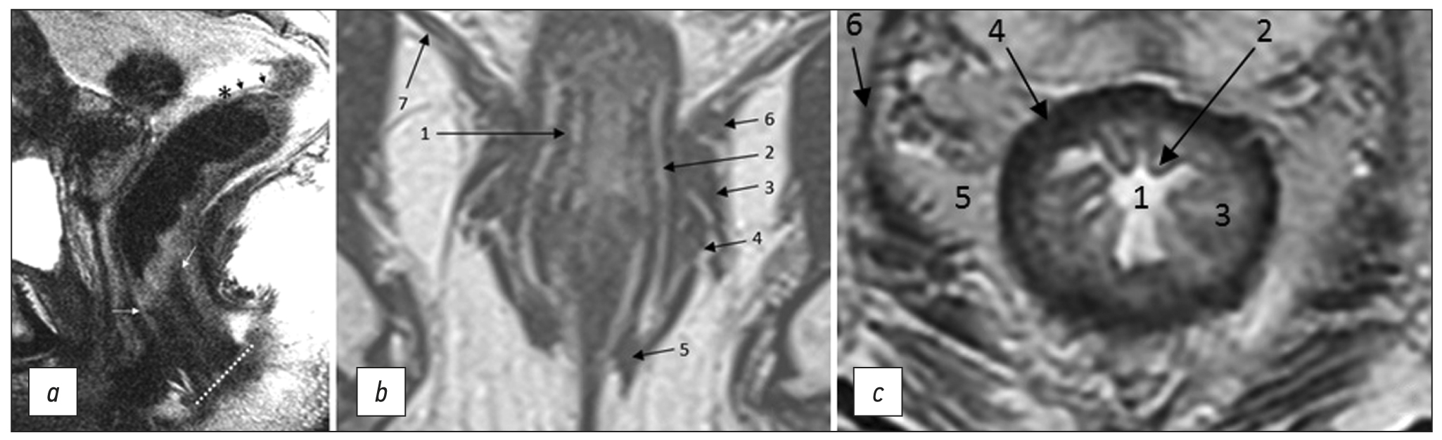
Unified terminology is a necessary condition for successful interdisciplinary communication within the field of oncology. The variety of anatomical, pathomorphological, and clinical terms used in rectal cancer is often accompanied by their ambiguous interpretation both in domestic and foreign scientific literature. This not only complicates the interaction between specialists, but also complicates the comparison of the results of rectal cancer treatment obtained in different medical institutions.
Based on the analysis of recent domestic and international scientific and methodological literature on rectal cancer, the key terms used in the diagnosis and treatment planning of rectal cancer were selected, followed by a two-time online discussion of their interpretations by experts from the Russian Society of Radiologists and Therapeutic Radiation Oncologists, the Association of Oncologists of Russia, and the Russian Association of Therapeutic Radiation Oncologists until reaching consensus (≥80%) of experts on all items. Terms that fail to attain consensus were excluded in the final list.
A list of anatomical, pathomorphological, and clinical terms used in the diagnosis, staging, and treatment planning of rectal cancer has been compiled and, based on expert consensus, their interpretation has been determined.
A lexicon recommended in the description and formulation of the conclusion of diagnostic studies in patients with rectal cancer is proposed.
Full Text
##article.viewOnOriginalSite##About the authors
Tatiana P. Berezovskaya
A.F. Tsyba Medical Radiological Research Center ― branch National Medical Research Radiological Center
Email: tberezovska@yahoo.com
ORCID iD: 0000-0002-3549-4499
SPIN-code: 5837-3465
MD, Dr. Sci. (Med.), Professor
Russian Federation, ObninskNatalia A. Rubtsova
P.A. Herzen Moscow Research Oncological Institute ― branch National Medical Research Radiological Center
Email: rna17@ya.ru
ORCID iD: 0000-0001-8378-4338
SPIN-code: 9712-9091
MD, Dr. Sci. (Med.)
Russian Federation, MoscowValentin E. Sinitsyn
Lomonosov Moscow State University
Email: vsin@mail.ru
ORCID iD: 0000-0002-5649-2193
SPIN-code: 8449-6590
MD, Dr. Sci. (Med.), Professor
Russian Federation, MoscowIrina V. Zarodnyuk
State Scientific Centre of Coloproctology
Email: zarodnyuk_iv@gnck.ru
ORCID iD: 0000-0002-9442-7480
SPIN-code: 8310-8989
MD, Dr. Sci. (Med.)
Russian Federation, MoscowNicolai V. Nudnov
Russian Scientific Center of Roentgenoradiology
Email: nudnov@mrrc.ru
ORCID iD: 0000-0001-5994-0468
SPIN-code: 3018-2527
MD, Dr. Sci. (Med.), Professor
Russian Federation, MoscowAndrei V. Mishchenko
N.N. Petrov National Medical Research Centre of Oncology
Email: dr.mishchenko@mail.ru
ORCID iD: 0000-0001-7921-3487
SPIN-code: 8825-4704
MD, Dr. Sci. (Med.)
Russian Federation, MoscowYuliya L. Trubacheva
State Scientific Centre of Coloproctology
Email: trubacheva_ul@gnck.ru
ORCID iD: 0000-0002-8403-195X
SPIN-code: 3427-9074
MD, Dr. Sci. (Med.)
Russian Federation, MoscowTatiana A. Bergen
E. Meshalkin National Medical Research Center
Email: tbergen@yandex.ru
ORCID iD: 0000-0003-1530-1327
SPIN-code: 5467-7347
MD, Dr. Sci. (Med.)
Russian Federation, MoscowPavel Yu. Grishko
N.N. Petrov National Medical Research Centre of Oncology
Email: dr.grishko@mail.ru
ORCID iD: 0000-0003-4665-6999
SPIN-code: 3109-1583
MD, Cand. Sci. (Med.)
Russian Federation, MoscowSvetlana S. Balyasnikova
N.N. Blokhin National Medical Research Center of Oncology
Email: Balyasnikova.Svetlana@gmail.com
ORCID iD: 0000-0002-9666-9301
SPIN-code: 3987-2336
MD, Cand. Sci. (Med.)
Russian Federation, MoscowYana A. Dayneko
A.F. Tsyba Medical Radiological Research Center ― branch National Medical Research Radiological Center
Email: vorobeyana@gmail.com
ORCID iD: 0000-0002-4524-0839
MD, Cand. Sci. (Med.)
Russian Federation, ObninskDarya V. Ryjkova
Almazov National Medical Research Centre
Email: d_ryjkova@mail.ru
ORCID iD: 0000-0002-7086-9153
MD, Dr. Sci. (Med.), Professor
Russian Federation, MoscowMalika M. Hodzhibekova
P.A. Herzen Moscow Research Oncological Institute ― branch National Medical Research Radiological Center
Email: malika_25@mail.ru
ORCID iD: 0000-0002-2172-5778
SPIN-code: 3999-7304
MD, Dr. Sci. (Med.)
Russian Federation, MoscowNataliya A. Rucheva
V.I. Shumakov National Medical Research Center of Transplantology and Artificial Organs
Email: rna1969@yandex.ru
ORCID iD: 0000-0002-8063-4462
SPIN-code: 2196-8300
MD, Cand. Sci. (Med.)
Russian Federation, MoscowIgor E. Turin
N.N. Blokhin National Medical Research Center of Oncology
Email: igortyurin@gmail.com
ORCID iD: 0000-0002-8587-4422
SPIN-code: 6499-2398
MD, Dr. Sci. (Med.), Professor
Russian Federation, MoscowSergey I. Achkasov
State Scientific Centre of Coloproctology
Email: achkasovy@mail.ru
ORCID iD: 0000-0001-9294-5447
SPIN-code: 5467-1062
MD, Dr. Sci. (Med.), Professor, Corresponding Member of the Academy of Sciences
Russian Federation, MoscowAlexey A. Nevolskikh
A.F. Tsyba Medical Radiological Research Center ― branch National Medical Research Radiological Center
Email: alexey.nevol@gmail.com
ORCID iD: 0000-0001-5961-2958
SPIN-code: 3787-6139
MD, Dr. Sci. (Med.)
Russian Federation, ObninskSergey S. Gordeev
N.N. Blokhin National Medical Research Center of Oncology
Email: ss.netoncology@gmail.com
ORCID iD: 0000-0002-9303-8379
SPIN-code: 6577-5540
MD, Dr. Sci. (Med.)
Russian Federation, MoscowInna V. Droshneva
P.A. Herzen Moscow Research Oncological Institute ― branch National Medical Research Radiological Center
Author for correspondence.
Email: droshnevainna@mail.ru
SPIN-code: 1908-2624
MD, Cand. Sci. (Med.)
Russian Federation, MoscowReferences
- Rectal cancer. Clinical recommendations. Approved at the meeting of the Scientific and Practical Council of the Ministry of Health of the Russian Federation. Moscow; 2022.(In Russ). Available from: https://cr.minzdrav.gov.ru/recomend/554_3. Accessed: 15.08.2023.
- Bogveradze N, Snaebjornsson P, Grotenhuis BA, et al. MRI anatomy of the rectum: Key concepts important for rectal cancer staging and treatment planning. Insights Imaging. 2023;14(1):13. doi: 10.1186/s13244-022-01348-8
- Gollub MJ, Arya S, Beets-Tan RG, et al. Use of magnetic resonance imaging in rectal cancer patients: Society of Abdominal Radiology (SAR) rectal cancer disease-focused panel (DFP) recommendations 2017. Abdom Radiol. 2018;43(11):2893–2902. doi: 10.1007/s00261-018-1642-9
- Nougaret S, Rousset P, Gormly K, et al. Structured and shared MRI staging lexicon and report of rectal cancer: A consensus proposal by the French Radiology Group (GRERCAR) and Surgical Group (GRECCAR) for rectal cancer. Diagn Interv Imaging. 2022;103(3):127–141. doi: 10.1016/j.diii.2021.08.003
- Grishko PY, Balyasnikova SS, Samsonov DV, et al. A modern view on the principles of diagnosis and treatment of rectal cancer according to MRI data (literature review). Medical Visualization. 2019;23(2):7–26.(In Russ). doi: 10.24835/1607-0763-2019-2-7-26
- Fernandes MC, Gollub MJ, Brown G. The importance of MRI for rectal cancer evaluation. Surg Oncol. 2022;(43):101739. doi: 10.1016/j.suronc.2022.101739
- Glynne-Jones R, Wyrwicz L, Tiret E, et al. Rectal cancer: ESMO clinical practice guidelines for diagnosis, treatment and follow-up. Ann Oncol. 2017;28(Suppl 4):22–40. doi: 10.1093/anonc/mdx22 4
- Beets-Tan R, Lambregts D, Maas M, et al. Magnetic resonance imaging for clinical management of rectal cancer: Updated recommendations from the 2016 European Society of Gastrointestinal and Abdominal Radiology (ESGAR) consensus meeting. Eur Radiol. 2018;28(4):1465–1475. doi: 10.1007/s0033 0-017-5026-2
- Oien K, Forsmo HM, Rösler C, et al. Endorectal ultrasound and magnetic resonance imaging for staging of early rectal cancers: How well does it work in practice? Acta Oncol. 2019;58(Sup1):49–54. doi: 10.1080/0284186X.2019.1569259
- Brierley JD, Gospodarowicz MK, Wittekind C. TNM Classification of Malignant Tumours. 8th ed. Wiley-Blackwell; 2017. 272 р.
- Kikuchi R, Takano M, Takagi K, et al. Management of early invasive colorectal cancer. Risk of recurrence and clinical guidelines. Dis Colon Rectum. 1995;38(12):1286–1295. doi: 10.1007/BF02049154
- Boot J, Gomez-Munoz F, Beets-Tan R. Imaging of rectal cancer. Radiologe. 2019;59(Suppl 1):46–50٠. doi: 10.1007/s00117-019-0579-5
- Lambregts D, Bogveradze N, Blomqvist L, et al. Current controversies in TNM for the radiological staging of rectal cancer and how to deal with them: Results of a global online survey and multidisciplinary expert consensus. Eur Radiol. 2022;32(7):4991–5003. doi: 10.1007/s00330-022-08591-z
- Mainovskaya OA, Rybakov EG, Chernyshov SV, et al. New morphological risk factors for metastasis to regional lymph nodes in rectal cancer with invasion of the submucosal base. Coloproctology. 2021;20(4):22–33. (In Russ). doi: 10.33878/2073-7556-2021-20-4-22-33
- Volkova SN, Stashuk GA, Chermensky GV, Naumov EK. The role of MRI in the detection of extramural vascular invasion as an indicator of the presence of regional and distant metastases of cancer of the lower ampullary rectum. Experimental Clin Gastroenterol. 2019;164(4):66–71. (In Russ). doi: 10.31146/1682-8658-ecg-164-4-66-71
- Lord AC, D’Souza N, Shaw A, et al. MRI-diagnosed tumor deposits and EMVI status have superior prognostic accuracy to current clinical TNM staging in rectal cancer. Ann Surg. 2022;276(2):334–344. doi: 10.1097/SLA.0000000000004499
- Rokan Z, Simillis C, Kontovounisios C, et al. Locally recurrent rectal cancer according to a standardized MRI classification system: A systematic review of the literature. J Clin Med. 2022;11(12):3511. doi: 10.3390/jcm11123511
- Grishko PY, Mishchenko AV, Ivko OV, et al. The possibilities of multiparametric magnetic resonance imaging in assessing the effectiveness of neoadjuvant treatment of rectal cancer. Radiation Diagnostics Therapy. 2019;10(4):49–56.(In Russ).
- Inoue A, Sheedy SP, Heiken JP, et al. MRI-detected extramural venous invasion of rectal cancer: Multimodality performance and implications at baseline imaging and after neoadjuvant therapy. Insights Imaging. 2021;(2):110. doi: 10.1186/s13244-021-01023-4
- Al-Sukhni E, Milot L, Fruitman M, et al. Diagnostic Accuracy of MRI for assessment of t category, lymph node metastases, and circumferential resection margin involvement in patients with rectal cancer: A systematic review and meta-analysis. Ann Sur Oncol. 2012;19(7):2212–2222. doi: 10.1245/s10434-011-2210-5
- Borgheresi A, De Muzio F, Agostini A, et al. Lymph nodes evaluation in rectal cancer: Where do we stand and future perspective. J Clin Med. 2022;11(9):2599. doi: 10.3390/jcm11092599
- Zhuang Z, Zhang Y, Wei M, et al. Magnetic resonance imaging evaluation of the accuracy of various lymph node staging criteria in rectal cancer: A systematic review and meta-analysis. Front Oncol. 2021;(11):709070. doi: 10.3389/fonc.2021.709070
- Li X, Sun Y, Tang L, et al. Evaluating local lymph node metastasis with magnetic resonance imaging, endoluminal ultrasound and computed tomography in rectal cancer: A meta-analysis. Color Dis. 2015;17(6):129–135. doi: 10.1111/codi.12909
- Weiser MR. AJCC 8th ed. Colorectal cancer. Ann Surg Oncol. 2018;25(6):1454–1455. doi: 10.1245/s10434-018-6462-1
- Ueno H, Nagtegaal ID, Quirke P, et al. Tumor deposits in colorectal cancer: Refining their definition in the TNM system. A G Surg. 2023;7(2):225–235. doi: 10.1002/ags3.12652
- Santiago I, Figueiredo N, Parés O, et al. MRI of rectal cancer: Relevant anatomy and staging key points. Insights Imaging. 2020;11(1):100. doi: 10.1186/s13244-020-00890-7
- Ogura A, Konishi T, Cunningham C, et al. Neoadjuvant (chemo)radiotherapy with total mesorectal excision only is not sufficient to prevent lateral local recurrence in enlarged nodes: Results of the multicenter lateral node study of patients with low cT3/4 rectal cancer. J Clin Oncol. 2019;37(1):33–43. doi: 10.1200/JCO.18.00032
- Gollub MJ, Costello JR, Ernst RD, et al. A primer on rectal MRI in patients on watch-and-wait treatment for rectal cancer. Abdom Radiol. 2023. doi: 10.1007/s00261-023-03900-6
- Berezovskaya TP, Daineko YA, Nevolskikh AA, et al. Prospective evaluation of the use of the MRTG system in determining the effectiveness of neoadjuvant chemoradiotherapy in patients with rectal cancer. Bulletin Radiol Radiol. 2021;102(1):6–17. (In Russ). doi: 10.20862/0042-4676-2021-102-1-6-17
- Almeida RR, Souza D, Matalon SA, et al. Rectal MRI after neoadjuvant chemoradiation therapy: A pictorial guide to interpretation. Abdom Radiol. 2021;46(7):3044–3057. doi: 10.1007/s00261-021-03007-w
- Shelygin YA, Chernyshov SV, Kazieva LY, et al. Comparative analysis of open and transanal total mesorectumectomy in rectal cancer. Coloproctology. 2018;(4):67–73. (In Russ).
- Maistrenko NA, Khvatov AA, Sazonov AA. Pelvic exenterations in the treatment of locally advanced tumors. Bulletin Surnamed after Grekov. 2014;173(6):37–43. (In Russ).
- Sidorov DV, Alekseev BY, Grishin NA, et al. Variants of pelvic exenteration in locally advanced primary and recurrent rectal cancer. Oncology J named after P.A. Herzen. 2013;(6):7–13. (In Russ).
Supplementary files


