

")


直肠癌术语:RSR、AOR和RATRO专家工作组达成的一致意见
- 作者: Berezovskaya T.P.1, Rubtsova N.A.2, Sinitsyn V.E.3, Zarodnyuk I.V.4, Nudnov N.V.5, Mishchenko A.V.6, Trubacheva Y.L.4, Bergen T.A.7, Grishko P.Y.6, Balyasnikova S.S.8, Dayneko Y.A.1, Ryjkova D.V.9, Hodzhibekova M.M.2, Rucheva N.A.10, Turin I.E.8, Achkasov S.I.4, Nevolskikh A.A.1, Gordeev S.S.8, Droshneva I.V.2
-
隶属关系:
- A.F. Tsyba Medical Radiological Research Center ― branch National Medical Research Radiological Center
- P.A. Herzen Moscow Research Oncological Institute ― branch National Medical Research Radiological Center
- Lomonosov Moscow State University
- State Scientific Centre of Coloproctology
- Russian Scientific Center of Roentgenoradiology
- N.N. Petrov National Medical Research Centre of Oncology
- E. Meshalkin National Medical Research Center
- N.N. Blokhin National Medical Research Center of Oncology
- Almazov National Medical Research Centre
- V.I. Shumakov National Medical Research Center of Transplantology and Artificial Organs
- 期: 卷 4, 编号 3 (2023)
- 页面: 306-321
- 栏目: 临床实践指南
- URL: https://journals.rcsi.science/DD/article/view/254071
- DOI: https://doi.org/10.17816/DD529668
- ID: 254071
如何引用文章

详细
统一术语是肿瘤学跨学科交流取得成功的必要条件。直肠癌的解剖学、病理形态学和临床术语多种多样,国内外科学文献对这些术语的解释往往模棱两可。这不仅使专科医生之间的交流变得复杂,而且难以比较不同医疗机构的直肠癌治疗效果。
根据对国内外关于直肠癌的现代科学和方法学文献的分析,选出了恶性肿瘤诊断和治疗计划中使用的关键术语,然后由俄罗斯放射科医师协会(Russian Society of Radiology,RSR)、 俄罗斯肿瘤科医生协会(Associations of Oncologists of Russia,AOR)和俄罗斯医疗放射肿瘤科医生协会(Russian Association of Therapeutic Radiation Oncologists,RATRO)的专家对这些术语的解释进行了两次在线讨论,直到专家们就所有项目达成共识(≥80%)。 未达成共识的术语没有被纳入最终清单。
这项研究产生了一份用于直肠癌诊断、分期和治疗计划的解剖学、病理形态学和临床术语清单,并通过专家共识确定了这些术语的释义。
建议一份术语表,用于描述和制定直肠癌患者诊断检查的结论。
作者简介
Tatiana P. Berezovskaya
A.F. Tsyba Medical Radiological Research Center ― branch National Medical Research Radiological Center
Email: tberezovska@yahoo.com
ORCID iD: 0000-0002-3549-4499
SPIN 代码: 5837-3465
MD, Dr. Sci. (Med.), Professor
俄罗斯联邦, ObninskNatalia A. Rubtsova
P.A. Herzen Moscow Research Oncological Institute ― branch National Medical Research Radiological Center
Email: rna17@ya.ru
ORCID iD: 0000-0001-8378-4338
SPIN 代码: 9712-9091
MD, Dr. Sci. (Med.)
俄罗斯联邦, MoscowValentin E. Sinitsyn
Lomonosov Moscow State University
Email: vsin@mail.ru
ORCID iD: 0000-0002-5649-2193
SPIN 代码: 8449-6590
MD, Dr. Sci. (Med.), Professor
俄罗斯联邦, MoscowIrina V. Zarodnyuk
State Scientific Centre of Coloproctology
Email: zarodnyuk_iv@gnck.ru
ORCID iD: 0000-0002-9442-7480
SPIN 代码: 8310-8989
MD, Dr. Sci. (Med.)
俄罗斯联邦, MoscowNicolai V. Nudnov
Russian Scientific Center of Roentgenoradiology
Email: nudnov@mrrc.ru
ORCID iD: 0000-0001-5994-0468
SPIN 代码: 3018-2527
MD, Dr. Sci. (Med.), Professor
俄罗斯联邦, MoscowAndrei V. Mishchenko
N.N. Petrov National Medical Research Centre of Oncology
Email: dr.mishchenko@mail.ru
ORCID iD: 0000-0001-7921-3487
SPIN 代码: 8825-4704
MD, Dr. Sci. (Med.)
俄罗斯联邦, MoscowYuliya L. Trubacheva
State Scientific Centre of Coloproctology
Email: trubacheva_ul@gnck.ru
ORCID iD: 0000-0002-8403-195X
SPIN 代码: 3427-9074
MD, Dr. Sci. (Med.)
俄罗斯联邦, MoscowTatiana A. Bergen
E. Meshalkin National Medical Research Center
Email: tbergen@yandex.ru
ORCID iD: 0000-0003-1530-1327
SPIN 代码: 5467-7347
MD, Dr. Sci. (Med.)
俄罗斯联邦, MoscowPavel Yu. Grishko
N.N. Petrov National Medical Research Centre of Oncology
Email: dr.grishko@mail.ru
ORCID iD: 0000-0003-4665-6999
SPIN 代码: 3109-1583
MD, Cand. Sci. (Med.)
俄罗斯联邦, MoscowSvetlana S. Balyasnikova
N.N. Blokhin National Medical Research Center of Oncology
Email: Balyasnikova.Svetlana@gmail.com
ORCID iD: 0000-0002-9666-9301
SPIN 代码: 3987-2336
MD, Cand. Sci. (Med.)
俄罗斯联邦, MoscowYana A. Dayneko
A.F. Tsyba Medical Radiological Research Center ― branch National Medical Research Radiological Center
Email: vorobeyana@gmail.com
ORCID iD: 0000-0002-4524-0839
MD, Cand. Sci. (Med.)
俄罗斯联邦, ObninskDarya V. Ryjkova
Almazov National Medical Research Centre
Email: d_ryjkova@mail.ru
ORCID iD: 0000-0002-7086-9153
MD, Dr. Sci. (Med.), Professor
俄罗斯联邦, MoscowMalika M. Hodzhibekova
P.A. Herzen Moscow Research Oncological Institute ― branch National Medical Research Radiological Center
Email: malika_25@mail.ru
ORCID iD: 0000-0002-2172-5778
SPIN 代码: 3999-7304
MD, Dr. Sci. (Med.)
俄罗斯联邦, MoscowNataliya A. Rucheva
V.I. Shumakov National Medical Research Center of Transplantology and Artificial Organs
Email: rna1969@yandex.ru
ORCID iD: 0000-0002-8063-4462
SPIN 代码: 2196-8300
MD, Cand. Sci. (Med.)
俄罗斯联邦, MoscowIgor E. Turin
N.N. Blokhin National Medical Research Center of Oncology
Email: igortyurin@gmail.com
ORCID iD: 0000-0002-8587-4422
SPIN 代码: 6499-2398
MD, Dr. Sci. (Med.), Professor
俄罗斯联邦, MoscowSergey I. Achkasov
State Scientific Centre of Coloproctology
Email: achkasovy@mail.ru
ORCID iD: 0000-0001-9294-5447
SPIN 代码: 5467-1062
MD, Dr. Sci. (Med.), Professor, Corresponding Member of the Academy of Sciences
俄罗斯联邦, MoscowAlexey A. Nevolskikh
A.F. Tsyba Medical Radiological Research Center ― branch National Medical Research Radiological Center
Email: alexey.nevol@gmail.com
ORCID iD: 0000-0001-5961-2958
SPIN 代码: 3787-6139
MD, Dr. Sci. (Med.)
俄罗斯联邦, ObninskSergey S. Gordeev
N.N. Blokhin National Medical Research Center of Oncology
Email: ss.netoncology@gmail.com
ORCID iD: 0000-0002-9303-8379
SPIN 代码: 6577-5540
MD, Dr. Sci. (Med.)
俄罗斯联邦, MoscowInna V. Droshneva
P.A. Herzen Moscow Research Oncological Institute ― branch National Medical Research Radiological Center
编辑信件的主要联系方式.
Email: droshnevainna@mail.ru
SPIN 代码: 1908-2624
MD, Cand. Sci. (Med.)
俄罗斯联邦, Moscow参考
- Rectal cancer. Clinical recommendations. Approved at the meeting of the Scientific and Practical Council of the Ministry of Health of the Russian Federation. Moscow; 2022.(In Russ). Available from: https://cr.minzdrav.gov.ru/recomend/554_3. Accessed: 15.08.2023.
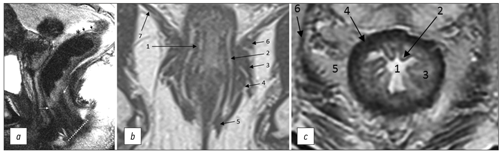
- Bogveradze N, Snaebjornsson P, Grotenhuis BA, et al. MRI anatomy of the rectum: Key concepts important for rectal cancer staging and treatment planning. Insights Imaging. 2023;14(1):13. doi: 10.1186/s13244-022-01348-8
- Gollub MJ, Arya S, Beets-Tan RG, et al. Use of magnetic resonance imaging in rectal cancer patients: Society of Abdominal Radiology (SAR) rectal cancer disease-focused panel (DFP) recommendations 2017. Abdom Radiol. 2018;43(11):2893–2902. doi: 10.1007/s00261-018-1642-9
- Nougaret S, Rousset P, Gormly K, et al. Structured and shared MRI staging lexicon and report of rectal cancer: A consensus proposal by the French Radiology Group (GRERCAR) and Surgical Group (GRECCAR) for rectal cancer. Diagn Interv Imaging. 2022;103(3):127–141. doi: 10.1016/j.diii.2021.08.003
- Grishko PY, Balyasnikova SS, Samsonov DV, et al. A modern view on the principles of diagnosis and treatment of rectal cancer according to MRI data (literature review). Medical Visualization. 2019;23(2):7–26.(In Russ). doi: 10.24835/1607-0763-2019-2-7-26
- Fernandes MC, Gollub MJ, Brown G. The importance of MRI for rectal cancer evaluation. Surg Oncol. 2022;(43):101739. doi: 10.1016/j.suronc.2022.101739
- Glynne-Jones R, Wyrwicz L, Tiret E, et al. Rectal cancer: ESMO clinical practice guidelines for diagnosis, treatment and follow-up. Ann Oncol. 2017;28(Suppl 4):22–40. doi: 10.1093/anonc/mdx22 4
- Beets-Tan R, Lambregts D, Maas M, et al. Magnetic resonance imaging for clinical management of rectal cancer: Updated recommendations from the 2016 European Society of Gastrointestinal and Abdominal Radiology (ESGAR) consensus meeting. Eur Radiol. 2018;28(4):1465–1475. doi: 10.1007/s0033 0-017-5026-2
- Oien K, Forsmo HM, Rösler C, et al. Endorectal ultrasound and magnetic resonance imaging for staging of early rectal cancers: How well does it work in practice? Acta Oncol. 2019;58(Sup1):49–54. doi: 10.1080/0284186X.2019.1569259
- Brierley JD, Gospodarowicz MK, Wittekind C. TNM Classification of Malignant Tumours. 8th ed. Wiley-Blackwell; 2017. 272 р.
- Kikuchi R, Takano M, Takagi K, et al. Management of early invasive colorectal cancer. Risk of recurrence and clinical guidelines. Dis Colon Rectum. 1995;38(12):1286–1295. doi: 10.1007/BF02049154
- Boot J, Gomez-Munoz F, Beets-Tan R. Imaging of rectal cancer. Radiologe. 2019;59(Suppl 1):46–50٠. doi: 10.1007/s00117-019-0579-5
- Lambregts D, Bogveradze N, Blomqvist L, et al. Current controversies in TNM for the radiological staging of rectal cancer and how to deal with them: Results of a global online survey and multidisciplinary expert consensus. Eur Radiol. 2022;32(7):4991–5003. doi: 10.1007/s00330-022-08591-z
- Mainovskaya OA, Rybakov EG, Chernyshov SV, et al. New morphological risk factors for metastasis to regional lymph nodes in rectal cancer with invasion of the submucosal base. Coloproctology. 2021;20(4):22–33. (In Russ). doi: 10.33878/2073-7556-2021-20-4-22-33
- Volkova SN, Stashuk GA, Chermensky GV, Naumov EK. The role of MRI in the detection of extramural vascular invasion as an indicator of the presence of regional and distant metastases of cancer of the lower ampullary rectum. Experimental Clin Gastroenterol. 2019;164(4):66–71. (In Russ). doi: 10.31146/1682-8658-ecg-164-4-66-71
- Lord AC, D’Souza N, Shaw A, et al. MRI-diagnosed tumor deposits and EMVI status have superior prognostic accuracy to current clinical TNM staging in rectal cancer. Ann Surg. 2022;276(2):334–344. doi: 10.1097/SLA.0000000000004499
- Rokan Z, Simillis C, Kontovounisios C, et al. Locally recurrent rectal cancer according to a standardized MRI classification system: A systematic review of the literature. J Clin Med. 2022;11(12):3511. doi: 10.3390/jcm11123511
- Grishko PY, Mishchenko AV, Ivko OV, et al. The possibilities of multiparametric magnetic resonance imaging in assessing the effectiveness of neoadjuvant treatment of rectal cancer. Radiation Diagnostics Therapy. 2019;10(4):49–56.(In Russ).
- Inoue A, Sheedy SP, Heiken JP, et al. MRI-detected extramural venous invasion of rectal cancer: Multimodality performance and implications at baseline imaging and after neoadjuvant therapy. Insights Imaging. 2021;(2):110. doi: 10.1186/s13244-021-01023-4
- Al-Sukhni E, Milot L, Fruitman M, et al. Diagnostic Accuracy of MRI for assessment of t category, lymph node metastases, and circumferential resection margin involvement in patients with rectal cancer: A systematic review and meta-analysis. Ann Sur Oncol. 2012;19(7):2212–2222. doi: 10.1245/s10434-011-2210-5
- Borgheresi A, De Muzio F, Agostini A, et al. Lymph nodes evaluation in rectal cancer: Where do we stand and future perspective. J Clin Med. 2022;11(9):2599. doi: 10.3390/jcm11092599
- Zhuang Z, Zhang Y, Wei M, et al. Magnetic resonance imaging evaluation of the accuracy of various lymph node staging criteria in rectal cancer: A systematic review and meta-analysis. Front Oncol. 2021;(11):709070. doi: 10.3389/fonc.2021.709070
- Li X, Sun Y, Tang L, et al. Evaluating local lymph node metastasis with magnetic resonance imaging, endoluminal ultrasound and computed tomography in rectal cancer: A meta-analysis. Color Dis. 2015;17(6):129–135. doi: 10.1111/codi.12909
- Weiser MR. AJCC 8th ed. Colorectal cancer. Ann Surg Oncol. 2018;25(6):1454–1455. doi: 10.1245/s10434-018-6462-1
- Ueno H, Nagtegaal ID, Quirke P, et al. Tumor deposits in colorectal cancer: Refining their definition in the TNM system. A G Surg. 2023;7(2):225–235. doi: 10.1002/ags3.12652
- Santiago I, Figueiredo N, Parés O, et al. MRI of rectal cancer: Relevant anatomy and staging key points. Insights Imaging. 2020;11(1):100. doi: 10.1186/s13244-020-00890-7
- Ogura A, Konishi T, Cunningham C, et al. Neoadjuvant (chemo)radiotherapy with total mesorectal excision only is not sufficient to prevent lateral local recurrence in enlarged nodes: Results of the multicenter lateral node study of patients with low cT3/4 rectal cancer. J Clin Oncol. 2019;37(1):33–43. doi: 10.1200/JCO.18.00032
- Gollub MJ, Costello JR, Ernst RD, et al. A primer on rectal MRI in patients on watch-and-wait treatment for rectal cancer. Abdom Radiol. 2023. doi: 10.1007/s00261-023-03900-6
- Berezovskaya TP, Daineko YA, Nevolskikh AA, et al. Prospective evaluation of the use of the MRTG system in determining the effectiveness of neoadjuvant chemoradiotherapy in patients with rectal cancer. Bulletin Radiol Radiol. 2021;102(1):6–17. (In Russ). doi: 10.20862/0042-4676-2021-102-1-6-17
- Almeida RR, Souza D, Matalon SA, et al. Rectal MRI after neoadjuvant chemoradiation therapy: A pictorial guide to interpretation. Abdom Radiol. 2021;46(7):3044–3057. doi: 10.1007/s00261-021-03007-w
- Shelygin YA, Chernyshov SV, Kazieva LY, et al. Comparative analysis of open and transanal total mesorectumectomy in rectal cancer. Coloproctology. 2018;(4):67–73. (In Russ).
- Maistrenko NA, Khvatov AA, Sazonov AA. Pelvic exenterations in the treatment of locally advanced tumors. Bulletin Surnamed after Grekov. 2014;173(6):37–43. (In Russ).
- Sidorov DV, Alekseev BY, Grishin NA, et al. Variants of pelvic exenteration in locally advanced primary and recurrent rectal cancer. Oncology J named after P.A. Herzen. 2013;(6):7–13. (In Russ).
补充文件


