

")


Cardiac magnetic resonance imaging in patients with history of COVID-19
- Authors: Maksimova A.S.1, Ryumshina N.I.1, Shelkovnikova T.A.1, Mochula O.V.1, Anfinogenova N.D.1, Ussov V.Y.1
-
Affiliations:
- Tomsk National Research Medical Center, Cardiology Research Institute
- Issue: Vol 4, No 3 (2023)
- Pages: 280-291
- Section: Original Study Articles
- URL: https://journals.rcsi.science/DD/article/view/254069
- DOI: https://doi.org/10.17816/DD494103
- ID: 254069
Cite item

Abstract
BACKGROUND: Myocarditis is among the most common complications arising from coronavirus infection (COVID-19).
AIM: This study aims to find the differences in the patterns of myocardial injury between patients who had COVID-19 and those from the pre-pandemic period, as determined by contrast-enhanced cardiac magnetic resonance imaging.
MATERIALS AND METHODS: The study encompassed a retrospective analysis of 47 patients who underwent contrast-enhanced cardiac magnetic resonance imaging to rule out acute myocarditis. Group 1 comprised 34 patients with a confirmed history of COVID-19 through PCR testing (nasal and/or throat swabs), while Group 2 comprised 13 individuals who underwent contrast-enhanced cardiac magnetic resonance imaging in 2017 prior to the onset of the COVID-19 pandemic. All patients enrolled in the study had clinical manifestation of cardiac injury without signs of coronary artery disease as an underlying cause of condition.
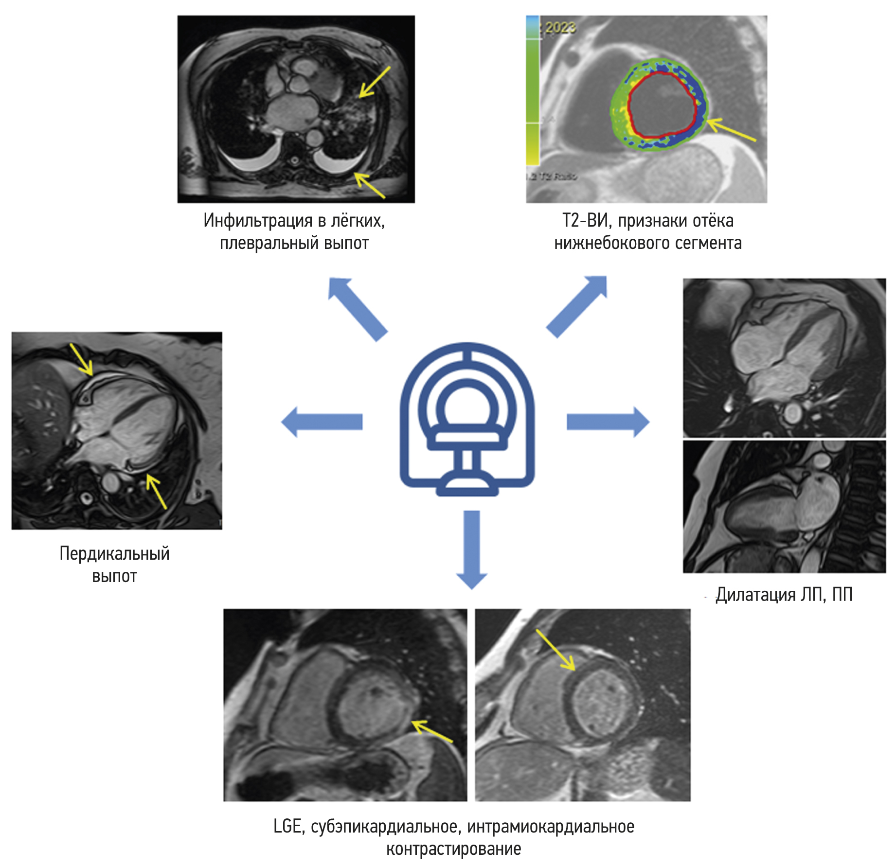
RESULTS: The mean time from the onset of heart symptoms to the administration of contrast-enhanced cardiac magnetic resonance imaging was 166 days. In group 1, a decrease in exercise tolerance was observed in 77% of patients, and 14 (42%), 30 (88%), and 28 (85%) of patients complained of chest pain, shortness of breath, and heart palpitations, respectively. In group 2, four patients (30%) had dyspnea, nine patients (69%) complained of chest pain, and six patients (46%) had heart palpitations and/or feeling of arrhythmia. Myocardial injury in group 1 was more generalized. The third of them had displayed preserved increased pulmonary vascularity and pleural effusion. Within group 1, men had significantly lower left ventricular ejection fraction, lower values of global longitudinal deformation, and higher values of left atrial function compared with the corresponding parameters in women. Differences in women were found only in the number of the affected segments in the left ventricular myocardium.
CONCLUSION: SARS-CoV-2 virus caused extended myocardial injury, affecting a significant number of myocardial segments. Men had more frequent postinflammatory complications, characterized by abnormal function of the left ventricle and left atrium. Obtained results require continuous efforts for further assessment of long-term consequences of previous COVID-19 to the cardiovascular system. In this regard, contrast-enhanced cardiac magnetic resonance imaging may represent a sensitive imaging tool for the assessment of cardiac injury severity.
Full Text
##article.viewOnOriginalSite##About the authors
Aleksandra S. Maksimova
Tomsk National Research Medical Center, Cardiology Research Institute
Email: asmaximova@yandex.ru
ORCID iD: 0000-0002-4871-3283
SPIN-code: 2879-9550
MD, Cand. Sci. (Med.)
Russian Federation, TomskNadezhda I. Ryumshina
Tomsk National Research Medical Center, Cardiology Research Institute
Author for correspondence.
Email: n.rumshina@list.ru
ORCID iD: 0000-0002-6158-026X
SPIN-code: 6555-8937
MD, Cand. Sci. (Med.)
Russian Federation, TomskTatiana A. Shelkovnikova
Tomsk National Research Medical Center, Cardiology Research Institute
Email: fflly@mail.ru
ORCID iD: 0000-0001-8089-2851
SPIN-code: 1826-7850
MD, Cand. Sci. (Med.)
Russian Federation, TomskOlga V. Mochula
Tomsk National Research Medical Center, Cardiology Research Institute
Email: mochula.olga@gmail.com
ORCID iD: 0000-0002-7502-7502
SPIN-code: 3712-8492
MD, Cand. Sci. (Med.)
Russian Federation, TomskNina D. Anfinogenova
Tomsk National Research Medical Center, Cardiology Research Institute
Email: cardio.intl@gmail.com
ORCID iD: 0000-0003-1106-0730
SPIN-code: 6784-5440
MD, Dr. Sci. (Med.)
Russian Federation, TomskVladimir Yu. Ussov
Tomsk National Research Medical Center, Cardiology Research Institute
Email: ussov1962@yandex.ru
ORCID iD: 0000-0002-7352-6068
SPIN-code: 1299-2074
MD, Dr. Sci. (Med.), Professor
Russian Federation, TomskReferences
- Ussov WY, Nudnov NV, Ignatenko GA, et al. Primary and prospective imaging of the chest using magnetic resonance imaging in patients with viral lung damage in COVID-19. Medical Imaging. 2020;24(4):11–26. (In Russ). doi: 10.24835/1607-0763-2020-4-11-26
- Srinivasan A, Wong F, Couch LS, Wang BX. Cardiac complications of COVID-19 in low-risk patients. Viruses. 2022;14(6):1322. doi: 10.3390/v14061322
- Cosyns B, Lochy S, Luchian ML, et al. The role of cardiovascular imaging for myocardial injury in hospitalized COVID-19 patients. Eur Heart J Cardiovasc Imaging. 2020;21(7):709–714. doi: 10.1093/ehjci/jeaa136
- Huang L, Zhao P, Tang D, et al. Cardiac involvement in patients recovered from COVID-2019 identified using magnetic resonance imaging. JACC Cardiovasc Imaging. 2020;13(11):2330–2339. doi: 10.1016/j.jcmg.2020.05.004
- Luetkens JA, Isaak A, Öztürk C, et al. Cardiac MRI in suspected acute COVID-19 myocarditis. Radiol Cardiothorac Imaging. 2021;3(2):e200628. doi: 10.1148/ryct.2021200628
- Puntmann VO, Carerj ML, Wieters I, et al. Outcomes of cardiovascular magnetic resonance imaging in patients recently recovered from coronavirus disease 2019 (COVID-19). JAMA Cardiol. 2020;5(11):1265–1273. doi: 10.1001/jamacardio.2020.3557
- Ferreira VM, Plein S, Wong TC, et al. Cardiovascular magnetic resonance for evaluation of cardiac involvement in COVID-19: Recommendations by the society for cardiovascular magnetic resonance. J Cardiovasc Magn Reson. 2023;25(1):21. doi: 10.1186/s12968-023-00933-0
- Yong SJ. Long COVID or post-COVID-19 syndrome: Putative pathophysiology, risk factors, and treatments. Infect Dis (Lond). 2021;53(10):737–754. doi: 10.1080/23744235.2021.1924397
- Lewis AJ, Burrage MK, Ferreira VM. Cardiovascular magnetic resonance imaging for inflammatory heart diseases. Cardiovascular Diagnosis Therapy. 2020;10(3):598–609. doi: 10.21037/cdt.2019.12.09
- Kokhan EV, Ozova M., Romanova VA, et al. Left atrial phasic function in patients with hypertension and recurrent atrial fibrillation: Gender differences of the relationship with diastolic dysfunction and central aortic pressure. Rational Pharmacotherapy Cardiology. 2019;15(5):622–633. (In Russ). doi: 10.20996/1819-6446-2019-15-5-622-633
- Kravchenko D, Isaak A, Zimmer S, et al. Cardiac MRI in patients with prolonged cardiorespiratory symptoms after mild to moderate COVID-19. Radiology. 2021;301(3):E419–E425. doi: 10.1148/radiol.2021211162
- Arutyunov GP, Paleev FN, Moiseeva OM, et al. 2020 Clinical practice guidelines for myocarditis in adults. Russ J Cardiol. 2021;26(11):4790. (In Russ). doi: 10.15829/1560-4071-2021-4790
- Feofanova TB, Zaletova TS, Abakarov RM, Zainudinov ZM. Assessment of the state of the cardiovascular system in patients with COVID-19. Int J Med Psychol. 2021;4(7):84–87. (In Russ).
- Shirokov NE, Yaroslavskaya EI, Krinochkin DV, et al. Relationship between latent left ventricular contractile dysfunction and signs of immune inflammation in patients with COVID-19 pneumonia. Cardiovascular Therapy Prevention. 2023;22(3):3434. (In Russ). doi: 10.15829/1728-8800-2023-3434
- Pozios I, Vouliotis AI, Dilaveris P, Tsioufis C. Electro-mechanical alterations in atrial fibrillation: Structural, electrical, and functional correlates. J Cardiovasc Dev Dis. 2023;10(4):149. doi: 10.3390/jcdd10040149
- Raisi-Estabragh Z, McCracken C, Condurache D, et al. Left atrial structure and function are associated with cardiovascular outcomes independent of left ventricular measures: A UK Biobank CMR study. Eur Heart J Cardiovasc Imaging. 2022;23(9):1191–1200. doi: 10.1093/ehjci/jeab266
- Floria M, Radu S, Gosav EM, et al. Left atrial structural remodelling in non-valvular atrial fibrillation: What have we learnt from CMR? Diagnostics (Basel). 2020;10(3):137. doi: 10.3390/diagnostics10030137
- Kim HD, Cho DH, Kim MN, et al. Left atrial dysfunction, fibrosis and the risk of thromboembolism in patients with paroxysmal and persistent atrial fibrillation. Int J Heart Fail. 2022;4(1):42–53. doi: 10.36628/ijhf.2021.0043
- Schönbauer R, Kammerlander AA, Duca F, et al. Prognostic impact of left atrial function in heart failure with preserved ejection fraction in sinus rhythm vs persistent atrial fibrillation. ESC Heart Fail. 2022;9(1):465–475. doi: 10.1002/ehf2.13723
- Chistyakova MV, Govorin AV, Mudrov VA, et al. Heart damage and endothelial dysfunction in patients with coronavirus infection. Therapists Bulletin. 2023;(1):1–7. (In Russ).
- Rienstra M, van Veldhuisen DJ, Hagens VE, et al. Gender-related differences in rhythm control treatment in persistent atrial fibrillation. J Am Coll Cardiol. 2005;46(7):1298–306. doi: 10.1016/j.jacc.2005.05.078
- Proietti M, Raparelli V, Basili S, et al. Relation of female sex to left atrial diameter and cardiovascular death in atrial fibrillation: The AFFIRM Trial. Int J Cardiol. 2016;(207):258–263. doi: 10.1016/j.ijcard.2016.01.169
- Yaroslavskaya EI, Krinochkin DV, Shirokov NE, et al. Clinical and echocardiographic profile of patients one year after COVID-19 pneumonia depending on the left ventricular global longitudinal strain. Siberian J Clin Experimental Med. 2022;37(4):52–62. (In Russ). doi: 10.29001/2073-8552-2022-37-4-52-62
- Wong GR, Nalliah CJ, Lee G, et al. Sex-Related differences in atrial remodeling in patients with atrial fibrillation: Relationship to ablation outcomes. Circ Arrhythm Electrophysiol. 2022;15(1):e009925. doi: 10.1161/CIRCEP.121.009925
- Bräuninger H, Stoffers B, Fitzek AD, et al. Cardiac SARS-CoV-2 infection is associated with pro-inflammatory transcriptomic alterations within the heart. Cardiovasc Res. 2022;118(2):542–555. doi: 10.1093/cvr/cvab322
- Wu L, Jiang Z, Meulendijks ER, et al. Atrial inflammation and microvascular thrombogenicity are increased in deceased COVID-19 patients. Cardiovasc Pathol. 2023;(64):107524. doi: 10.1016/j.carpath.2023.107524
Supplementary files


