

")


Frequency of various cardiac complications in children with repaired tetralogy of Fallot identified by computer tomography
- Authors: Kabdullina A.M.1, Sinitsyn V.E.2, Rakhimzhanova R.I.1, Dautov T.B.3, Saduakassova A.B.4, Kaliyev B.B.1, Bastarbekova L.A.1, Moldakhanova Z.A.1
-
Affiliations:
- Astana Medical University
- Lomonosov Moscow State University
- Department of the Radiology of National Research Cardiac Surgery Center
- Medical Centre Hospital of President’s Affairs Administration of the Republic of Kazakhstan
- Issue: Vol 4, No 3 (2023)
- Pages: 268-279
- Section: Original Study Articles
- URL: https://journals.rcsi.science/DD/article/view/254068
- DOI: https://doi.org/10.17816/DD375285
- ID: 254068
Cite item

Abstract
BACKGROUND: Tetralogy of Fallot represents 7–10% of all cases of congenital heart disease, as it occurs in approximately 0.5 per 1,000 live births and is the second most common form of complex congenital heart disease. Advances in diagnosis, surgical techniques, and postoperative treatment have led to an increasing number of patients reaching adulthood, with a dramatic increase in the survival rate to almost 90% at 30 years, thereby creating a need for long-term monitoring of certain anatomic parameters to identify complications in a timely manner. This study aimed to investigate the frequency of computed tomography detected complications after radical correction of Tetralogy of Fallot in pediatric patients.
AIM: to identify markers between the most frequency computed tomography detected complications after repair of Tetralogy of Fallot in pediatric patients.
MATERIALS AND METHODS: A retrospective analysis was conducted on 613 patients with Tetralogy of Fallot from October 2011 to June 2020. The study included a total of 116 patients (69 men and 47 women) who experienced complications after a repair of Tetralogy of Fallot, as identified by computed tomography. At the time of repair of Tetralogy of Fallot, the patient’s average age ranged from 10 to 36 months (mean: 12 months), average body weight was 21 kg, average height was 105.4 cm, and average body surface area was 0.74 m2. The patients’ median age at the time of the computed tomography examination was 17.5 years (age range: 7–36 years).
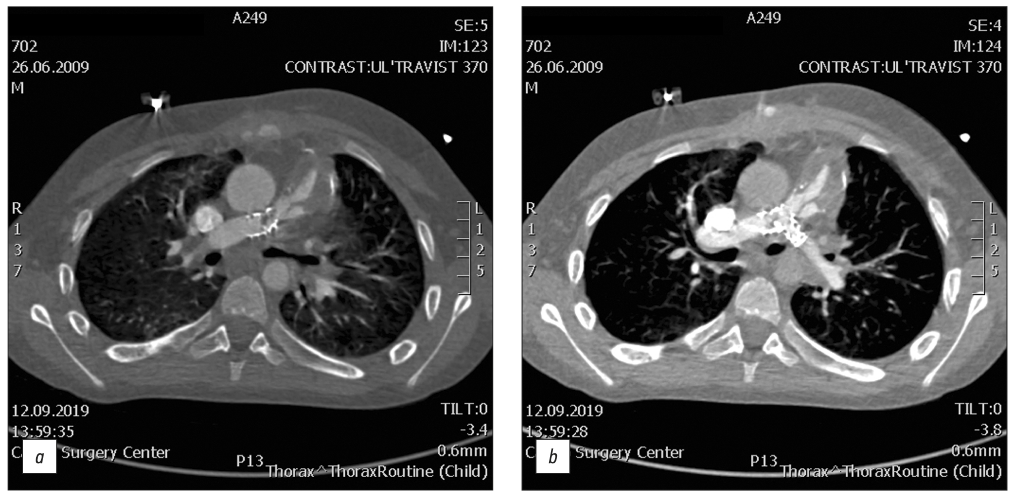
RESULTS: Among the 116 patients who exhibited complications after an repair of Tetralogy of Fallot, 49 had a pulmonary artery stenosis, 92 had a pulmonary artery branch stenosis (56 of them of the left main pulmonary artery branch, and 36 of them of the right main pulmonary artery branch), 8 had a right ventricular outflow tract stenosis, 32 had a ventricular septal defect, 1 had a shunt thrombosis, 12 had a postoperative deformation of the pulmonary artery, 10 exhibited a marked right ventricular dilatation, 2 had an right ventricular outflow tract aneurysm, and 6 suffered from conduit calcification and stenosis. Moreover, patients with left main pulmonary artery branch stenosis had a 6.5 times greater chance of developing an right main pulmonary artery branch stenosis in (p <0.001).
CONCLUSION: The most frequently computed tomography detected complications after a repair of Tetralogy of Fallot were pulmonary artery stenosis and pulmonary artery branch stenosis. Patients with pulmonary artery stenosis and pulmonary artery branch stenosis exhibit no significant differences in terms of age, anthropometric parameters (height, weight, and body surface area), and gender distribution in the presence or absence of different stenosis types (pulmonary artery, right main pulmonary artery branch, or left main pulmonary artery branch). However, an right main pulmonary artery branch stenosis increases the chances of developing an left main pulmonary artery branch stenosis.
Full Text
##article.viewOnOriginalSite##About the authors
Azhar M. Kabdullina
Astana Medical University
Author for correspondence.
Email: azharazh@mail.ru
ORCID iD: 0000-0003-0521-5484
SPIN-code: 4169-1761
Kazakhstan, Astana
Valentin E. Sinitsyn
Lomonosov Moscow State University
Email: vsini@mail.ru
ORCID iD: 0000-0002-5649-2193
SPIN-code: 8449-6590
MD, Dr. Sci. (Med.), Professor
Russian Federation, MoscowRaushan I. Rakhimzhanova
Astana Medical University
Email: rakhimzhanova01@rambler.ru
ORCID iD: 0000-0002-3490-6324
MD, Dr. Sci. (Med.), Professor
Kazakhstan, AstanaTairkhan B. Dautov
Department of the Radiology of National Research Cardiac Surgery Center
Email: tairkhan.dautov@mail.ru
ORCID iD: 0000-0002-5267-0108
MD, Dr. Sci. (Med.), Professor
Kazakhstan, AstanaAigul B. Saduakassova
Medical Centre Hospital of President’s Affairs Administration of the Republic of Kazakhstan
Email: sadik.a@mail.ru
ORCID iD: 0000-0001-7089-5696
MD, Dr. Sci. (Med.)
Kazakhstan, AstanaBaurzhan B. Kaliyev
Astana Medical University
Email: Baur233113@mail.ru
ORCID iD: 0000-0003-4825-749X
Kazakhstan, Astana
Lyazzat A. Bastarbekova
Astana Medical University
Email: lbastarbekova@mail.ru
ORCID iD: 0000-0001-8246-4754
SPIN-code: 8634-6601
Kazakhstan, Astana
Zhanar A. Moldakhanova
Astana Medical University
Email: moldahanova1991@mail.ru
ORCID iD: 0000-0002-5980-9563
Kazakhstan, Astana
References
- Apostolopoulou SC, Manginas A, Kelekis NL, Noutsias M. Cardiovascular imaging approach in pre and postoperative tetralogy of Fallot. BMC Cardiovasc Dis. 2019;19(1):7. doi: 10.1186/s12872-018-0996-9
- Shaaban M, Tantawy S, Elkafrawy F, et al. Multi-detector computed tomography in the assessment of tetralogy of Fallot patients: Is it a must? Egyptian Heart J. 2020;72(1):17. doi: 10.1186/s43044-020-00047-3
- Goo HW. Changes in right ventricular volume, volume load, and function measured with cardiac computed tomography over the entire time course of tetralogy of Fallot. Korean J Radiol. 2019;20(6):956–966. doi: 10.3348/kjr.2018.0891
- Stout KK, Daniels CJ, Aboulhosn JA, et al. 2018 AHA/ACC Guideline for the management of adults with congenital heart disease: Executive summary: A report of the American College of Cardiology / American Heart Association Task Force on Clinical Practice Guidelines. J Am College Cardiol. 2019;73(12):1494–1563. doi: 10.1016/j.jacc.2018.08.1028
- Chelliah A, Shah AM, Farooqi KM, Einstein AJ. Cardiovascular CT in cyanotic congenital heart disease. Curr Cardiovasc Imaging Rep. 2019;12(7):30. doi: 10.1007/s12410-019-9507-3
- Goo HW. Comparison of chest pain protocols for electrocardiography-gated dual-source cardiothoracic CT in children and adults: The effect of tube current saturation on radiation dose reduction. Korean J Radiol. 2018;19(1):23–31. doi: 10.3348/kjr.2018.19.1.23
- Siripornpitak S, Goo HW. CT and MRI for repaired complex adult congenital heart diseases. Korean J Radiol. 2021;22(3):308–323. doi: 10.3348/kjr.2020.0895
- Singh R, Jain N, Kumar S, Garg N. Multi-detector computed tomography angiographic evaluation of right ventricular outflow tract obstruction and other associated cardiovascular anomalies in tetralogy of Fallot patients. Polish J Radiol. 2019;84:511–516. doi: 10.5114/pjr.2019.91203
- Kossaify A. Echocardiographic assessment of the right ventricle, from the conventional approach to speckle tracking and three-dimensional imaging, and insights into the “Right Way” to explore the forgotten chamber. Clin Med Insights Cardiol. 2015;9:65–75. doi: 10.4137/CMC.S27462
- Pushparajah K, Duong P, Mathur S, Babu-Narayan SV. Cardiovascular MRI and CT in congenital heart disease. Echo Res Pract. 2019;6(4):R121–138. doi: 10.1530/ERP-19-0048
Supplementary files


