

")


电子计算机断层扫描确定的法洛四联症修复患儿各种心脏并发症的发生率
- 作者: Kabdullina A.M.1, Sinitsyn V.E.2, Rakhimzhanova R.I.1, Dautov T.B.3, Saduakassova A.B.4, Kaliyev B.B.1, Bastarbekova L.A.1, Moldakhanova Z.A.1
-
隶属关系:
- Astana Medical University
- Lomonosov Moscow State University
- Department of the Radiology of National Research Cardiac Surgery Center
- Medical Centre Hospital of President’s Affairs Administration of the Republic of Kazakhstan
- 期: 卷 4, 编号 3 (2023)
- 页面: 268-279
- 栏目: 原创性科研成果
- URL: https://journals.rcsi.science/DD/article/view/254068
- DOI: https://doi.org/10.17816/DD375285
- ID: 254068
如何引用文章

详细
论证。法洛四联症(Tetralogy of Fallot,ToF)占所有先天性心脏病(congenital heart disease,CHD)病例的7-10%,每1000例活产中约有0.5例发生,是第二种最常见的复杂先天性心脏病。随着诊断、手术技术和术后治疗的进步,越来越多的患者长大成人,30岁时的存活率急剧上升到近 90%,因此需要对某些解剖参数进行长期监测,以便及时发现并发症。本研究旨在调查儿童患者患儿根治性矫正ToF后计算机断层扫描(computed tomography,CT)发现并发症的发生率。
该研究的目的是确定小儿ToF修复术(repair of ToF,rToF)后CT检测到的最常见并发症之间的标记。
材料和方法。我们对2011年10月至2020年6月期间的613例ToF患者进行了回顾性分析。116名患者(69名男性和47名女性)被纳入该研究,这些患者在接受rToF后出现了通过CT发现的并发症。患者接受rToF时的平均年龄为10至36个月(平均值为12个月),平均体重为21kg,平均身高为105.4cm,平均体表面积(body surface area,BSA)为0.74m2。患者接受CT检查时的中位年龄为17.5岁(年龄范围:7至36岁)。
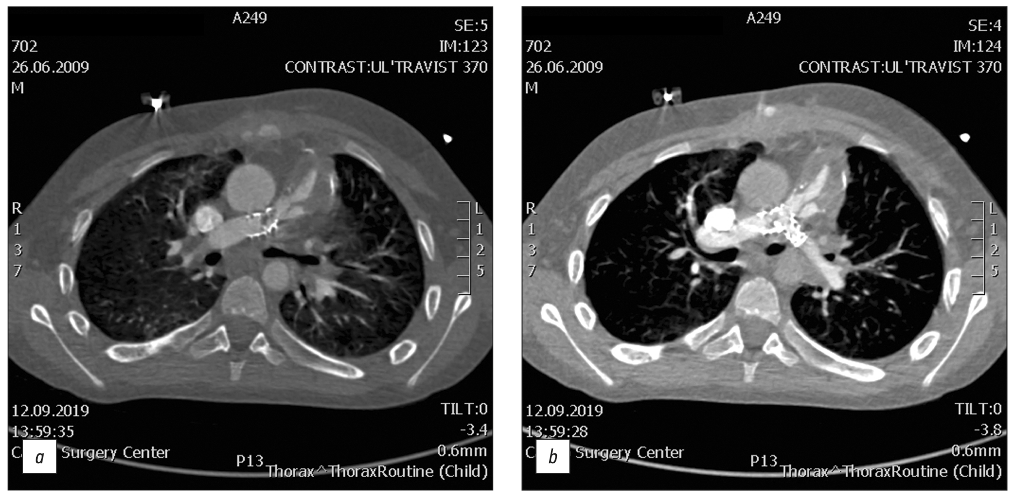
结果。在116例rToF后出现并发症的患者中,49例有肺动脉(pulmonary artery,PA)狭窄,92例有PA分支狭窄(其中56例有PA左主分支(left main PA branch,LPA),36例有PA右主分支(right main PA branch,RPA)),8例有右室流出道(right ventricular outflow tract,RVOT)狭窄、32例有室间隔缺损,1例有分流道血栓形成,12例有术后PA变形,10例有明显的右心室扩张,2例有RVOT动脉瘤,6例有导管钙化和狭窄。此外,对于LPA狭窄患者来说,发生RPA狭窄的几率比正常人高出6.5倍(p<0.001)。
结论。rToF后最常在CT上发现的并发症是PA狭窄和PA分支狭窄。PA狭窄和PA分支狭窄患者在年龄、人体测量数(身高、体重和BSA)和性别分布方面与是否存在不同狭窄类型(PA、RPA或LPA)无明显差异。然而,RPA狭窄会增加发生LPA狭窄的几率。
作者简介
Azhar M. Kabdullina
Astana Medical University
编辑信件的主要联系方式.
Email: azharazh@mail.ru
ORCID iD: 0000-0003-0521-5484
SPIN 代码: 4169-1761
哈萨克斯坦, Astana
Valentin E. Sinitsyn
Lomonosov Moscow State University
Email: vsini@mail.ru
ORCID iD: 0000-0002-5649-2193
SPIN 代码: 8449-6590
MD, Dr. Sci. (Med.), Professor
俄罗斯联邦, MoscowRaushan I. Rakhimzhanova
Astana Medical University
Email: rakhimzhanova01@rambler.ru
ORCID iD: 0000-0002-3490-6324
MD, Dr. Sci. (Med.), Professor
哈萨克斯坦, AstanaTairkhan B. Dautov
Department of the Radiology of National Research Cardiac Surgery Center
Email: tairkhan.dautov@mail.ru
ORCID iD: 0000-0002-5267-0108
MD, Dr. Sci. (Med.), Professor
哈萨克斯坦, AstanaAigul B. Saduakassova
Medical Centre Hospital of President’s Affairs Administration of the Republic of Kazakhstan
Email: sadik.a@mail.ru
ORCID iD: 0000-0001-7089-5696
MD, Dr. Sci. (Med.)
哈萨克斯坦, AstanaBaurzhan B. Kaliyev
Astana Medical University
Email: Baur233113@mail.ru
ORCID iD: 0000-0003-4825-749X
哈萨克斯坦, Astana
Lyazzat A. Bastarbekova
Astana Medical University
Email: lbastarbekova@mail.ru
ORCID iD: 0000-0001-8246-4754
SPIN 代码: 8634-6601
哈萨克斯坦, Astana
Zhanar A. Moldakhanova
Astana Medical University
Email: moldahanova1991@mail.ru
ORCID iD: 0000-0002-5980-9563
哈萨克斯坦, Astana
参考
- Apostolopoulou SC, Manginas A, Kelekis NL, Noutsias M. Cardiovascular imaging approach in pre and postoperative tetralogy of Fallot. BMC Cardiovasc Dis. 2019;19(1):7. doi: 10.1186/s12872-018-0996-9
- Shaaban M, Tantawy S, Elkafrawy F, et al. Multi-detector computed tomography in the assessment of tetralogy of Fallot patients: Is it a must? Egyptian Heart J. 2020;72(1):17. doi: 10.1186/s43044-020-00047-3
- Goo HW. Changes in right ventricular volume, volume load, and function measured with cardiac computed tomography over the entire time course of tetralogy of Fallot. Korean J Radiol. 2019;20(6):956–966. doi: 10.3348/kjr.2018.0891
- Stout KK, Daniels CJ, Aboulhosn JA, et al. 2018 AHA/ACC Guideline for the management of adults with congenital heart disease: Executive summary: A report of the American College of Cardiology / American Heart Association Task Force on Clinical Practice Guidelines. J Am College Cardiol. 2019;73(12):1494–1563. doi: 10.1016/j.jacc.2018.08.1028
- Chelliah A, Shah AM, Farooqi KM, Einstein AJ. Cardiovascular CT in cyanotic congenital heart disease. Curr Cardiovasc Imaging Rep. 2019;12(7):30. doi: 10.1007/s12410-019-9507-3
- Goo HW. Comparison of chest pain protocols for electrocardiography-gated dual-source cardiothoracic CT in children and adults: The effect of tube current saturation on radiation dose reduction. Korean J Radiol. 2018;19(1):23–31. doi: 10.3348/kjr.2018.19.1.23
- Siripornpitak S, Goo HW. CT and MRI for repaired complex adult congenital heart diseases. Korean J Radiol. 2021;22(3):308–323. doi: 10.3348/kjr.2020.0895
- Singh R, Jain N, Kumar S, Garg N. Multi-detector computed tomography angiographic evaluation of right ventricular outflow tract obstruction and other associated cardiovascular anomalies in tetralogy of Fallot patients. Polish J Radiol. 2019;84:511–516. doi: 10.5114/pjr.2019.91203
- Kossaify A. Echocardiographic assessment of the right ventricle, from the conventional approach to speckle tracking and three-dimensional imaging, and insights into the “Right Way” to explore the forgotten chamber. Clin Med Insights Cardiol. 2015;9:65–75. doi: 10.4137/CMC.S27462
- Pushparajah K, Duong P, Mathur S, Babu-Narayan SV. Cardiovascular MRI and CT in congenital heart disease. Echo Res Pract. 2019;6(4):R121–138. doi: 10.1530/ERP-19-0048
补充文件


