

")


Evaluation of fetal absorbed doses from computed tomography examinations of pregnant patients: A systematic review
- Authors: Vodovatov A.V.1, Golchenko O.A.2, Mashchenko I.A.3, Alekseeva D.V.3, Chipiga L.A.1,3, Khutornoy I.V.4, Kozlova P.V.3, Trufanov G.E.3, Druzhinina P.S.1, Ryzhov S.A.5, Soldatov I.V.5
-
Affiliations:
- Saint-Petersburg Research Institute of Radiation Hygiene after Professor P.V. Ramzaev
- City polyclinic № 19
- Almazov National Medical Research Centre
- Lomonosov Moscow State University
- Research and Practice Center of Diagnostics and Telemedicine Technologies
- Issue: Vol 4, No 2 (2023)
- Pages: 170-184
- Section: Systematic reviews
- URL: https://journals.rcsi.science/DD/article/view/146884
- DOI: https://doi.org/10.17816/DD472150
- ID: 146884
Cite item

Abstract
BACKGROUND: Currently, no systematic data are available on fetal radiation exposure as a result of radiographic studies during pregnancy. Consequently, there are no approved methods of its calculation that can be used in clinical practice. It is especially relevant for computed tomography scans as it is a widely used and highly informative method of diagnostic imaging associated with high exposure levels.
AIM: to systematize currently available data on radiation dose absorbed by the fetus from computed tomography scans in pregnant women.
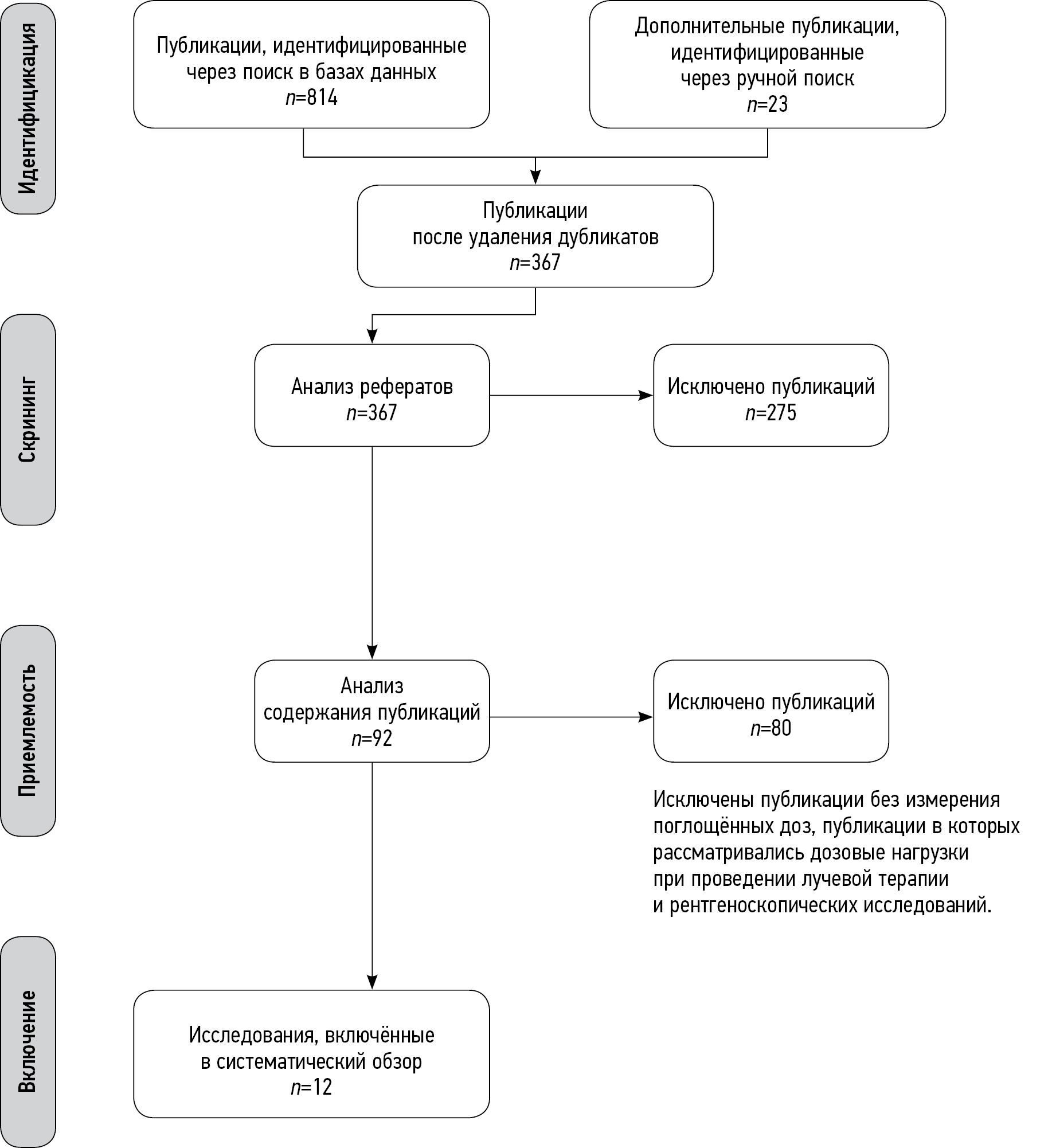
MATERIALS AND METHODS: The search for publications in Russian and English was conducted in PubMed/Medline, Google Scholar and еLibrary. The final analysis included 12 papers including 8 studies using human body phantoms, 3 retrospective studies and one prospective clinical study.
RESULTS: Abdominal and pelvic computed tomography scans as well as whole-body scans were found to be associated with the highest fetal radiation exposure. However, in none of the publications the fetal exposure limit was exceeded.
CONCLUSION: Clinically indicated non-contrast-enhanced computed tomography scans in pregnant women are not likely to be associated with the fetal absorbed doses that exceed the limit of 100 mGy regardless of the scanned area. However, this limit might be exceeded in case of performing multiple studies or if multiphase abdominal or pelvic computed tomography scans, or whole-body computed tomography scans are performed in patients with multiple trauma. In these cases, a decision regarding the need for these investigations should be made by a multi-disciplinary team (including radiation safety specialists, diagnostic radiologists and clinicians) based on the results of additional risk assessment.
Full Text
##article.viewOnOriginalSite##About the authors
Aleksandr V. Vodovatov
Saint-Petersburg Research Institute of Radiation Hygiene after Professor P.V. Ramzaev
Email: vodovatoff@gmail.com
ORCID iD: 0000-0002-5191-7535
SPIN-code: 4560-8978
Cand. Sci. (Biol.)
Russian Federation, Saint PetersburgOlga A. Golchenko
City polyclinic № 19
Email: breakerxolyga@yandex.ru
ORCID iD: 0000-0003-4614-9241
Russian Federation, Saint Petersburg
Irina A. Mashchenko
Almazov National Medical Research Centre
Email: mashchenko_ia@almazovcentre.ru
ORCID iD: 0000-0002-4949-8829
SPIN-code: 5154-7080
MD, Cand. Sci. (Med.), Leading Researcher, Assistant Professor
Russian Federation, Saint PetersburgDarya V. Alekseeva
Almazov National Medical Research Centre
Email: darja-karpova@yandex.ru
ORCID iD: 0000-0001-9528-9377
SPIN-code: 6484-4327
Assistant Lecturer
Russian Federation, Saint PetersburgLarisa A. Chipiga
Saint-Petersburg Research Institute of Radiation Hygiene after Professor P.V. Ramzaev; Almazov National Medical Research Centre
Email: larisa.chipiga@gmail.com
ORCID iD: 0000-0001-9153-3061
SPIN-code: 3920-7798
Cand. Sci. (Engin.), Research Associate, Assistant Professor
Russian Federation, Saint Petersburg; Saint PetersburgIvan V. Khutornoy
Lomonosov Moscow State University
Email: mcdm.avk@gmail.com
ORCID iD: 0000-0002-5405-603X
SPIN-code: 8020-0222
Graduate Student
Russian Federation, MoscowPolina V. Kozlova
Almazov National Medical Research Centre
Email: apollinaria@bk.ru
ORCID iD: 0000-0002-3240-7038
SPIN-code: 3555-0410
Assistant Lecturer
Russian Federation, Saint PetersburgGennady E. Trufanov
Almazov National Medical Research Centre
Email: trufanovge@mail.ru
ORCID iD: 0000-0002-1611-5000
SPIN-code: 3139-3581
MD, Dr. Sci. (Med.)
Russian Federation, Saint PetersburgPolina S. Druzhinina
Saint-Petersburg Research Institute of Radiation Hygiene after Professor P.V. Ramzaev
Email: druzhininapauline@gmail.com
ORCID iD: 0000-0003-2921-067X
SPIN-code: 9003-3234
Junior Research Associate
Russian Federation, Saint PetersburgSergey A. Ryzhov
Research and Practice Center of Diagnostics and Telemedicine Technologies
Email: mosrg@mail.ru
ORCID iD: 0000-0002-0640-7368
SPIN-code: 6595-4011
Research Associate
Russian Federation, MoscowIlia V. Soldatov
Research and Practice Center of Diagnostics and Telemedicine Technologies
Author for correspondence.
Email: SoldatovIV2@zdrav.mos.ru
ORCID iD: 0000-0002-4867-0746
SPIN-code: 4065-6048
Russian Federation, Moscow
References
- Shtentsel RE, Semenova ES, Mashchenko IA, et al. The history of the formation and development of methods of radiation diagnostics in perinatology. Translational Med. 2021;8(3):29–36. (In Russ). doi: 10.18705/2311-4495-2021-8-3-29-36
- Sadro C, Bernstein MP, Kanal KM. Imaging of trauma. Part 2, Abdominal trauma and pregnancy: A radiologist’s guide to doing what is best for the mother and baby. Am J Roentgenol. 2012;199(6):1207–1219. doi: 10.2214/AJR.12.9091
- Wang PI, Chong ST, Kielar AZ, et al. Imaging of pregnant and lactating patients: Part 1, evidence-based review and recommendations. Am J Roentgenol. 2012;198(4):778–784. doi: 10.2214/AJR.11.7405
- Committee Opinion No. 723: Guidelines for diagnostic imaging during pregnancy and lactation. Obstet Gynecol. 2017;130(4):210–216. doi: 10.1097/AOG.0000000000002355
- ICRP. Recommendations International Commission on Radiation Protection 2007. Publication ICRP No. 103. Transl. from English. Ed. by M.F. Kiselev and N.K. Shandala. Moscow: Alana; 2009. 312 р.
- ACR-SPR practice parameter for imaging pregnant or potentially pregnant adolescents and women with ionizing radiation. 2018. 23 p. Available from: https://www.acr.org/-/media/acr/files/practice-parameters/pregnant-pts.pdf. Accessed: 17.05.2023.
- Frush D. The cumulative radiation dose paradigm in pediatric imaging. Br J Radiol. 2021;94(1126):20210478. doi: 10.1259/bjr.20210478
- Vodovatov AV, Chipiga LA, Piven PA, et al. Assessment of the absorbed doses in the fetus from the computed tomography of the chest for the pregnant women. Radiatsionnaya Gygiena. 2021;14(3):126–135. (In Russ). doi: 10.21514/1998-426X-2021-14-3-126-135
- Liu H, Liu F, Li J, et al. Clinical and CT imaging features of the COVID-19 pneumonia: Focus on pregnant women and children. J Infect. 2020;80(5):7–13. doi: 10.1016/j.jinf.2020.03.007
- Dehan L, Lin L, Xin W, et al. Pregnancy and perinatal outcomes of women with coronavirus disease (COVID-19) pneumonia: A preliminary analysis. Am J Roentgenol. 2020;215(1):127–132. doi: 10.2214/AJR.20.23072
- Garcia EM, Camacho MA, Karolyi DR, et al.; Expert Panel on Gastrointestinal Imaging. ACR appropriateness criteria right lower quadrant pain-suspected appendicitis. Am J Roentgenol. 2018;15(11):373–387. doi: 10.1016/j.jacr.2018.09.033
- Sazhin AV, Kirienko AI, Kurtser MA, et al. Acute appendicitis during pregnancy (in Russian only). Pirogov Russian Journal of Surgery. 2019;(1):70–77. (In Russ).
- Gu J, Bednarz B, Caracappa PF, Xu XG. The development, validation and application of a multi-detector CT (MDCT) scanner model for assessing organ doses to the pregnant patient and the fetus using Monte Carlo simulations. Phys Med Biol. 2009;54(9):2699–2717. doi: 10.1088/0031-9155/54/9/007
- Kelaranta A, Mäkelä T, Kaasalainen T, Kortesniemi M. Fetal radiation dose in three common CT examinations during pregnancy: Monte Carlo study. Phys Med. 2017;(43):199–206. doi: 10.1016/j.ejmp.2017.09.120
- Angel E, Wellnitz CV, Goodsitt MM, et al. Radiation dose to the fetus for pregnant patients undergoing multidetector CT imaging: Monte Carlo simulations estimating fetal dose for a range of gestational age and patient size. Radiology. 2008;249(1):220–227. doi: 10.1148/radiol.2491071665
- Goldberg-Stein S, Liu B, Hahn PF, Lee SI. Body CT during pregnancy: Utilization trends, examination indications, and fetal radiation doses. Am J Roentgenol. 2011;196(1):146–151. doi: 10.2214/AJR.10.4271
- Vandecaveye V, Amant F, Lecouvet F, et al. Imaging modalities in pregnant cancer patients. Int J Gynecol Cancer. 2021;31(3):423–431. doi: 10.1136/ijgc-2020-001779
- Kwan ML, Miglioretti DL, Marlow EC, et al.; Radiation-Induced Cancers Study Team. Trends in medical imaging during pregnancy in the United States and Ontario, Canada, 1996 to 2016. JAMA Network Open. 2019(7):197–249. doi: 10.1001/jamanetworkopen.2019.7249
- Prokop M, Galanski M. Spiral and multilayered computed tomography. Moscow; 2011. 440 р. (In Russ).
- Arablinskiy AV, Magdebura YA. CT in the diagnosis of nontraumatic acute abdomen. REJR. 2018;8(2):58–71. (In Russ). doi: 10.21569/2222-7415-2018-8-2-58-71
- Kirsch J, Brown RK, Henry TS, et al.; Expert Panels on Cardiac and Thoracic Imaging. ACR appropriateness criteria acute chest pain-suspected pulmonary embolism. J Am Coll Radiol. 2017;14(5):2–12. doi: 10.1016/j.jacr.2017.02.027
- Shyu JY, Khurana B, Soto JA, et al. Expert Panel on Major Trauma Imaging. ACR appropriateness criteria major blunt trauma. J Am Coll Radiol. 2020;17(5):160–174. doi: 10.1016/j.jacr.2020.01.024
- Panchenko EP, Balahonova TV, Danilov NM, et al. Diagnosis and management of pulmonary embolism Eurasian Association of Cardiology (EAC) clinical practice guidelines (2021). Eurasian Heart J. 2021;(1):44–77. (In Russ). doi: 10.381092225-1685-2021-1-44-77
- Ria F, D’Ercole L, Origgi D, et al.; Association of Medical Physics Task Group. Statement of the Italian Association of Medical Physics (AIFM) task group on radiation dose monitoring systems. Insights Imaging. 2022;13(1):23. doi: 10.1186/s13244-022-01155-1
- Sensakovic WF, Royall I, Hough M, et al. Fetal dosimetry at CT: A primer. Radiographics. 2020;40(4):1061–1070. doi: 10.1148/rg.2020190166
- Jaffe TA, Yoshizumi TT, Toncheva GI, et al. Early first-trimester fetal radiation dose estimation in 16-MDCT without and with automated tube current modulation. Am J Roentgenol. 2008;190(4):860–864. doi: 10.2214/AJR.07.2925
- Huda W, Randazzo W, Tipnis S, et al. Embryo dose estimates in body CT. Am J Roentgenol. 2010;194(4):874–880. doi: 10.2214/AJR.09.4032
- Damilakis J, Perisinakis K, Tzedakis A, et al. Radiation dose to the conceptus from multidetector CT during early gestation: A method that allows for variations in maternal body size and conceptus position. Radiology. 2010;257(2):483–489. doi: 10.1148/radiol.10092397
- Lazarus E, Debenedectis C, North D, et al. Utilization of imaging in pregnant patients: 10-year review of 5270 examinations in 3285 patients 1997–2006. Radiology. 2009;251(2):517–524. doi: 10.1148/radiol.2512080736
- Lazarus E, Mayo-Smith WW, Mainiero MB, Spencer PK. CT in the evaluation of nontraumatic abdominal pain in pregnant women. Radiology. 2007;244(3):784–790. doi: 10.1148/radiol.2443061634
- Goldberg-Stein SA, Liu B, Hahn PF, Lee SI. Radiation dose management: Part 2, estimating fetal radiation risk from CT during pregnancy. Am J Roentgenol. 2012;198(4):352–356. doi: 10.2214/AJR.11.7458
- Litmanovich D, Boiselle PM, Bankier AA, et al. Dose reduction in computed tomographic angiography of pregnant patients with suspected acute pulmonary embolism. J Comput Assist Tomogr. 2009;33(6):961–966. doi: 10.1097/RCT.0b013e318198cd18
- Begano D, Söderberg M, Bolejko A. To use or not use patient shielding on pregnant women undergoing CT pulmonary angiography: A phantom study. Radiation Protection Dosimetry. 2020;189(4):458–465.
- Ryckx N, Sans-Merce M, Schmidt S, et al. The use of out-of-plane high Z patient shielding for fetal dose reduction in computed tomography: Literature review and comparison with Monte-Carlo calculations of an alternative optimisation technique. Phys Med. 2018;(48):156–161. doi: 10.1016/j.ejmp.2018.03.017
- Tack D, Kalra MK, Gevenois PA. Radiation dose from multidetector CT (2nd ed.). Springer; 2012. doi: 10.1007/978-3-642-24535-0
- Doshi SK, Negus IS, Oduko JM. Fetal radiation dose from CT pulmonary angiography in late pregnancy: A phantom study. Br J Radiol. 2008;81(968):653–658. doi: 10.1259/bjr/22775594
- Gilet AG, Dunkin JM, Fernandez TJ, et al. Fetal radiation dose during gestation estimated on an anthropomorphic phantom for three generations of CT scanners. Am J Roentgenol. 2011;196(5):1133–1137. doi: 10.2214/AJR.10.4497
Supplementary files


