

")


Low-dose computed tomography in COVID-19: systematic review
- Authors: Blokhin I.A.1, Rumyantsev D.А.1, Suchilova M.M.1, Gonchar A.P.1, Omelyanskaya O.V.1
-
Affiliations:
- Moscow Center for Diagnostics and Telemedicine
- Issue: Vol 4, No 1 (2023)
- Pages: 25-37
- Section: Systematic reviews
- URL: https://journals.rcsi.science/DD/article/view/146873
- DOI: https://doi.org/10.17816/DD119870
- ID: 146873
Cite item

Abstract
BACKGROUND: The increased number of computed tomography scans during the COVID-19 pandemic has emphasized the task of decreasing radiation exposure of patients, since it is known to be associated with an elevated risk of cancer development. The ALARA (as low as reasonably achievable) principle, proposed by the International Commission on Radiation Protection, should be adhered to in the operation of radiation diagnostics departments, even during the pandemic.
AIM: To systematize data on the appropriateness and effectiveness of low-dose computed tomography in the diagnosis of lung lesions in COVID-19.
MATERIALS AND METHODS: Relevant national and foreign literature in scientific libraries PubMed and eLIBRARY, using English and Russian queries “low-dose computed tomography” and “COVID-19,” published between 2020 and 2022 were analyzed. Publications were evaluated after assessing the relevance to the review topic by title and abstract analysis. The references were further analyzed to identify articles omitted during the search that may meet the inclusion criteria.
RESULTS: Published studies summarized the current data on the imaging of COVID-19 lung lesions and the use of computed tomography scans and identified possible options for reducing the effective dose.
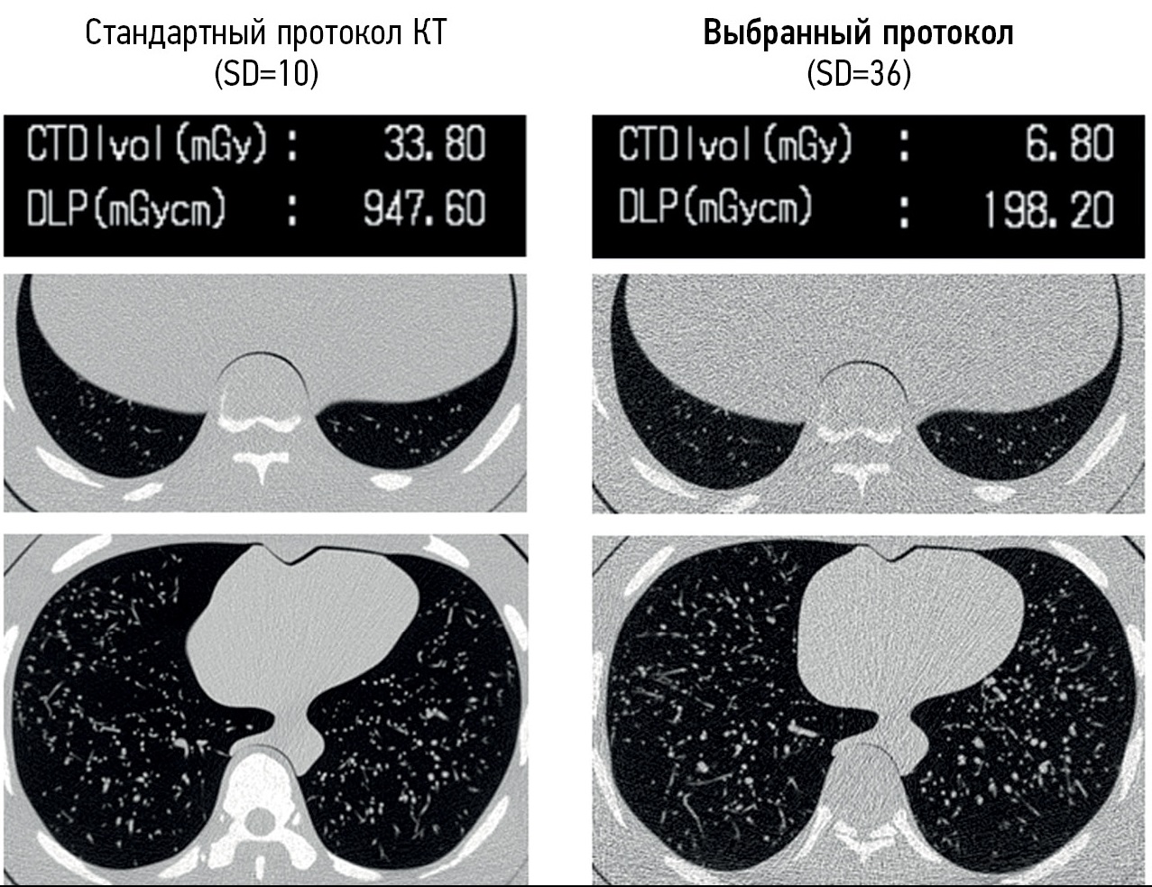
CONCLUSION: We present techniques to reduce radiation exposure during chest computed tomography and preserve high-quality diagnostic images potentially sufficient for reliable detection of COVID-19 signs. Reducing radiation dose is a valid approach to obtain relevant diagnostic information, preserving opportunities for the introduction of advanced computational analysis technologies in clinical practice.
Full Text
##article.viewOnOriginalSite##About the authors
Ivan A. Blokhin
Moscow Center for Diagnostics and Telemedicine
Author for correspondence.
Email: BlokhinIA@zdrav.mos.ru
Russian Federation, Moscow
Denis А. Rumyantsev
Moscow Center for Diagnostics and Telemedicine
Email: x.radiology@mail.ru
ORCID iD: 0000-0001-7670-7385
SPIN-code: 8734-2085
Russian Federation, Moscow
Maria M. Suchilova
Moscow Center for Diagnostics and Telemedicine
Email: SuchilovaMM@zdrav.mos.ru
ORCID iD: 0000-0003-1117-0294
SPIN-code: 4922-1894
Russian Federation, Moscow
Anna P. Gonchar
Moscow Center for Diagnostics and Telemedicine
Email: GoncharAP@zdrav.mos.ru
ORCID iD: 0000-0001-5161-6540
SPIN-code: 3513-9531
Russian Federation, Moscow
Olga V. Omelyanskaya
Moscow Center for Diagnostics and Telemedicine
Email: OmelyanskayaOV@zdrav.mos.ru
ORCID iD: 0000-0002-0245-4431
SPIN-code: 8948-6152
Russian Federation, Moscow
References
- Yang Y, Yang M, Shen C, et al. Evaluating the accuracy of different respiratory specimens in the laboratory diagnosis and monitoring the viral shedding of 2019-nCoV infections. medRxiv. 2020. doi: 10.1101/2020.02.11.20021493
- Rubin GD, Ryerson CJ, Haramati LB, et al. The role of chest imaging in patient management during the COVID-19 pandemic: A multinational consensus statement from the fleischner society. Radiology. 2020;296(1):172–180. doi: 10.1148/radiol.2020201365
- Temporary methodological recommendations prevention, diagnosis and treatment of new coronavirus infection (COVID-19). Version 12 (09/21/2021). Moscow; 2021. 232 p.
- Ng M, Lee EY, Yang J, et al. Imaging profile of the COVID-19 infection: Radiologic findings and literature review. Radiology: Cardiothoracic Imaging. 2020;2(1):e200034. doi: 10.1148/ryct.2020200034
- Ai T, Yang Z, Hou H, et al. Correlation of chest CT and RT-PCR testing for coronavirus disease 2019 (COVID-19) in China: A report of 1014 cases. Radiology. 2020;296(2):E32–E40. doi: 10.1148/radiol.2020200642
- Kang Z, Li X, Zhou S. Recommendation of low-dose CT in the detection and management of COVID-2019. Eur Radiolohy. 2020;30(8):4356–4357. doi: 10.1007/s00330-020-06809-6
- Morozov SP, Kuzmina ES, Ledekhova NV, et al. Mobilization of the scientific and practical potential of the radiation diagnostics service of Moscow in the COVID-19 pandemic. Digital Diagnostics. 2020;1(1):5–12. (In Russ). doi: 10.17816/DD51043
- Pan F, Ye T, Sun P, et al. Time course of lung changes on chest CT during recovery from 2019 novel coronavirus (COVID-19) pneumonia. Radiology. 2020;295(3):715–721. doi: 10.1148/radiol.2020200370
- Lei DP, Fan B, Mao J, et al. The progression of computed tomographic (CT) images in patients with coronavirus disease (COVID-19) pneumonia: Running title: the CT progression of COVID-19 pneumonia. J Infect. 2020;80(6):e30–e31. doi: 10.1016/j.jinf.2020.03.020
- Power SP, Moloney F, Twomey M, et al. Computed tomography and patient risk: Facts, perceptions and uncertainties. World J Radiol. 2016;8(12):902–915. doi: 10.4329/wjr.v8.i12.902
- Yeung AW. The “As low as reasonably achievable” (ALARA) principle: A brief historical overview and a bibliometric analysis of the most cited publications. Radioprotection. 2019;54(2):103–109. doi: 10.1051/radiopro/2019016
- Kalra MK, Homayounieh F, Arru C, et al. Chest CT practice and protocols for COVID-19 from radiation dose management perspective. Eur Radiol. 2020;30(12):6554–6560. doi: 10.1007/s00330-020-07034-x
- Krasnov AS, Kabanov DO, Tereshchenko GV. Fundamentals of dosimetry and dose load optimization during multispiral computed tomography. Issues Hematology Oncology Immunopathology Pediatrics. 2018;17(3):127–132. (In Russ).
- Singh S, Kalra MK, Thrall JH, Mahesh M. CT radiation dose reduction by modifying primary factors. J Am Coll Radiol. 2011;8(5):369–372. doi: 10.1016/j.jacr.2011.02.001
- Zarb F, Rainford L, McEntee MF. Developing optimized CT scan protocols: Phantom measurements of image quality. Radiography. 2011;17(2):109–114. doi: 10.1016/j.radi.2010.10.004
- Hilts M, Duzenli C. Image noise in X-ray CT polymer gel dosimetry. J Physics: Conference Series. 2004;3(1):252. doi: 10.1088/1742-6596/3/1/040
- Lira D, Padole A, Kalra MK, Singh S. Tube potential and CT radiation dose optimization. Am J Roentgenol. 2015;204(1):W4–W10. doi: 10.2214/AJR.14.13281
- Reid J, Gamberoni J, Dong F, Davros W. Optimization of kVp and mAs for pediatric low-dose simulated abdominal CT: Is it best to base parameter selection on object circumference? AJR Am J Roentgenol. 2010;195(4):1015–1020. doi: 10.2214/AJR.09.3862
- Khoramian D, Sistani S, Firouzjah RA. Assessment and comparison of radiation dose and image quality in multi-detector CT scanners in non-contrast head and neck examinations. Paul J Radiol. 2019;84:61–67. doi: 10.5114/pjr.2019.82743
- Mahesh M, Scatarige JC, Cooper J, Fishman EK. Dose and pitch relationship for a particular multislice CT scanner. AJR Am J Roentgenol. 20011;77(6):1273–1275. doi: 10.2214/ajr.177.6.1771273
- Tack D, Gevenois PA, Abada H. Radiation dose from adult and pediatric multidetector computed tomography. Springer. 2007. doi: 10.1007/978-3-540-68575-3
- Greffier J, Pereira F, Hamard A, et al. Effect of tin filter-based spectral shaping CT on image quality and radiation dose for routine use on ultralow-dose CT protocols: A phantom study. Diagnostic Interventional Imaging. 2020;101(6):373–381. doi: 10.1016/j.diii.2020.01.002
- Paul J, Krauss B, Banckwitz R, et al. Relationships of clinical protocols and reconstruction kernels with image quality and radiation dose in a 128-slice CT scanner: Study with an anthropomorphic and water phantom // Eur J Radiology. 2012;81(5):e699–e703. doi: 10.1016/j.ejrad.2011.01.078
- Hashemi S, Mehrez H, Cobbold RS, Paul NS. Optimal image reconstruction for detection and characterization of small pulmonary nodules during low-dose CT. Eur Radiol. 2014;24(6):1239–1250. doi: 10.1007/s00330-014-3142-9
- Beister M, Kolditz D, Kalender WA. Iterative reconstruction methods in X-ray CT. Physica Medica. 2012;28(2):94–108. doi: 10.1016/j.ejmp.2012.01.003
- Shiri I, Akhavanallaf A, Sanaat A, et al. Ultra-low-dose chest CT imaging of COVID-19 patients using a deep residual neural network. Eur Radiology. 2021;31(3):1420–1431. doi: 10.1007/s00330-020-07225-6
- Shan H, Padole A, Homayounieh F, et al. Competitive performance of a modularized deep neural network compared to commercial algorithms for low-dose CT image reconstruction. Nat Machine Intelligence. 2019;1(6):269–276. doi: 10.1038/s42256-019-0057-9
- Blokhin I, Gombolevskiy V, Chernina V, et al. Inter-observer agreement between low-dose and standard-dose CT with soft and sharp convolution kernels in COVID-19 pneumonia. J Clin Med. 2022;11:669. doi: 10.3390/jcm11030669
- Filatova DA, Sinitsyn VE, Mershina EA. The possibilities of reducing radiation exposure during computed tomography to assess changes in the lungs characteristic of COVID-19: The use of adaptive statistical iterative reconstruction. Digital Diagnostics. 2021;2(2):94–104. (In Russ). doi: 10.17816/DD62477
- Afshar P, Rafiee MJ, Naderkhani F, et al. Human-level COVID-19 diagnosis from low-dose CT scans using a two-stage time-distributed capsule network. Sci Rep. 2022;12(1):4827. doi: 10.1038/s41598-022-08796-8
- Fukumoto W, Nakamura Y, Yoshimura K, et al. Triaging of COVID-19 patients using low dose chest CT: Incidence and factor analysis of lung involvement on CT images. J Infect Chemother. 2022;28(6):797–801. doi: 10.1016/j.jiac.2022.02.025
- Bieba CM, Desmet JN, Dubbeldam A, et al. Radiological findings in low-dose CT for COVID-19 pneumonia in 182 patients: Correlation of signs and severity with patient outcome. Medicine (Baltimore). 2022;101(9):e28950. doi: 10.1097/MD.0000000000028950
- Piqueras BM, Casajús EA, Iriarte UC, et al. Low-dose chest CT for preoperative screening for SARS-CoV-2 infection. Radiologia (Engl Ed). 2022;64(4):317–323. doi: 10.1016/j.rxeng.2021.11.004
- Thieß HM, Bressem KK, Adams L, et al. Do submillisievert-chest CT protocols impact diagnostic quality in suspected COVID-19 patients? Acta Radiol Open. 2022;11(1):20584601211073864. doi: 10.1177/20584601211073864
- Greffier J, Hoballah A, Sadate A, et al. Ultra-low-dose chest CT performance for the detection of viral pneumonia patterns during the COVID-19 outbreak period: A monocentric experience. Quant Imaging Med Surg. 2021;11(7):3190–3199. doi: 10.21037/qims-20-1176
- Karakaş HM, Yıldırım G, Çiçek ED. The reliability of low-dose chest CT for the initial imaging of COVID-19: Comparison of structured findings, categorical diagnoses and dose levels. Diagn Interv Radiol. 2021;27(5):607–614. doi: 10.5152/dir.2021.20802
- Finance J, Zieleskewicz L, Habert P, et al. Low dose chest CT and lung ultrasound for the diagnosis and management of COVID-19. J Clinic Med. 2021;10(10):2196. doi: 10.3390/jcm10102196
- Desmet J, Biebaû C, De Wever W, et al. Performance of low-dose chest CT as a triage tool for suspected COVID-19 patients. J Belgian Society Radiology. 2021;105(1):9. doi: 10.5334/jbsr.2319
- Aslan S, Bekçi T, Çakır İM, et al. Diagnostic performance of low-dose chest CT to detect COVID-19: A Turkish population study. Diagn Interv Radiol. 2021;27(2):181–187. doi: 10.5152/dir.2020.20350
- Stoleriu MG, Gerckens M, Obereisenbuchner F, et al. Automated quantitative thin slice volumetric low dose CT analysis predicts disease severity in COVID-19 patients. Clin Imaging. 2021;79:96–101. doi: 10.1016/j.clinimag.2021.04.008
- Bai L, Zhou J, Shen C, et al. Assessment of radiation doses and image quality of multiple low-dose CT exams in COVID-19 clinical management. Chin J Acad Radiol. 2021;4(4):257–261. doi: 10.1007/s42058-021-00083-1
- Agostini A, Borgheresi A, Carotti M, et al. Third-generation iterative reconstruction on a dual-source, high-pitch, low-dose chest CT protocol with tin filter for spectral shaping at 100 kV: A study on a small series of COVID-19 patients. Radiol Med. 2021;126(3):388–398. doi: 10.1007/s11547-020-01298-5
- Zali A, Sohrabi MR, Mahdavi A, et al. Correlation between low-dose chest computed tomography and RT-PCR results for the diagnosis of COVID-19: A report of 27,824 cases in Tehran, Iran. Acad Radiol. 2021;28(12):1654–1661. doi: 10.1016/j.acra.2020.09.003
- Argentieri G, Bellesi L, Pagnamenta A, et al. Diagnostic yield, safety, and advantages of ultra-low dose chest CT compared to chest radiography in early stage suspected SARS-CoV-2 pneumonia: A retrospective observational study. Medicine (Baltimore). 2021;100(21):e26034. doi: 10.1097/MD.0000000000026034
- Leger T, Jacquier A, Barral PA, et al. Low-dose chest CT for diagnosing and assessing the extent of lung involvement of SARS-CoV-2 pneumonia using a semi quantitative score. PLoS One. 2020;15(11):e0241407. doi: 10.1371/journal.pone.0241407
- Hamper CM, Fleckenstein FN, Büttner L, et al. Submillisievert chest CT in patients with COVID-19: experiences of a German Level-I center. Eur J Radiol Open. 2020;7:100283. doi: 10.1016/j.ejro.2020.100283
- Li J, Wang X, Huang X, et al. Application of Care Dose 4D combined with Karl 3D technology in the low dose computed tomography for the follow-up of COVID-19. BMC Med Imaging. 2020;20(1):56. doi: 10.1186/s12880-020-00456-5
- Dangis A, Gieraerts C, De Bruecker Y, et al. Accuracy and reproducibility of low-dose submillisievert chest CT for the diagnosis of COVID-19. Radiol Cardiothorac Imaging. 2020;2(2):e200196. doi: 10.1148/ryct.2020200196
- Radpour A, Bahrami-Motlagh H, Taaghi MT, et al. COVID-19 evaluation by low-dose high resolution CT scans protocol. Acad Radiol. 2020;27(6):901. doi: 10.1016/j.acra.2020.04.016
- Tofighi S, Najafi S, Johnston SK, Gholamrezanezhad A. Low-dose CT in COVID-19 outbreak: Radiation safety, image wisely, and image gently pledge. Emerg Radiol. 2020;27(6):601–605. doi: 10.1007/s10140-020-01784-3
- Tabatabaei SM, Talari H, Gholamrezanezhad A, et al. A low-dose chest CT protocol for the diagnosis of COVID-19 pneumonia: A prospective study. Emerg Radiol. 2020;27(6):607–615. doi: 10.1007/s10140-020-01838-6
- Schulze-Hagen M, Hübel C, Meier-Schroers M, et al. Low-dose chest CT for the diagnosis of COVID-19: A systematic, prospective comparison with PCR. Dtsch Arztebl Int. 2020;117(22-23):389–395. doi: 10.3238/arztebl.2020.0389
- Zhao Y, Wang Y, Duan W, et al. Low-dose chest CT presentation and dynamic changes in patients with novel coronavirus disease 2019. Radiol Infect Dis. 2020;7(4):186–194. doi: 10.1016/j.jrid.2020.08.001
- Castelli M, Maurin A, Bartoli A, et al. Prevalence and risk factors for lung involvement on low-dose chest CT (LDCT) in a paucisymptomatic population of 247 patients affected by COVID-19. Insights Imaging. 2020;11(1):117. doi: 10.1186/s13244-020-00939-7
- Morozov SP, Kuzmina ES, Vetsheva NN, et al. Moscow screening: screening of lung cancer using low-dose computed tomography. Problems Social Hygiene Healthcare History Med. 2019;27(S):630–636. (In Russ). doi: 10.32687/0869-866X-2019-27-si1-630-636
- Patent RUS No. 2701922 C1. Gombolevsky VA, Morozov SP, Chernina VYu, et al. A method for screening lung cancer using ultra-low-dose computed tomography in patients with a body weight of up to 69 kg. mode: Available from: https://patents.google.com/patent/RU2701922C1/ru. Accessed: 15.01.2023.
- Gombolevskiy V, Morozov S, Chernina V, et al. A phantom study to optimise the automatic tube current modulation for chest CT in COVID-19. Eur Radiol Exp. 2021;5(1):21. doi: 10.1186/s41747-021-00218-0
- Kim YK, Lee BE, Lee SJ, et al. Ultra-low-dose CT of the thorax using iterative reconstruction: Evaluation of image quality and radiation dose reduction. Am J Roentgenol. 2015;204(6):1197–1202. doi: 10.2214/AJR.14.13629
- Blokhin IA, Gonchar AP, Kotenko MR, et al. The influence of body mass index on the reliability of the 0–4 CT scale: Comparison of computed tomography protocols. Digital Diagnostics. 2022;3(2):108–118. (In Russ). doi: 10.17816/DD104358
- Gierada DS, Bierhals AJ, Choong CK, et al. Effects of CT section thickness and reconstruction kernel on emphysema quantification. Academic Radiology. 2010;17(2):146–156. doi: 10.1016/j.acra.2009.08.007
- Gao Y, Hua M, Lv J, et al. Reproducibility of radiomic features of pulmonary nodules between low-dose CT and conventional-dose CT. Quant Imaging Med Surg. 2022;12(4):2368–2377. doi: 10.21037/qims-21-609
- Blokhin IA, Solovev AV, Vladzymyrskiy AV, et al. Automated analysis of lung lesions in COVID-19: Comparison of standard and low-dose CT. SJCEM. 2023;37(4):114–123. (In Russ). doi: 10.29001/2073-8552-2022-37-4-114-123
- Bak SH, Kim JH, Jin H, et al. Emphysema quantification using low-dose computed tomography with deep learning-based kernel conversion comparison. Eur Radiol. 2020;30(12):6779–6787. doi: 10.1007/s00330-020-07020-3
Supplementary files


