

")


“Superior Pectus Carinatum” (Currarino–Silverman Syndrome) in a 66-year-old woman: a case report
- Authors: Mannatrizio D.1, Fascia G.1, Guglielmi G.1,2
-
Affiliations:
- Department of Clinical and Experimental Medicine, Foggia University School of Medicine
- Radiology Unit, Barletta University Hospital
- Issue: Vol 3, No 2 (2022)
- Pages: 141-148
- Section: Case reports
- URL: https://journals.rcsi.science/DD/article/view/104865
- DOI: https://doi.org/10.17816/DD104865
- ID: 104865
Cite item

Abstract
The premature fusion of some of the sternal ossification centers and the obliteration of the manubrio-sternal joint caused a rare deformity called Currarino–Silverman syndrome. Patients present an abnormally short sternum with a forward angulation at the manubrio-sternal junction. Cardiopulmonary diseases and spinal deformities are the most frequent related disorders. It was also described as a component of Turner’s and Noonan’s syndromes.
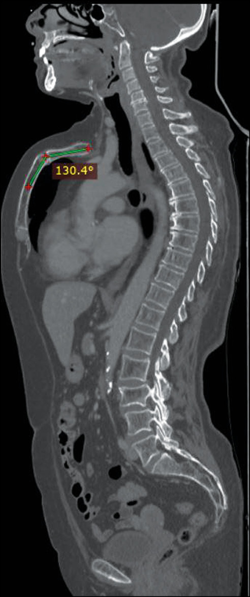
Herein, we present the case of a 66-year-old woman who presented to our clinic for follow-up computed tomography after surgery and chemotherapy for breast cancer with frequent episodes of dyspnea, wheezing, bronchitis, and mild dyspnea annually, which was more frequent during childhood. Computed tomography showed the absence of metastatic lesions and other accompanying diseases, except for a rare deformity of the anterior chest wall, the so-called, a “superior” pectus carinatum, a chondromanubrial deformity with a dorsal-open angle of 130º, and a sternum body length of 9 cm, which is not depressed in the lower third.
Keywords
Full Text
##article.viewOnOriginalSite##About the authors
Domenico Mannatrizio
Department of Clinical and Experimental Medicine, Foggia University School of Medicine
Email: dr.mannatrizio@gmail.com
ORCID iD: 0000-0003-3365-7132
MD
Italy, FoggiaGiacomo Fascia
Department of Clinical and Experimental Medicine, Foggia University School of Medicine
Email: giacomo.fascia@unifg.it
ORCID iD: 0000-0001-5244-5093
MD
Italy, FoggiaGiuseppe Guglielmi
Department of Clinical and Experimental Medicine, Foggia University School of Medicine; Radiology Unit, Barletta University Hospital
Author for correspondence.
Email: giuseppe.guglielmi@unifg.it
ORCID iD: 0000-0002-4325-8330
MD, Professor
Italy, Foggia; BarlettaReferences
- Shamberger RC, Welch KJ. Surgical correction of pectus carinatum. J Pediatr Surg. 1987;22(1):48–53. doi: 10.1016/s0022-3468(87)80014-3
- Muntean A, Stoica I, Saxena AK. Pigeon chest: comparative analysis of surgical techniques in minimal access repair of pectus carinatum (MARPC). World J Pediatr. 2018;14(1):18–25. doi: 10.1007/s12519-018-0121-2
- Emil S. Current options for the treatment of pectus carinatum: when to brace and when to operate? Eur J. 2018;28(4):347–354. doi: 10.1055/s-0038-1667297
- Currarino G, Silverman F. Premature obliteration of the sternal sutures and pigeon-breast deformity. Radiology. 1958;70(4):532–540. doi: 10.1148/70.4.532
- Fokin A, Steuerwald NM, Ahrens WA, Allen KE. Anatomical, histologic, and genetic characteristics of congenital chest wall deformities. Semin Thorac Cardiovasc Surg. 2009;21(1):44–57. doi: 10.1053/j.semtcvs.2009.03.001
- Fokin A. Pouter pigeon breast. Chest Surg Clin N Am. 2000;10(2):377–391.
- Gabrielsen T, Ladyman G. Early closure of the sternal sutures and congenital heart disease. Am J Roentgenol Radium Ther Nucl Med. 1963;89:975–983.
- Chidambaram B, Mehta AV. Currarino-Silverman syndrome (pectus carinatum type 2 deformity) and mitral valve disease. Chest. 1992;102(3):780–782. doi: 10.1378/chest.102.3.780
- Regier DS, Oetgen M, Tanpaiboon P. Mucopolysaccharidosis type IVA. 2013 Jul 11 [updated 2021 Jun 17]. In: Adam MP, Ardinger HH, Pagon RA, et al., ed. GeneReviews [Internet]. Seattle (WA): University of Washington, Seattle; 1993–2022.
- Martinez-Ferro M, Bellia-Munzon G, Schewitz IA, Toselli L. Pectus carinatum: when less is more. Afr J Thorac Crit Care Med. 2019;25(3):10.7196/AJTCCM.2019.v25i3.019. doi: 10.7196/AJTCCM.2019.v25i3.019
- Lester C. Pigeon breast (pectus carinatum) and other protrusion deformities of the chest of developmental origin. Ann Surg. 137(4):482–489. doi: 10.1097/00000658-195304000-00008
- Welch KJ, Vos A. Surgical correction of pectus carinatum (pigeon breast). J Pediatr Surg. 1973; 8(5):659–667. doi: 10.1016/0022-3468(73)90404-1
- Coelho MS, Santos A, Pizarro L, et al. “Pectus excavatum/pectus carinatum”: tratamento cirúrgico. J Pneumol. 1983;10(Supрl):47.
- Ramadan S, Wilde J, Tabard-Fougère A, et al. Cardiopulmonary function in adolescent patients with pectus excavatum or carinatum. BMJ Open Respir Res. 2021;8(1):e001020. doi: 10.1136/bmjresp-2021-001020
- Buziashvili D, Gopman JM, Weissler H, et al. An evidence-based approach to management of pectus excavatum and carinatum. Ann Plast Surg. 2019;82(3):352–358. doi: 10.1097/SAP.0000000000001654
- Szafer D, Taylor JS, Pei A, et al. A simplified method for three-dimensional optical imaging and measurement of patients with chest wall deformities. J Laparoendosc Adv Surg Tech A. 2019;29(2):267–271. doi: 10.1089/lap.2018.0191
- Geraedts TC, Daemen JH, Vissers YL, et al. Minimally invasive repair of pectus carinatum by the Abramson method: a systematic review. J Pediatr Surg. 2021;5:S0022-3468(21)00829-0. doi: 10.1016/j.jpedsurg.2021.11.028
- Brichon PY, Wihlm JM. Correction of a severe pouter pigeon breast by triple sternal osteotomy with a novel titanium rib bridge fixation. Ann Thorac Surg. 2010;90(6):e97–99. doi: 10.1016/j.athoracsur.2010.08.068
- Tarhan T, Meurer A, Tarhan O. Combined extra-/intrathoracic correction of pectus carinatum and other asymmetric chest wall deformities: A novel technique. Oper Orthop Traumatol. 2018;30(6):469–478. doi: 10.1007/s00064-018-0567-3
- McHam B, Winkler L. Pectus Carinatum. 2021 Aug 9. In: StatPearls [Internet]. Treasure Island (FL): StatPearls Publishing; 2022.
- Rea G, Sezen CB. Chest wall deformities. 2021 Aug 11. In: StatPearls [Internet]. Treasure Island (FL): StatPearls Publishing; 2022.
- Ramadan S, Wilde J, Tabard-Fougère A, et al. Cardiopulmonary function in adolescent patients with pectus excavatum or carinatum. BMJ. 2021;8(1):e001020. doi: 10.1136/bmjresp-2021-001020
Supplementary files


