


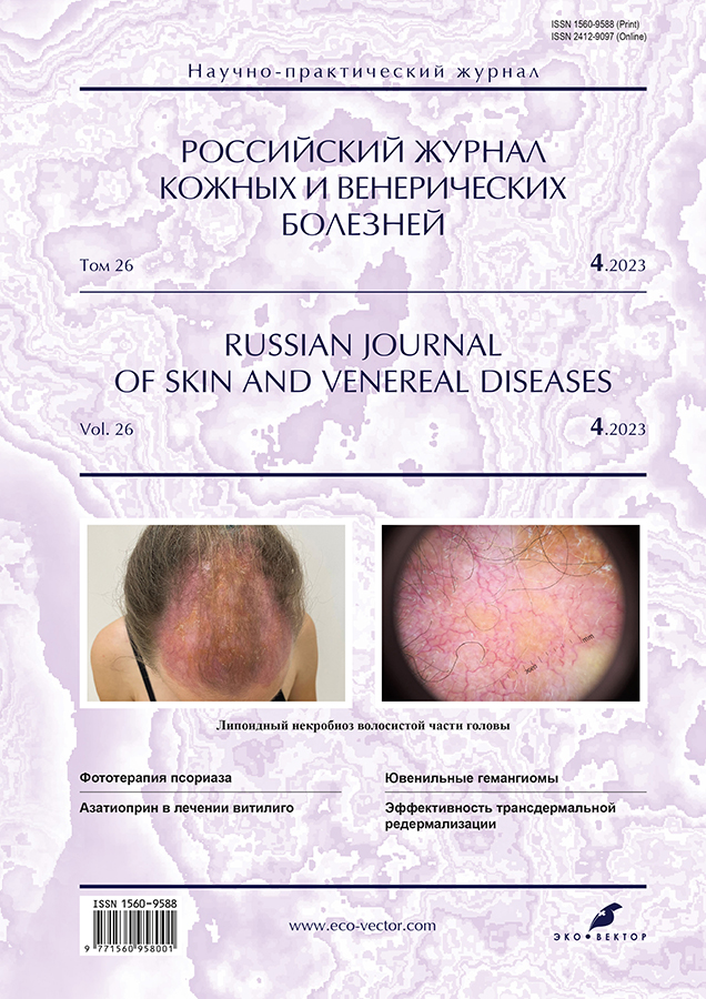
Necrobiosis lipoidica of the scalp
- Authors: Teplyuk N.P.1, Grabovskaya O.V.1, Rogozina V.A.1
-
Affiliations:
- I.M. Sechenov First Moscow State Medical University of the Ministry of Health of the Russian Federation (Sechenov University)
- Issue: Vol 26, No 4 (2023)
- Pages: 405-412
- Section: DERMATOLOGY
- URL: https://journals.rcsi.science/1560-9588/article/view/132561
- DOI: https://doi.org/10.17816/dv450869
- ID: 132561
Cite item

Abstract
Necrobiosis lipoidica is a chronic granulomatous skin disease of a vascular-exchange nature from the group of localized skin lipoidosis, accompanied by the development of degenerative changes in the connective tissue.
The reason for the development of necrobiosis lipoidica is unknown, but the most common theory remains the theory of vascular disorders, including the deposition of immune complexes (IgM, complement component C3), microangiopathic changes (deposition of a glycoprotein in the walls of blood vessels), as well as other combinations of inflammatory and structural changes leading to collagen degeneration and decreased perfusion and oxygenation of the skin. Necrobiosis lipoidica occurs in association with systemic diseases (sarcoidosis, autoimmune thyroiditis, inflammatory bowel disease, such as ulcerative colitis, and rheumatoid arthritis) and can also occur in otherwise healthy individuals. Necrobiosis lipoidica is closely associated with diabetes mellitus.
The predominant localization of necrobiosis lipoidica is tibial surface of the legs. Atypical localization is: skin of the scalp and face.
Clinical polymorphism of necrobiosis lipoidica, possibility of transition from one form to another, its combination with other dermatoses can pose difficult diagnostic questions for the doctor.
The article describes our own clinical observation of a patient with complaints of rashes on the skin of the legs and scarring alopecia in the scalp. The focus on the scalp had a tendency to grow and signs of development of scarring alopecia. According to the results of a biopsy performed at the Rakhmanov Department of Skin and Veneral Diseases, I.M. Sechenov First Moscow State Medical University, was diagnosed with "Necrobiosis Lipoidica". The case was interesting due to the extreme rarity of rashes on the skin of the scalp and the absence of dermatoscopic features of lesions in this area. Due to ongoing therapy for a month, there was a positive effect in the form of a lack of growth of existing plaques and the appearance of new ones, a decrease in the brightness of the color of the rash, a partial regression with an outcome in residual hyperpigmentation.
Thus, despite the absence of a gold standard in the treatment of necrobiosis lipoidica, the most well-studied group of drugs for the treatment of the disease at the moment are glucocorticoids, which confirms our experience. Therapy of patients with necrobiosis lipoidica should be carried out by an interdisciplinary team (dermatovenereologist, endocrinologist, therapist).
Full Text
##article.viewOnOriginalSite##About the authors
Natalya P. Teplyuk
I.M. Sechenov First Moscow State Medical University of the Ministry of Health of the Russian Federation (Sechenov University)
Email: Teplyukn@gmail.com
ORCID iD: 0000-0002-5800-4800
SPIN-code: 8013-3256
MD, Dr. Sci. (Med.), Professor
Russian Federation, MoscowOlga V. Grabovskaya
I.M. Sechenov First Moscow State Medical University of the Ministry of Health of the Russian Federation (Sechenov University)
Email: olgadoctor2013@yandex.ru
ORCID iD: 0000-0002-5259-7481
SPIN-code: 1843-1090
MD, Cand. Sci. (Med.), Associate Professor
Russian Federation, MoscowVarvara A. Rogozina
I.M. Sechenov First Moscow State Medical University of the Ministry of Health of the Russian Federation (Sechenov University)
Author for correspondence.
Email: varvara.rgzn@gmail.com
ORCID iD: 0000-0002-5471-6130
SPIN-code: 3505-7408
Russian Federation, Moscow
References
- Olisova OY, Teplyuk NP. Illustrated guide to dermatology. Moscow: GEOTAR-Media; 2022. 376 р. (In Russ).
- Smirnova LM, Semenchuk YA, Panchenko LA. Lipoid necrobiosis: Review article. Russ J Skin Venereal Dis. 2018;21(1):40–44. (In Russ). doi: 10.18821/1560-9588-2018-21-1-1-40-44
- Goldsmith WN. Necrobiosis lipoidica. Proc R Soc Med. 1935;28(4):363.
- Rollins TG, Winkelmann RK. Necrobiosis lipoidica granulomatosis necrobiosis lipoidica diabeticorum in the nondiabetic. Arch Dermatol. 1960;(82):537–543. doi: 10.1001/archderm.1960.01580040055010
- Jockenhöfer F, Kröger K, Klode J, et al. Cofactors and comorbidities of necrobiosis lipoidica: Analysis of the German DRG data from 2012. J Dtsch Dermatol Ges. 2016;14(3):277–284. doi: 10.1111/ddg.12749
- Erfurt-Berge C, Seitz AT, Rehse C, et al. Update on clinical and laboratory features in necrobiosis lipoidica: A retrospective multicentre study of 52 patients. Eur J Dermatol. 2012;22(6):770–775. doi: 10.1684/ejd.2012.1839
- Hashemi DA, Brown-Joel ZO, Tkachenko E, et al. Clinical features and comorbidities of patients with necrobiosis lipoidica with or without diabetes. JAMA Dermatology. 2019;155(4):455–459. doi: 10.1001/jamadermatol.2018.5635
- Lepe K, Riley CA, Salazar FJ. Necrobiosis lipoidica. Treasure Island: StatPearls; 2021.
- Butov YS, Ilyina TA, Vavilov AM. Clinical and histological signs of lipid necrobiosis. Russ J Skin Venereal Dis. 2003;(4):38–42. (In Russ).
- Ngo BT, Hayes KD, DiMiao DJ, et al. Manifestations of cutaneous diabetic microangiopathy. Am J Clin Dermatol. 2005;6(4):225–237. doi: 10.2165/00128071-200506040-00003
- Samsonov VA, Khachukova LM. Lipoid necrobiosis: Pathogenesis, clinic, treatment. Bulletin Dermatol. 2002;(1):13–19. (In Russ).
- Petunina VV, Shmakova AS, Khamaganova IV, Kashevarov DF. Clinical case of long-term undiagnosed lipid necrobiosis on the background of endocrine pathology. Vestnik SurGU. Medicina. 2021;(4):70–72. (In Russ). doi: 10.34822/2304-9448-2021-4-70-73
- Forman L. Necrobiosis lipoidica diabeticorum of the scalp. Proc R Soc Med. 1954;47(8):658–659.
- Yaşar Ş, Kaynak E, Güneş P, et al. Atypical localization of necrobiosis lipoidica: Involvement of the face and scalp. Skin Appendage Disord. 2017;3(2):92–94. doi: 10.1159/000462980
- Reid SD, Ladizinski B, Lee K, et al. Update on necrobiosis lipoidica: A review of etiology, diagnosis, and treatment options. J Am Academy Dermatol. 2013;69(5):Е:783–791. doi: 10.1016/j.jaad.2013.05.034
- Erfurt-Berge C, Heusinger V, Reinboldt-Jockenhöfer F, et al. Comorbidity and therapeutic approaches in patients with necrobiosis lipoidica. Dermatology. 2022;238(1):148–155. doi: 10.1159/000514687
Supplementary files


