


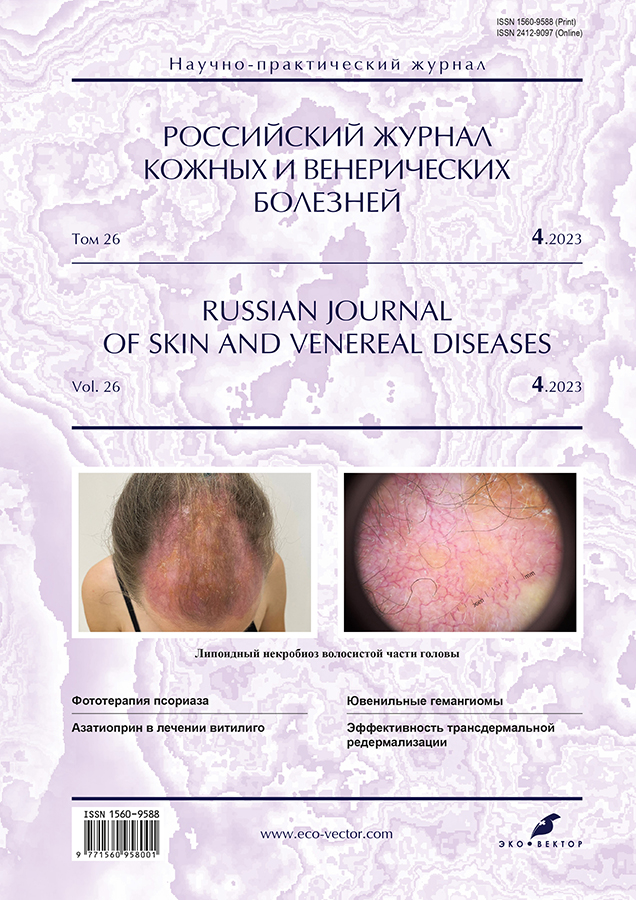
Липоидный некробиоз волосистой части головы
- Авторы: Теплюк Н.П.1, Грабовская О.В.1, Рогозина В.А.1
-
Учреждения:
- Первый Московский государственный медицинский университет имени И.М. Сеченова (Сеченовский Университет)
- Выпуск: Том 26, № 4 (2023)
- Страницы: 405-412
- Раздел: ДЕРМАТОЛОГИЯ
- URL: https://journals.rcsi.science/1560-9588/article/view/132561
- DOI: https://doi.org/10.17816/dv450869
- ID: 132561
Цитировать

Аннотация
Липоидный некробиоз ― хроническое гранулематозное заболевание кожи сосудисто-обменного характера из группы локализованных липоидозов кожи, сопровождающееся дегенеративными изменениями соединительной ткани.
Причина развития липоидного некробиоза неизвестна, однако наиболее распространённой остаётся теория сосудистых нарушений, включающих отложение иммунных комплексов (IgМ, С3-компонент комплемента), микроангиопатические изменения (отложение гликопротеина в стенках сосудов), а также другие комбинации воспалительных и структурных изменений, приводящие к дегенерации коллагена и снижению перфузии и оксигенации кожи. Липоидный некробиоз сочетается с системными заболеваниями (саркоидоз; аутоиммунный тиреоидит; воспалительные заболевания кишечника, например язвенный колит, ревматоидный артрит), в том числе может развиваться у соматически здоровых лиц. Часто липоидный некробиоз ассоциируется с сахарным диабетом.
Преимущественная локализация очагов липоидного некробиоза ― область голеней. Атипичной локализацией считаются кожа волосистой части головы и лицо.
Клинический полиморфизм липоидного некробиоза, возможность перехода одной формы в другую, его сочетание с другими дерматозами могут ставить перед врачом сложные диагностические вопросы.
В статье приводится описание собственного клинического наблюдения пациента с жалобами на высыпания на коже голеней и рубцовую алопецию в области волосистой части головы. Очаг на волосистой части головы имел тенденцию к росту и признаки развития рубцовой алопеции. По результатам биопсии был выставлен диагноз липоидного некробиоза. Случай был интересен крайней редкостью высыпаний на коже волосистой части головы и отсутствием дерматоскопических особенностей очагов в этой области. На фоне проводимой терапии в течение месяца наблюдалась положительная динамика.
Таким образом, несмотря на отсутствие золотого стандарта в терапии липоидного некробиоза, наиболее хорошо изученной группой препаратов для лечения заболевания на данный момент являются глюкокортикоиды, что и подтверждает наш опыт. Терапия пациентов с липоидным некробиозом должна осуществляться междисциплинарной командой (дерматовенеролог, эндокринолог, терапевт).
Ключевые слова
Полный текст
Открыть статью на сайте журналаОб авторах
Наталия Павловна Теплюк
Первый Московский государственный медицинский университет имени И.М. Сеченова (Сеченовский Университет)
Email: Teplyukn@gmail.com
ORCID iD: 0000-0002-5800-4800
SPIN-код: 8013-3256
д-р мед. наук, профессор
Россия, МоскваОльга Валентиновна Грабовская
Первый Московский государственный медицинский университет имени И.М. Сеченова (Сеченовский Университет)
Email: olgadoctor2013@yandex.ru
ORCID iD: 0000-0002-5259-7481
SPIN-код: 1843-1090
канд. мед. наук, доцент
Россия, МоскваВарвара Андреевна Рогозина
Первый Московский государственный медицинский университет имени И.М. Сеченова (Сеченовский Университет)
Автор, ответственный за переписку.
Email: varvara.rgzn@gmail.com
ORCID iD: 0000-0002-5471-6130
SPIN-код: 3505-7408
Россия, Москва
Список литературы
- Олисова О.Ю., Теплюк Н.П. Иллюстрированное руководство по дерматологии. Москва: ГЭОТАР-Медиа, 2022. 376 с.
- Смирнова Л.М., Семенчак Ю.А., Панченко Л.А. Липоидный некробиоз: обзорная статья // Российский журнал кожных и венерических болезней. 2018. Т. 21, № 1. С. 40–44. doi: 10.18821/1560-9588-2018-21-1-1-40-44
- Goldsmith W.N. Necrobiosis lipoidica // Proc R Soc Med. 1935. Vol. 28, N 4. Р. 363.
- Rollins T.G., Winkelmann R.K. Necrobiosis lipoidica granulomatosis necrobiosis lipoidica diabeticorum in the nondiabetic // Arch Dermatol. 1960. N 82. Р. 537–543. doi: 10.1001/archderm.1960.01580040055010
- Jockenhöfer F., Kröger K., Klode J., et al. Cofactors and comorbidities of necrobiosis lipoidica: Analysis of the German DRG data from 2012 // J Dtsch Dermatol Ges. 2016. Vol. 14, N 3. Р. 277–284. doi: 10.1111/ddg.12749
- Erfurt-Berge C., Seitz A.T., Rehse C., et al. Update on clinical and laboratory features in necrobiosis lipoidica: A retrospective multicentre study of 52 patients // Eur J Dermatol. 2012. Vol. 22, N 6. Р. 770–775. doi: 10.1684/ejd.2012.1839
- Hashemi D.A., Brown-Joel Z.O., Tkachenko E., et al. Clinical features and comorbidities of patients with necrobiosis lipoidica with or without diabetes // JAMA Dermatology. 2019. Vol. 155, N 4. Р. 455–459. doi: 10.1001/jamadermatol.2018.5635
- Lepe K., Riley C.A., Salazar F.J. Necrobiosis lipoidica. Treasure Island: StatPearls, 2021.
- Бутов Ю.С., Ильина Т.А., Вавилов А.М. Клинико-гистологические признаки липоидного некробиоза // Российский журнал кожных и венерических болезней. 2003. № 4. С. 38–42.
- Ngo B.T., Hayes K.D., DiMiao D.J., et al. Manifestations of cutaneous diabetic microangiopathy // Am J Clin Dermatol. 2005. Vol. 6, N 4. Р. 225–237. doi: 10.2165/00128071-200506040-00003
- Самсонов В.А., Хачукова Л.М. Липоидный некробиоз: патогенез, клиника, лечение // Вестник дерматологии. 2002. № 1. С. 13–19.
- Петунина В.В., Шмакова А.С., Хамаганова И.В., Кашеваров Д.Ф. Клинический случай длительно не диагностированного липоидного некробиоза на фоне эндокринной патологии // Вестник СурГУ. Медицина. 2021. № 4. С. 70–72. doi: 10.34822/2304-9448-2021-4-70-73
- Forman L. Necrobiosis lipoidica diabeticorum of the scalp // Proc R Soc Med. 1954. Vol. 47, N 8. Р. 658–659.
- Yaşar Ş., Kaynak E., Güneş P., et al. Atypical localization of necrobiosis lipoidica: Involvement of the face and scalp // Skin Appendage Disord. 2017. Vol. 3, N 2. Р. 92–94. doi: 10.1159/000462980
- Reid S.D., Ladizinski B., Lee K., et al. Update on necrobiosis lipoidica: A review of etiology, diagnosis, and treatment options // J Am Academy Dermatol. 2013. Vol. 69, N 5. Р. 783–791. doi: 10.1016/j.jaad.2013.05.034
- Erfurt-Berge C., Heusinger V., Reinboldt-Jockenhöfer F., et al. Comorbidity and therapeutic approaches in patients with necrobiosis lipoidica // Dermatology. 2022. Vol. 238, N 1. Р. 148–155. doi: 10.1159/000514687
Дополнительные файлы


