

")


Наблюдения доплеровского мерцающего артефакта: база данных радиочастотных ультразвуковых сигналов
- Авторы: Леонов Д.В.1,2, Решетников Р.В.1,3, Кульберг Н.С.1,4, Насибуллина А.А.2, Громов А.И.5
-
Учреждения:
- Научно-практический клинический центр диагностики и телемедицинских технологий Департамента здравоохранения г. Москвы
- Национальный исследовательский университет МЭИ
- Первый Московский государственный медицинский университет имени И.М. Сеченова (Сеченовский Университет)
- Федеральный исследовательский центр «Информатика и управление» Российской академии наук
- Московский государственный медико-стоматологический университет им. А.И. Евдокимова
- Выпуск: Том 2, № 3 (2021)
- Страницы: 261-276
- Раздел: Оригинальные исследования
- URL: https://journals.rcsi.science/DD/article/view/76511
- DOI: https://doi.org/10.17816/DD76511
- ID: 76511
Цитировать

Аннотация
Обоснование. Мерцающий артефакт в доплеровских режимах ультразвукового исследования проявляется быстрой хаотической сменой окрашенных пикселей на экране прибора. Явление, которое можно использовать в качестве полезного диагностического признака, исследовано недостаточно. Большинство предположений о причинах артефакта сделаны на основании изображений с экрана ультразвукового прибора без глубокого изучения свойств принимаемых сигналов.
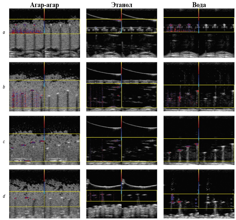
Материалы и методы. Радиочастотные ультразвуковые сигналы были записаны при исследовании фантомов. Исследовались как объекты, приводящие к появлению мерцающего артефакта на экране прибора, так и имитации сосудов и мягких тканей. Сбор данных проводился с июля 2016 по март 2021 г. Данные получены при помощи исследовательского ультразвукового прибора «Сономед-500» с датчиками 7,5 L38 и 3,4 C60.
Содержимое базы данных. Представлена база данных, содержащая радиочастотные сигналы, полученные с выхода формирователя луча из приёмного тракта ультразвукового медицинского диагностического прибора в режиме цветового доплеровского картирования и В-режиме. Представленные в базе данных сигналы содержат признаки мерцающего артефакта. База состоит из исследований пяти различных фантомов общим объёмом 10,5 ГБ. Радиочастотные данные сохранены в бинарном виде. Настройки сканирования, необходимые для анализа радиочастотных данных, содержатся в текстовых файлах. Каждое исследование сопровождается примером характерной сонограммы в графическом формате. База данных доступна по адресу: https://mosmed.ai/datasets/ultrasound_doppler_twinkling_artifact.
Доступность кода. Для просмотра и анализа базы данных к архиву прилагаем разработанную нами программу TwinklingDatasetDisplay. Доступен исходный код программы: https://github.com/Center-of-Diagnostics-and-Telemedicine/TwinklingDatasetDisplay.git.
Условия использования. База данных может быть использована для разработки и тестирования алгоритмов обработки ультразвуковых сигналов. Доступ к базе данных и коду для её просмотра открыт для всех желающих.
Полный текст
Открыть статью на сайте журналаОб авторах
Денис Владимирович Леонов
Научно-практический клинический центр диагностики и телемедицинских технологий Департамента здравоохранения г. Москвы; Национальный исследовательский университет МЭИ
Email: strat89@mail.ru
ORCID iD: 0000-0003-0916-6552
SPIN-код: 5510-4075
Scopus Author ID: 56781375200
ResearcherId: P-5266-2017
кандидат технических наук
Россия, 127051, Москва, ул. Петровка, д.24, стр.1; 109029, Москва, ул. Средняя Калитниковская, д. 28, стр. 1Роман Владимирович Решетников
Научно-практический клинический центр диагностики и телемедицинских технологий Департамента здравоохранения г. Москвы; Первый Московский государственный медицинский университет имени И.М. Сеченова (Сеченовский Университет)
Email: reshetnikov@fbb.msu.ru
ORCID iD: 0000-0002-9661-0254
SPIN-код: 8592-0558
кандидат физико-математических наук
Россия, 127051, Москва, ул. Петровка, д.24, стр.1; 119991, Москва, ул. Трубецкая, д. 8, стр. 2Николай Сергеевич Кульберг
Научно-практический клинический центр диагностики и телемедицинских технологий Департамента здравоохранения г. Москвы; Федеральный исследовательский центр «Информатика и управление» Российской академии наук
Email: kulberg@npcmr.ru
ORCID iD: 0000-0001-7046-7157
SPIN-код: 2135-9543
кандидат физико-математических наук
Россия, 127051, Москва, ул. Петровка, д.24, стр.1; 119333, Москва, ул. Вавилова, д. 44 , кор. 2Анастасия Александровна Насибуллина
Национальный исследовательский университет МЭИ
Email: nastya.nasibullina@yandex.ru
ORCID iD: 0000-0003-1695-7731
SPIN-код: 2482-3372
Студентка
Россия, 109029, Москва, ул. Средняя Калитниковская, д. 28, стр. 1Александр Игоревич Громов
Московский государственный медико-стоматологический университет им. А.И. Евдокимова
Автор, ответственный за переписку.
Email: gromov.ai@medsigroup.ru
ORCID iD: 0000-0002-9014-9022
SPIN-код: 6842-8684
доктор медицинских наук, профессор
Россия, 127473, Москва, улица Делегатская, дом 20, строение 1Список литературы
- Masch W.R., Cohan R.H., Ellis J.H., et al. Clinical effectiveness of prospectively reported sonographic twinkling artifact for the diagnosis of renal calculus in patients without known urolithiasis//Am J Roentgenol. 2016. Vol. 206. P. 326–331. doi: 10.2214/ajr.15.14998
- Fujimoto Y., Shimono C., Shimoyama N., Osaki M. Twinkling artifact of microcalcifications in breast ultrasound//Ultrasound Med Biol. 2017. Vol. 43, Suppl. 1. P. S21. doi: 10.1016/j.ultrasmedbio.2017.08.1010
- Bennett J.M., Estrada J.C., Shoemaker M.B., Pretorius M. Twinkling artifact associated with guidewire placement//Anesth Analg. 2015. Vol. 121, N 1. P. 69–71. doi: 10.1213/ane.0000000000000683
- Sen V., Imamoglu C., Kucukturkmen I., et al. Can Doppler ultrasonography Twinkling artifact be used as an alternative imaging modality to non-contrast-enhanced computed tomography in patients with ureteral stones? A prospective clinical study//Urolithiasis. 2017. Vol. 45, N 2. P. 215–219. doi: 10.1007/s00240-016-0891-8
- Winkel R.R., Kalhauge A., Fredfeldt K.E. The usefulness of ultrasound colour-Doppler twinkling artefact for detecting urolithiasis compared with low dose nonenhanced computerized tomography//Ultrasound Med Biol. 2012. Vol. 38, N 7. P. 1180–1187. doi: 10.1016/j.ultrasmedbio.2012.03.003
- Yavuz A., Ceken K., Alimoglu E., Kabaalioglu A. The reliability of color Doppler "twinkling" artifact for diagnosing millimetrical nephrolithiasis: comparison with B-Mode US and CT scanning results//J Med Ultrasonics. 2015. Vol. 42, N 2. P. 215–222. doi: 10.1007/s10396-014-0599-8
- Tian J., Xu L. Color Doppler Twinkling artifact in diagnosis of tuberculous pleuritis: A comparison with gray-scale ultrasonography and computed tomography//Ultrasound Med Biol. 2018. Vol. 44, N 6. P. 1291–1295. doi: 10.1016/j.ultrasmedbio.2018.01.003
- Relea A., Alonso J.A., González M., et al. Usefulness of the twinkling artifact on Doppler ultrasound for the detection of breast microcalcifications//Radiología. 2018. Vol. 60, N 5. P. 413–423. doi: 10.1016/j.rx.2018.04.004
- Lu W., Sapozhnikov O.A., Bailey M.R., et al. Evidence for trapped surface bubbles as the cause for the twinkling artifact in ultrasound imaging//Ultrasound Med. 2013. Vol. 39, N 6. P. 1026–1038. doi: 10.1016/j.ultrasmedbio.2013.01.011
- Aytac S.K., Ozcan H. Effect of color Doppler system on the «twinkling» sign associated with urinary tract calculi//J Clin Ultrasound. 1999. Vol. 27, N 8. P. 433–439. doi: 10.1002/(sici)1097-0096(199910)27:8<433::aid-jcu4>3.0.co;2-1
- Rahmouni A., Bargoin R., Herment A., et al. Color doppler twinkling artifactin hyperechoic regions//Radiology. 1996. Vol. 199, N 1. P. 269–271. doi: 10.1148/radiology.199.1.8633158
- Kamaya A., Tuthill T., Rubin J.M. Twinkling artifact on color Doppler sonography: dependence on machine parameters and underlying cause//Am J Roentgenol. 2003. Vol. 180, N 1. P. 215–222. doi: 10.2214/ajr.180.1.1800215
- Weinstein S.P., Seghal C., Conant E.F., Patton J.A. Microcalcifications in breast tissue phantoms visualized with acoustic resonance coupled with power doppler US: initial observations//Radiology. 2002: Vol. 4, N 1. P. 265–269. doi: 10.1148/radiol.2241010511
- Seghal C. Apparatus for imaging an element within a tissue and method therefor//United States Patent. 1999. Vol. 477, N 5. Р. 997.
- Li T., Khokhlova T.D., Sapozhnikov O.A., et al. A new active cavitation mapping technique for pulsed HIFU applications–bubble Doppler//IEEE Trans Ultrason Ferroelectr Freq Control. 2014. Vol. 61, N 10. P. 1698–708. doi: 10.1109/TUFFC.2014.006502
- Simon J.C., Sapozhnikov O.A., Kreider W., et al. The role of trapped bubbles in kidney stone detection with the color Doppler ultrasound twinkling artifact//Phys Med Biol. 2018. Vol. 63, N 2. P. 025011. doi: 10.1088/1361-6560/aa9a2f
- Leonov D.V., Kulberg N.S., Gromov A.I., et al. Diagnostic mode detecting solid mineral inclusions in medical ultrasound imaging//Acoust Phys. 2018. Vol. 64, N 5. P. 624–636. doi: 10.1134/S1063771018050068
- Yu A.C., Johnston K.W., Cobbold R.S. Frequency-based signal processing for ultrasound color flow imaging//Canadian Acoustics. 2007. Vol. 35, N 2. P. 11–23.
- Yu A.C., Løvstakken L. Eigen-based clutter filter design for ultrasound color flow imaging: a review//IEEE Trans Ultrason Ferroelectr Freq Control. 2010. Vol. 57, N 5. P. 1096–1111. doi: 10.1109/TUFFC.2010.1521
- Yu A.C., Cobbold R.S. Single-Ensemble-based eigen-processing methods for color flow imaging ― Part I. The Hankel-SVD filter//IEEE Trans Ultrason Ferroelectr Freq Control. 2008. Vol. 55, N 3. P. 559–572. doi: 10.1109/TUFFC.2008.682
- Shen Z., Feng N., Shen Y., Lee C.H. An improved parametric relaxation approach to blood flow signal estimation with single-ensemble in color flow imaging//J Med Biomed Engineering. 2013. Vol. 33, N 3. P. 309–318. doi: 10.5405/jmbe.1368
- Yoo Y.M., Managuli R., Kim Y. Adaptive clutter filtering for ultrasound color flow imaging//Ultrasound Med Biol. 2003. Vol. 29, N 9. P. 1311–1320. doi: 10.1016/S0301-5629(03)01014-7
- Torp H. Clutter rejection filters in color flow imaging: a theoretical approach//IEEE Trans Ultrason Ferroelectr Freq Control. 1997. Vol. 44, N 2. P. 417–424. doi: 10.1109/58.585126
- Wang P.D., Shen Y., Feng N.Z. A novel clutter rejection scheme in color flow imaging//Ultrasonics. 2006. Vol. 44, Suppl 1. P. e303–305. doi: 10.1016/j.ultras.2006.06.017
- Bjærum S., Torp H. Statistical evaluation of clutter filters in color flow imaging//Ultrasonics. 2000. Vol. 38, N 1-8. P. 376–380. doi: 10.1016/s0041-624x(99)00153-5
- Kargel C., Hoebenreich G., Plevnik G., et al. Velocity estimation and adaptive clutter filtering for color flow imaging//WSEAS. 2002. Р. 1711–1716.
- Kargel C., Höbenreich G., Trummer B., Insana M.F. Adaptive clutter rejection filtering in ultrasonic strain-flow imaging//IEEE Trans Ultrason Ferroelectr Freq Control. 2003. Vol. 50, N 7. P. 824–835. doi: 10.1109/tuffc.2003.1214502
- Lo M.T., Hu K., Peng C.K., Novak V. Multimodal pressure flow analysis: application of hilbert huang transform in carabral blood flow regulation//EURASIP J Adv Signal Process. 2008. Vol. 2008. P. 785243. doi: 10.1155/2008/785243
- Gerbands J.J. On the relationships between SVD, KLT and PCA//Pattern Recognition. 1981. Vol. 14. P. 375–381. doi: 10.1016/0031-3203(81)90082-0
- Løvstakken L. Signal processing in diagnostic ultrasound: algorithms for real-time estimation and visualization of blood flow velocity. Doctoral thesis, norwegian university of science and technology. Trondheim; 2007. Режим доступа: https://pdfslide.net/ documents/signal-processing-in-diagnostic-ultrasound-algorithms-for-real-time-.html. Дата обращения: 14.08.2021.
- XRAD C++ software library. Режим доступа: https://github.com/ Center-of-Diagnostics-and-Telemedicine/xrad.git. Дата обращения: 14.08.2021.
- Leonov D.V., Kulberg N.S., Gromov A.I., et al. Causes of ultrasound doppler twinkling artifact//Acoust Phys. 2018. Vol. 64, N 1. P. 105–114. doi: 10.1134/S1063771018010128
- Mari J.M., Cachard C. Acquire real-time RF digital ultrasound data from a commercial scanner//Electronic J Technical Acoustics. 2007. Vol. 3. P. 28–43.
- Leonov D.V., Kulberg N.S., Gromov A.I., Morozov S.P. Detection of microcalcifications using the ultrasound Doppler twinkling artifact//Biomedical Engineering. 2020. Vol. 54, N 3. P. 174–178. doi: 10.1007/s10527-020-09998-y
- Leonov D.V., Kulberg N.S., Fin V.A., et al. Clutter filtering for diagnostic ultrasound color flow imaging//Biomedical Engineering. 2019. Vol. 53, N 3. P. 217–221. doi: 10.1007/s10527-019-09912-1
- Leonov D.V., Kulberg N.S., Fin V.A., et al. Comparison of filtering techniques in ultrasound color flow imaging//Biomedical Engineering. 2019. Vol. 53, N 2. P. 97–101. doi: 10.1007/s10527-019-09885-1
- Song P., Manduca A., Trzasko J.D., Chen S. Ultrasound small vessel imaging with block-wise adaptive local clutter filtering//IEEE Trans Med Imaging. 2017. Vol. 36, N 1. P. 251–262. doi: 10.1109/TMI.2016.2605819
- Li Y.L., Hyun D., Abou-Elkacem L., et al. Visualization of small-diameter vessels by reduction of incoherent reverberation with coherent flow power doppler//IEEE Trans Ultrason Ferroelectr Freq Control. 2016. Vol. 63, N 11. P. 1878–1889. doi: 10.1109/TUFFC.2016.2616112
- Chee A.J., Alfred C.H. Receiver operating characteristic analysis of eigen-based clutter filters for ultrasound color flow imaging//IEEE Trans Ultrason Ferroelectr Freq Control. 2017. Vol. 65, N 3. P. 390–399. doi: 10.1109/TUFFC.2017.2784183
- Chee A.J., Yiu B.Y., Alfred C.H. A GPU-Parallelized eigen-based clutter filter framework for ultrasound color flow imaging//IEEE Trans Ultrason Ferroelectr Freq Control. 2017. Vol. 64, N 1. P. 150–163. doi: 10.1109/TUFFC.2016.2606598
Дополнительные файлы


