

")


Doppler twinkling artifact observations: an open-access database of raw ultrasonic signals
- Авторлар: Leonov D.V.1,2, Reshetnikov R.V.1,3, Kulberg N.S.1,4, Nasibullina A.A.2, Gromov A.I.5
-
Мекемелер:
- Moscow Center for Diagnostics and Telemedicine
- National Research University Moscow Power Engineering Institute
- The First Sechenov Moscow State Medical University (Sechenov University)
- Federal Research Center Computer Science and Control of the Russian Academy of Sciences
- A.I. Yevdokimov Moscow State University of Medicine and Dentistry
- Шығарылым: Том 2, № 3 (2021)
- Беттер: 261-276
- Бөлім: Original Study Articles
- URL: https://journals.rcsi.science/DD/article/view/76511
- DOI: https://doi.org/10.17816/DD76511
- ID: 76511
Дәйексөз келтіру

Аннотация
BACKGROUND: Doppler twinkling artifact is a rapid change of colors seen in CFI-mode in the presence of kidney stones and calculi. Therefore, numerous researchers use the twinkling artifact as a diagnostic sign. However, this phenomenon is under-researched, because most assumptions concerning its causes are made based on pure visual observations of the scanner’s screen leaving the important steps of signal transformation hidden behind the “black box” curtains of ultrasound machines.
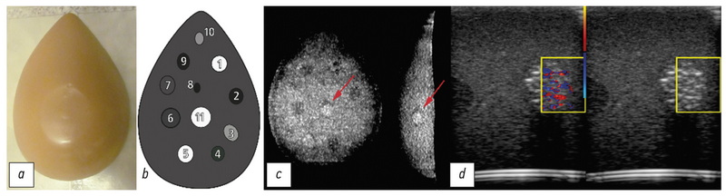
MATERIALS AND METHODS: Raw radiofrequency ultrasound signals were recorded in the phantom studies. The recorded echoes were received from objects that create the Doppler twinkling artifact and artificial blood vessels and soft tissues imitators. The data were collected between June 2016 and March 2021. Sonomed-500 with the 7.5 L38 and 3.4 C60 probes served as the research machine for the signal capture.
Data records: We present the database containing raw radiofrequency ultrasound signals from the beam former output of the research ultrasound machine. The dataset consists of CFI and B-mode echoes recorded from twinkling objects. Therefore, this database can be useful for those who test, develop and study ultrasound signal processing algorithms. Furthermore, the database is freely available online. The 10.5 GB database consists of echoes received from five phantoms. Raw radiofrequency signals were stored in the binary files; scanning parameters were stored in text files. The database is available at: https://mosmed.ai/datasets/ultrasound_doppler_twinkling_artifact.
Code availability: The public can visualize the database content with the specially written program TwinklingDatasetDisplay available at: https://github.com/Center-of-Diagnostics-and-Telemedicine/TwinklingDatasetDisplay.git.
Usage notes: The database can be used to test and develop signal-processing algorithms, such as wall filtration, velocity estimation, feature extraction, speckle reduction, etc. Furthermore, the public is free to share (copy, distribute, and transmit) and remix (adapt and do derivative works) the dataset considering appropriate credit is given.
Толық мәтін
##article.viewOnOriginalSite##Авторлар туралы
Denis Leonov
Moscow Center for Diagnostics and Telemedicine; National Research University Moscow Power Engineering Institute
Email: strat89@mail.ru
ORCID iD: 0000-0003-0916-6552
SPIN-код: 5510-4075
Scopus Author ID: 56781375200
ResearcherId: P-5266-2017
Cand. Sci. (Tech)
Ресей, 24 bld.1, Petrovka street,127051 Moscow; 28-1, Srednyaya Kalitnikovskaya street, Moscow, 109029Roman Reshetnikov
Moscow Center for Diagnostics and Telemedicine; The First Sechenov Moscow State Medical University (Sechenov University)
Email: reshetnikov@fbb.msu.ru
ORCID iD: 0000-0002-9661-0254
SPIN-код: 8592-0558
Cand. Sci. (Phys.-Math.)
Ресей, 24 bld.1, Petrovka street,127051 Moscow; 127051 Moscow, Russia; 8 bld.2Nikolay Kulberg
Moscow Center for Diagnostics and Telemedicine; Federal Research Center Computer Science and Control of the Russian Academy of Sciences
Email: kulberg@npcmr.ru
ORCID iD: 0000-0001-7046-7157
SPIN-код: 2135-9543
Cand. Sci. (Phys.-Math.)
Ресей, 24 bld.1, Petrovka street,127051 Moscow; 44, buil. 2, st. Vavilova, Moscow 119333Anastasia Nasibullina
National Research University Moscow Power Engineering Institute
Email: nastya.nasibullina@yandex.ru
ORCID iD: 0000-0003-1695-7731
SPIN-код: 2482-3372
Student
Ресей, 28-1, Srednyaya Kalitnikovskaya street, Moscow, 109029Alexandr Gromov
A.I. Yevdokimov Moscow State University of Medicine and Dentistry
Хат алмасуға жауапты Автор.
Email: gromov.ai@medsigroup.ru
ORCID iD: 0000-0002-9014-9022
SPIN-код: 6842-8684
MD, Dr. Sci. (Med.), Professor
Ресей, Delegatskaya st., 20, p. 1, 127473, MoscowӘдебиет тізімі
- Masch WR, Cohan RH, Ellis JH, et al. Clinical effectiveness of prospectively reported sonographic twinkling artifact for the diagnosis of renal calculus in patients without known urolithiasis. Am J Roentgenol. 2016;206:326–331. doi: 10.2214/ajr.15.14998
- Fujimoto Y, Shimono C, Shimoyama N, Osaki M. Twinkling artifact of microcalcifications in breast ultrasound. Ultrasound Med Biol. 2017;43(Suppl. 1):S21. doi: 10.1016/j.ultrasmedbio.2017.08.1010
- Bennett J.M., Estrada J.C., Shoemaker M.B., Pretorius M. Twinkling Artifact Associated with Guidewire Placement. Anesth Analg. 2015;121(1):69–71. doi: 10.1213/ane.0000000000000683
- Sen V, Imamoglu C, Kucukturkmen I, et al. Can Doppler ultrasonography Twinkling artifact be used as an alternative imaging modality to non-contrast-enhanced computed tomography in patients with ureteral stones? A prospective clinical study. Urolithiasis. 2017;45(2):215–219. doi: 10.1007/s00240-016-0891-8
- Winkel RR, Kalhauge A, Fredfeldt KE. The usefulness of ultrasound colour-Doppler twinkling artefact for detecting urolithiasis compared with low dose nonenhanced computerized tomography. Ultrasound Med Biol. 2012;38(7):1180–1187. doi: 10.1016/j.ultrasmedbio.2012.03.003
- Yavuz A, Ceken K, Alimoglu E, Kabaalioglu A. The reliability of color Doppler "twinkling" artifact for diagnosing millimetrical nephrolithiasis: comparison with B-Mode US and CT scanning results. J Med Ultrasonics. 2015;42(2):215–222. doi: 10.1007/s10396-014-0599-8
- Tian J, Xu L. Color Doppler Twinkling artifact in diagnosis of tuberculous pleuritis: A comparison with gray-scale ultrasonography and computed tomography. Ultrasound Med Biol. 2018;44(6): 1291–1295. doi: 10.1016/j.ultrasmedbio.2018.01.003
- Relea A, Alonso JA, González M, et al. Usefulness of the twinkling artifact on Doppler ultrasound for the detection of breast microcalcifications. Radiología. 2018;60(5):413–423. doi: 10.1016/j.rx.2018.04.004
- Lu W, Sapozhnikov OA, Bailey MR, et al. Evidence for trapped surface bubbles as the cause for the twinkling artifact in ultrasound imaging. Ultrasound Med. 2013;39(6):1026–1038. doi: 10.1016/j.ultrasmedbio.2013.01.011
- Aytac SK, Ozcan H. Effect of color Doppler system on the «twinkling» sign associated with urinary tract calculi. J Clin Ultrasound. 1999;27(8):433–439. doi: 10.1002/(sici)1097-0096(199910)27:8<433::aid-jcu4>3.0.co;2-1
- Rahmouni A, Bargoin R, Herment A, et al. Color Doppler Twinkling artifactin hyperechoic regions. Radiology. 1996;199(1):269–271. doi: 10.1148/radiology.199.1.8633158
- Kamaya A, Tuthill T, Rubin JM. Twinkling artifact on color Doppler sonography: dependence on machine parameters and underlying cause. Am J Roentgenol. 2003;180(1):215–222. doi: 10.2214/ajr.180.1.1800215
- Weinstein SP, Seghal C, Conant EF, Patton JA. Microcalcifications in breast tissue phantoms visualized with acoustic resonance coupled with power doppler US: initial observations. Radiology. 2002:224(1):265–269. doi: 10.1148/radiol.2241010511
- Seghal C. Apparatus for imaging an element within a tissue and method therefor. United States Patent. 1999;477(5):997.
- Li T, Khokhlova TD, Sapozhnikov OA, et al. A new active cavitation mapping technique for pulsed HIFU applications–bubble Doppler. IEEE Trans Ultrason Ferroelectr Freq Control. 2014;61(10): 1698–1708. doi: 10.1109/TUFFC.2014.006502
- Simon JC, Sapozhnikov OA, Kreider W, et al. The role of trapped bubbles in kidney stone detection with the color Doppler ultrasound twinkling artifact. Phys Med Biol. 2018;63(2):025011. doi: 10.1088/1361-6560/aa9a2f
- Leonov DV, Kulberg NS, Gromov AI, et al. Diagnostic mode detecting solid mineral inclusions in medical ultrasound imaging. Acoust Phys. 2018;64(5):624–636. doi: 10.1134/S1063771018050068
- Yu AC, Johnston KW, Cobbold RS. Frequency-based signal processing for ultrasound color flow imaging. Canadian Acoustics. 2007;35(2):11–23.
- Yu AC, Løvstakken L. Eigen-based clutter filter design for ultrasound color flow imaging: a review. IEEE Trans Ultrason Ferroelectr Freq Control. 2010;57(5):1096–1111. doi: 10.1109/TUFFC.2010.1521
- Yu AC, Cobbold RS. Single-ensemble-based eigen-processing methods for color flow imaging ― Part I. The Hankel-SVD filter. IEEE Trans Ultrason Ferroelectr Freq Control. 2008;55(3):559–572. doi: 10.1109/TUFFC.2008.682
- Shen Z, Feng N, Shen Y, Lee CH. An improved parametric relaxation approach to blood flow signal estimation with single-ensemble in color flow imaging. J Med Biomed Engineering. 2013;33(3):309–318. doi: 10.5405/jmbe.1368
- Yoo YM, Managuli R, Kim Y. Adaptive clutter filtering for ultrasound color flow imaging. Ultrasound Med Biol. 2003;29(9): 1311–1320. doi: 10.1016/S0301-5629(03)01014-7
- Torp H. Clutter rejection filters in color flow imaging: a theoretical approach. IEEE Trans Ultrason Ferroelectr Freq Control. 1997;44(2):417–424. doi: 10.1109/58.585126
- Wang PD, Shen Y, Feng NZ. A novel clutter rejection scheme in color flow imaging. Ultrasonics. 2006;44( Suppl 1):e303–305. doi: 10.1016/j.ultras.2006.06.017
- Bjærum S, Torp H. Statistical evaluation of clutter filters in color flow imaging. Ultrasonics. 2000;38(1-8):376–380. doi: 10.1016/s0041-624x(99)00153-5
- Kargel C, Hoebenreich G, Plevnik G, et al. Velocity estimation and adaptive clutter filtering for color flow imaging. WSEAS. 2002. Р. 1711–1716.
- Kargel C, Höbenreich G, Trummer B, Insana MF. Adaptive clutter rejection filtering in ultrasonic strain-flow imaging. IEEE Trans Ultrason Ferroelectr Freq Control. 2003;50(7):824–835. doi: 10.1109/tuffc.2003.1214502
- Lo MT, Hu K, Peng CK, Novak V. Multimodal pressure flow analysis: application of hilbert huang transform in carabral blood flow regulation. EURASIP J Adv Signal Process. 2008;2008:785243. doi: 10.1155/2008/785243
- Gerbands JJ. On the relationships between SVD, KLT and PCA. Pattern Recognition. 1981;14:375–381. doi: 10.1016/0031-3203(81)90082-0
- Løvstakken L. Signal processing in diagnostic ultrasound: algorithms for real-time estimation and visualization of blood flow velocity. Doctoral thesis, norwegian university of science and technology. Trondheim; 2007. Available from: https://pdfslide.net/ documents/signal-processing-in-diagnostic-ultrasound-algorithms-for-real-time-.html. Accessed: 14.08.2021.
- XRAD C++ software library. Available from: https://github.com/ Center-of-Diagnostics-and-Telemedicine/xrad.git. Accessed: 14.08.2021.
- Leonov DV, Kulberg NS, Gromov AI., et al. Causes of ultrasound doppler twinkling artifact. Acoust Phys. 2018;64(1):105–114. doi: 10.1134/S1063771018010128
- Mari JM, Cachard C. Acquire real-time RF digital ultrasound data from a commercial scanner. Electronic J Technical Acoustics. 2007;3:28–43.
- Leonov DV, Kulberg NS, Gromov AI, Morozov SP. Detection of microcalcifications using the ultrasound Doppler twinkling artifact. Biomedical Engineering. 2020;54(3):174–178. doi: 10.1007/s10527-020-09998-y
- Leonov DV, Kulberg NS, Fin VA, et al. Clutter filtering for diagnostic ultrasound color flow imaging. Biomedical Engineering. 2019;53(3):217–221. doi: 10.1007/s10527-019-09912-1
- Leonov DV, Kulberg NS, Fin VA, et al. Comparison of filtering techniques in ultrasound color flow imaging. Biomedical Engineering. 2019;53(2):97–101. doi: 10.1007/s10527-019-09885-1
- Song P, Manduca A, Trzasko JD, Chen S. Ultrasound small vessel imaging with block-wise adaptive local clutter filtering. IEEE Trans Med Imaging. 2017;36(1):251–262. doi: 10.1109/TMI.2016.2605819
- Li YL, Hyun D, Abou-Elkacem L, et al. Visualization of small-diameter vessels by reduction of incoherent reverberation with coherent flow power doppler. IEEE Trans Ultrason Ferroelectr Freq Control. 2016;63(11):1878–1889. doi: 10.1109/TUFFC.2016.2616112
- Chee AJ, Alfred CH. Receiver operating characteristic analysis of eigen-based clutter filters for ultrasound color flow imaging. IEEE Trans Ultrason Ferroelectr Freq Control. 2017;65(3):390–399. doi: 10.1109/TUFFC.2017.2784183
- Chee AJ, Yiu BY, Alfred CH. A GPU-Parallelized eigen-based clutter filter framework for ultrasound color flow imaging. IEEE Trans Ultrason Ferroelectr Freq Control. 2017;64(1):150–163. doi: 10.1109/TUFFC.2016.2606598
Қосымша файлдар

