

")


正电子发射断层扫描和计算机断层扫描相结合对EGFR基因突变的非小细胞肺癌患者的各种靶向治疗方案效果的比较评估
- 作者: Strutynsky V.A.1,2, Sinitsyn V.E.1, Platonova O.E.2
-
隶属关系:
- Lomonosov Moscow State University
- JSC “Medicine”
- 期: 卷 5, 编号 3 (2024)
- 页面: 394-406
- 栏目: 原创性科研成果
- URL: https://journals.rcsi.science/DD/article/view/310026
- DOI: https://doi.org/10.17816/DD624504
- ID: 310026
如何引用文章
全文:

详细
论证。在俄罗斯的医学文献中,还没有专门研究正电子发射断层扫描与计算机断层扫描相结合,对非小细胞肺癌和EGFR基因患者使用酪氨酸激酶抑制剂进行各种靶向治疗方案的效果进行比较评估。
目的 — 研究使用RECIST1.1标准和代谢指数SUVmax和 SUVmean的变化比较评估非小细胞肺癌和EGFR基因突变患者对酪氨酸激酶抑制剂靶向单药治疗和联合治疗肿瘤反应的可能性。
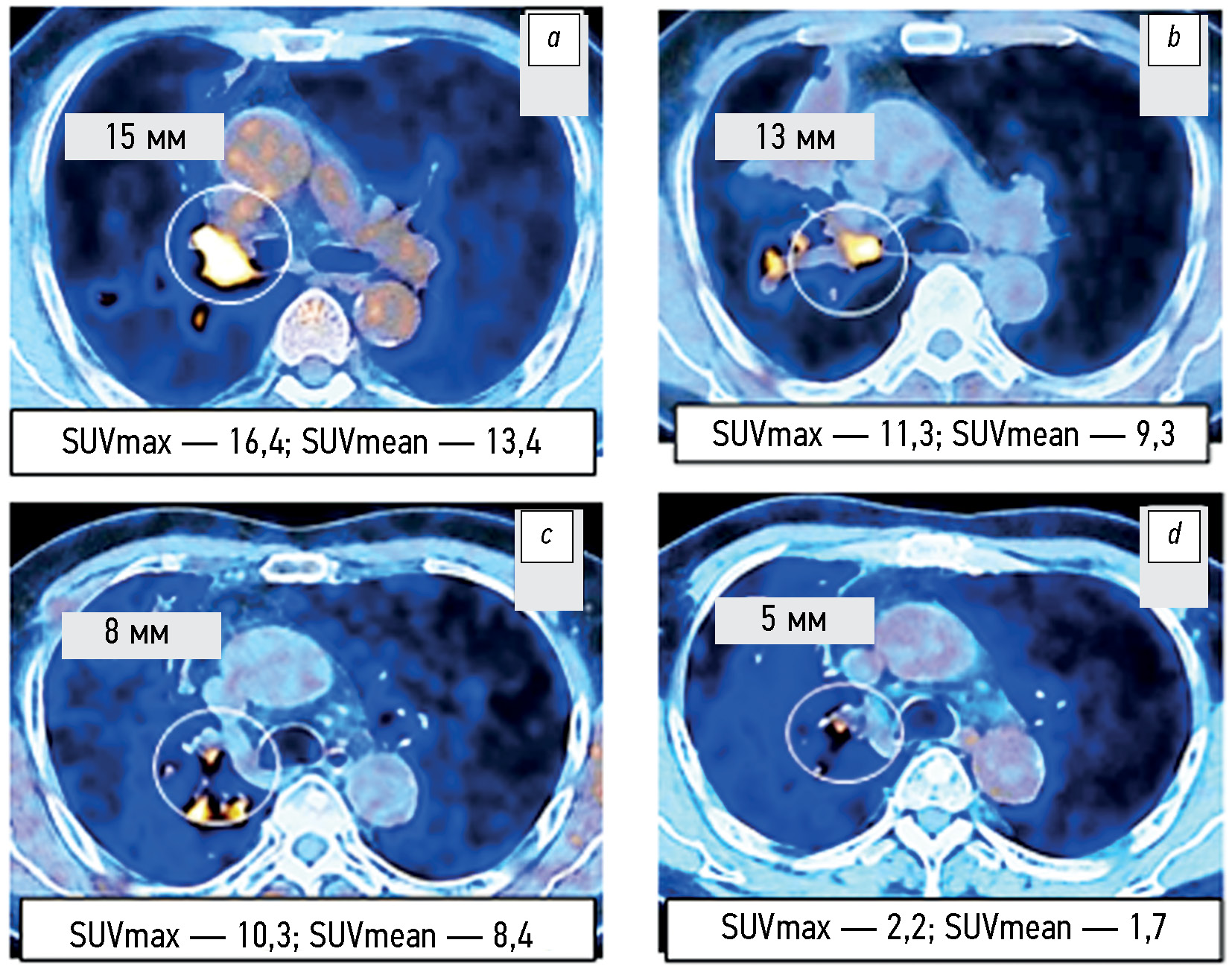
材料和方法。分析了2019年至2022年期间105名非小细胞肺癌患者的18F-氟脱氧葡萄糖(18F-FDG)正电子发射和计算机断层扫描相结合的研究方案,其中包括75名EGFR基因活化突变的患者。辐射负载因人而异,范围为45至90mSv。18F-FDG放射性药物的容积活性为260-500MBq。 评估了治疗前和治疗开始后1.5-2.0个月内最大靶灶直径总和以及代谢指数SUVmax和 SUVmean的变化。在17名非小细胞肺癌患者中,正电子发射断层扫描和计算机断层扫描相结合变化的观察持续时间至少为12个月。
结果。根据接受酪氨酸激酶抑制剂贝伐单抗或贝化疗结合综合治疗的第2组和第3组EGFR基因突变非小细胞肺癌患者的正电子发射和计算机断层扫描相结合的数据和代谢指数SUVmax和SUVmean的变化,发现疾病进展的频率(P=0.043和P=0.029)明显低于第1组患者和对照组(4.2% vs 20.0-21.8%)。部分治疗反应(P=0.092)检测率较高的趋势(58.3% vs 40.0%)也不明显。 治疗早期最大靶灶直径之和的类似变化在统计学上并不显著(p=0.187)。研究表明,在对部分非小细胞肺癌患者的长期观察中,至少有50%的病例中,最大直径总和的变化重复了第一次对照研究中发现的SUVmax和SUVmean的相应变化。
结论。根据正电子发射和计算机断层扫描相结合的数据和代谢指数SUVmax和SUVmean的变化表明,对照组患者酪氨酸激酶抑制剂贝伐珠单抗或化疗的单药靶向结合的综合治疗早期反应,与酪氨酸激酶抑制剂靶向单药治疗或化疗的反应相比,尽管根据RECIST1.1标准靶灶最大直径的变化趋势无统计意义,但疾病代谢进展的频率明显降低。至少有50%的病例在治疗早期代谢指数SUVmax和SUVmean的变化超过了靶向病灶最大直径总和的类似变化,根据 RECIST1.1标准,这些变化可用于及时识别有进一步发展的高风险患者群体。
关键词
作者简介
Vladislav A. Strutynsky
Lomonosov Moscow State University; JSC “Medicine”
编辑信件的主要联系方式.
Email: Rammen2@yandex.ru
SPIN 代码: 6810-5644
俄罗斯联邦, Moscow; Moscow
Valentin E. Sinitsyn
Lomonosov Moscow State University
Email: Vsini@mail.ru
ORCID iD: 0000-0002-5649-2193
SPIN 代码: 8449-6590
MD, Dr. Sci. (Medicine), Professor
俄罗斯联邦, MoscowOksana E. Platonova
JSC “Medicine”
Email: Platonova@medicina.ru
ORCID iD: 0000-0003-0093-7285
MD, Cand. Sci. (Medicine)
俄罗斯联邦, Moscow参考
- Kaprin AD, Starinskii VV, Shakhzadova AO. Malignant neoplasms in Russia in 2019 (morbidity and mortality). Moscow: Moskovskii gosudarstvennyi nauchno–issledovatel’skii meditsinskii institut im. P.A. Gertsena; 2020. (In Russ.)
- Sakaeva DD, Reutova EV. Targeted therapy for metastatic non-small cell lung cancer. In: Laktionov KK, Breder VV, editors. Lung cancer. Moscow: “Granat”; 2020. P:75–88. (In Russ.)
- Tyulyandin SA. Targeted therapy: twenty years of success and failures. Practical oncology. 2019;20(4):274–288. doi: 10.31917/2004274
- Deng W, Wang K, Jiang Y, et al. Erlotinib plus bevacizumab versus erlotinib alone in patients with EGFR-positive advanced non-small-cell lung cancer: a systematic review and meta-analysis of randomised controlled trials. BMJ Open. 2022;12(8):e062036. doi: 10.1136/bmjopen-2022-062036
- Landre T, Des Guetz G, Chouahnia R, et al. First-line angiogenesis inhibitor plus erlotinib versus erlotinib alone for advanced non-small-cell lung cancer harboring an EGFR mutation. Cancer Res Clin Oncol. 2020;146(12):3333–3339. doi: 10.1007/s00432-020-03311-w
- Maemondo M, Fukuhara T, Saito H, et al. NEJ026: Final overall survival analysis of bevacizumab plus erlotinib treatment for NSCLC patients harboring activating EGFR-mutations. Journal of Clinical Oncology. 2020;38(15):9506–9506. doi: 10.1200/JCO.2020.38.15_suppl.9506
- Rocco D, Della Gravara L, Palazzolo G, et al. The role of antiangiogenic monoclonal antibodies combined to EGFR-TKIs in the treatment of advanced non-small cell lung cancer with activating EGFR mutations: acquired resistance mechanisms and strategies to overcome them. Cancer Drug Resist. 2022;5(4):1016–1024. doi: 10.20517/cdr.2022.77
- Saito H, Fukuhara T, Furuya N, et al. Erlotinib plus bevacizumab versus erlotinib alone in patients with EGFR-positive advanced non-squamous non-small-cell lung cancer (NEJ026): interim analysis of an open-label, randomised, multicentre, phase 3 trial. Lancet Oncol. 2019;20(5):625–635. doi: 10.1016/S1470-2045(19)30035-X
- Stinchcombe TE, Jänne PA, Wang X, et al. Effect of Erlotinib Plus Bevacizumab vs Erlotinib Alone on Progression-Free Survival in Patients With Advanced EGFR-Mutant Non-Small Cell Lung Cancer: A Phase 2 Randomized Clinical Trial. JAMA Oncol. 2019;5(10):1448–1455. doi: 10.1001/jamaoncol.2019.1847
- Meshcheryakova NA. Positron emission tomography combined with computed tomography in the diagnosis and evaluation of treatment effectiveness of non-small cell lung cancer [dissertation]. Moscow; 2018.
- Meshcheryakova NA, Dolgushin MB, Borisova TN, Davydov MM, Laktionov KK. Efficacy of 18F-FDG and 18F-FLT PET/CT for Assessment of Chemoradiotherapy in Patient with Non-Small Cell Lung Cancer (Clinical Observation). Medical Visualization. 2017;(1):53–56. doi: 10.24835/1607-0763-2017-1-53-56
- Wahl R, Jacene Н, Kasamon Y, Lodge M. From RECIST to PERCIST: Evolving Considerations for PET Response Criteria in Solid Tumors. J Nucl Med. 2009;50(5):4–11. doi: 10.2967/jnumed.111.093443
- Gelezhe PB, Morozov SP, Shavladze N. Comparison of the accuracy of evaluating the attenuation correction and tumor size during sequential performance of breast 18F-FDG PET CT and PET/ MRI. Vestnik of the Russian scientific center of roentgenoradiology. 2019;19(4):48–62. EDN: LWGHUU
- Ding Q, Chen X, Yang L, et al. PET/CT evaluation of response to chemotherapy in non-small cell lung cancer: PET response criteria in solid tumors (PERCIST) versus response evaluation criteria in solid tumors (RECIST). J Thorac Dis. 2014;6(6):677–683. doi: 10.3978/j.issn.2072-1439.2014.05.10
- Pierson C, Grinchak T, Sokolovic C, et al. Response criteria in solid tumors (PERCIST/RECIST) and SUVmax in early-stage non-small cell lung cancer patients treated with stereotactic body radiotherapy. Radiat Oncol. 2018;13(1):34. doi: 10.1186/s13014-018-0980-7
- Beer L, Hochmair M, Haug AR, et al. Comparison of RECIST and PERCIST for the Evaluation of Response to PD-1/PD-L1 Blockade Therapy in Patients With Non-Small Cell Lung Cancer. Clin Nucl Med. 2019;44(7):535–543. doi: 10.1097/RLU.0000000000002603
- Kaira K, Higuchi T, Naruse I, et al. Metabolic activity by 18 F–FDG-PET/ CT is predictive of early response after nivolumab in previously treated NSCLC. Eur J Nucl Med Mol Imaging. 2018;45(1):56–66. doi: 10.1007/s00259-017-3806-1
- Khodzhibekova MM. Value of combined PET/CT in the diagnosis and monitoring of treatment of lymphoma patients [dissertation]. Moscow; 2019. (In Russ.)
- Koopman D, Jager PL, Slump CH, et al. SUV variability in EARL-accredited conventional and digital PET. EJNMMI Res. 2019;9(1):106. doi: 10.1186/s13550-019-0569-7
- Xie X, Chen H, Yang H, Lin H. Predictive value of positron emission tomography for the prognosis of molecularly targeted therapy in solid tumors. Z Onco Targets Ther. 2018;7(11):8885–8899. doi: 10.2147/OTT.S178076
- Kamiyoshihara M, Igai H, Ohsawa F, Numajiri K, et al. Gefitinib Monotherapy Afforded Long-Term Survival of an Octogenarian Patient with a Postoperative Recurrence of a Pulmonary Adenocarcinoma — A Case Report. Gan To Kagaku Ryoho. 2023;50(2):187–189. (In Japanese)
- Stinchcombe TE. Foreword: Gefitinib in Non-Small-Cell Lung Cancer: The IDEAL 1 Trial. J Clin Oncol. 2023;41(6):1159–1160. doi: 10.1200/JCO.22.02660
- Zhong WZ, Wang Q, Mao WM, et al. Gefitinib versus vinorelbine plus cisplatin as adjuvant treatment for stage II-IIIA (N1-N2) EGFR-mutant NSCLC (ADJUVANT/CTONG1104): a randomised, open-label, phase 3 study. Lancet Oncol. 2018;19(1):139–148. doi: 10.1016/S1470-2045(17)30729-5
- Liu M, Luo N, Fang Z, Liu Q, et al. The efficacy and toxicity of maintenance therapy with bevacizumab plus pemetrexed versus bevacizumab/pemetrexed alone for stage IIIB/IV nonsquamous non-small cell lung cancer: A meta-analysis of randomized controlled trials. Clin Pharm Ther. 2022;47(2):157–167. doi: 10.1111/jcpt.13534
- Chen Z, Shen S. Intercalated combination of chemotherapy and erlotinib for stage IIIA non-small-cell lung cancer: a multicenter, open-label, single-arm, phase II study. Cancer Manag Res. 2019;11:6543–6552. doi: 10.2147/CMAR.S189287 DOI: https://doi.org/10.17816/DD624504
补充文件


