

")


使用人工智能对乳腺Ⅹ线摄影结果进行两次 查看:一种组织大规模预防性研究的新模式
- 作者: Vasilev Y.A.1, Tyrov I.A.2, Vladzymyrskyy A.V.1, Arzamasov K.M.1, Shulkin I.M.1, Kozhikhina D.D.1, Pestrenin L.D.1
-
隶属关系:
- Moscow Center for Diagnostics and Telemedicine
- Moscow Health Care Department
- 期: 卷 4, 编号 2 (2023)
- 页面: 93-104
- 栏目: 原创性科研成果
- URL: https://journals.rcsi.science/DD/article/view/146879
- DOI: https://doi.org/10.17816/DD321423
- ID: 146879
如何引用文章

详细
论证。近年来,医疗数据集和人工智能软件技术开发的可达性,使得医疗诊断,特别是乳腺Ⅹ线摄影的解决方案激增。这种登记为医疗设备的软件可被用于描述数乳腺Ⅹ线摄影,这允许提供医疗服务时在很大程度上节省时间、物质和人力的资源,同时确保乳房预防性检查的质量。
该研究的目的是证明使用基于人工智能技术的软件对数字乳房X光照片进行第一次解读的可用性和有效性,同时保持放射科医生对X射线图像进行第二次描述的做法。
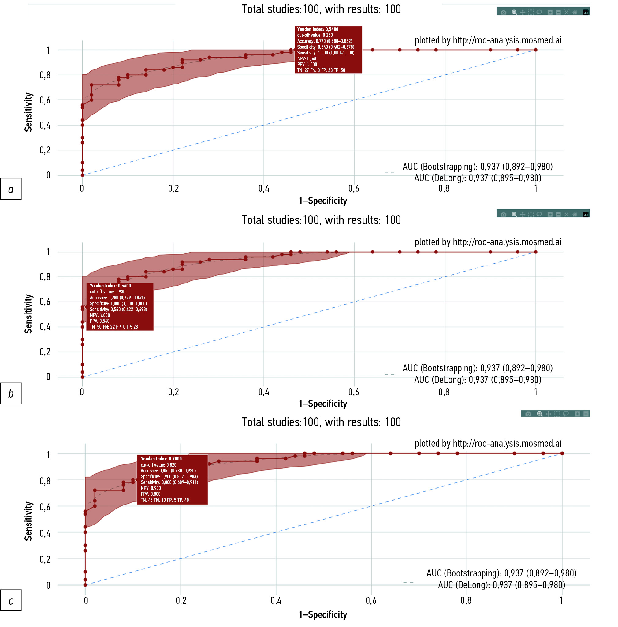
材料和方法。我们用在俄罗斯联邦登记为医疗设备的基于人工智能技术的软件处理了100张数字乳房X光照片的数据集,其中50张为“无目标病变”,50张为“存在目标病变”(有恶性肿瘤症状)。进行了ROC分析。研究局限性:诊断准确性度量值是基于人工智能技术的软件版本获得的,是在2022年底有效的。
结果。当设置为80.0%的灵敏度时,人工智能显示出90.0%的特异度(95% CI 81.7-98.3)和85.0%的准确性(95% CI 78.0-92.0)。当设置为100%的特异度时,人工智能显示出56.0%的灵敏度(95% CI 42.2-69.8)和78.0%的准确性(95% CI 69.9-86.1)。当设置为100%灵敏度时,人工智能的特异度为54.0%(95% CI 40.2-67.8),准确性为77.0%(95% CI 68.8-85.2)。提出了两种方法,涉及通过人工智能对数字乳腺Ⅹ线摄影进行的第一次自主解读。第一种方法是利用人工智能评估X射线图像,其灵敏度高于放射科医生进行的双重乳腺Ⅹ线摄影描述,特异度水平相当。第二种方法是基于人工智能技术的软件将对乳腺Ⅹ线摄影进行分类(“无目标病变”或“存在目标病变”),表明其对结果的“信心”程度,取决于预测值所处的“走廊”。
结论。使用基于人工智能技术的软件对数字乳房X光照片进行第一次自主描述的两种提出方案都能提供与放射科医生对图像进行的双重描述相同甚至更高的诊断质量。在全国范围内在实践中实现这种方法的经济效益可能是每年6亿至55亿卢布。
作者简介
Yuriy A. Vasilev
Moscow Center for Diagnostics and Telemedicine
Email: npcmr@zdrav.mos.ru
ORCID iD: 0000-0002-0208-5218
SPIN 代码: 4458-5608
MD, Cand. Sci. (Med)
俄罗斯联邦, MoscowIlya A. Tyrov
Moscow Health Care Department
Email: npcmr@zdrav.mos.ru
ORCID iD: 0000-0001-9337-624X
SPIN 代码: 8625-3458
俄罗斯联邦, Moscow
Anton V. Vladzymyrskyy
Moscow Center for Diagnostics and Telemedicine
Email: npcmr@zdrav.mos.ru
ORCID iD: 0000-0002-2990-7736
SPIN 代码: 3602-7120
MD, Dr. Sci. (Med), Professor
俄罗斯联邦, MoscowKirill M. Arzamasov
Moscow Center for Diagnostics and Telemedicine
Email: npcmr@zdrav.mos.ru
ORCID iD: 0000-0001-7786-0349
SPIN 代码: 3160-8062
MD, Cand. Sci. (Med)
俄罗斯联邦, MoscowIgor M. Shulkin
Moscow Center for Diagnostics and Telemedicine
Email: npcmr@zdrav.mos.ru
ORCID iD: 0000-0002-7613-5273
SPIN 代码: 5266-0618
俄罗斯联邦, Moscow
Daria D. Kozhikhina
Moscow Center for Diagnostics and Telemedicine
Email: npcmr@zdrav.mos.ru
ORCID iD: 0000-0001-7690-8427
SPIN 代码: 5869-3854
俄罗斯联邦, Moscow
Lev D. Pestrenin
Moscow Center for Diagnostics and Telemedicine
编辑信件的主要联系方式.
Email: PestreninLD@zdrav.mos.ru
ORCID iD: 0000-0002-1786-4329
SPIN 代码: 7193-7706
Junior Research Associate
俄罗斯联邦, Moscow参考
- Malignant neoplasms in Russia in 2021 (morbidity and mortality). Ed by A.D. Kaprin, V.V. Starinsky, A.O. Shahzadova. Мoscow; 2022. 252 р. (In Russ).
- The state of oncological assistance to the population of Russia in 2021. Ed by A.D. Kaprin, V.V. Starinsky, A.O. Shahzadova. Мoscow; 2022. 239 р. (In Russ).
- Chen Y, James JJ, Michalopoulou E, et al. Performance of radiologists and radiographers in double reading mammograms: The UK national health service breast screening program. Radiology. 2023;306(1):102–109. doi: 10.1148/radiol.212951
- Euler-Chelpin MV, Lillholm M, Napolitano G, et al. Screening mammography: Benefit of double reading by breast density. Breast Cancer Res Treat. 2018;171(3):767–776. doi: 10.1007/s10549-018-4864-1
- Hickman SE, Woitek R, Le EP, et al. Machine learning for workflow applications in screening mammography: Systematic review and meta-analysis. Radiology. 2022;302(1):88–104. doi: 10.1148/radiol.2021210391
- Liu J, Lei J, Ou Y, et al. Mammography diagnosis of breast cancer screening through machine learning: A systematic review and meta-analysis. Clin Exp Med. 2022. doi: 10.1007/s10238-022-00895-0
- Rozhkova NI, Rojtberg PG, Varfolomeeva AA, et al. Neural network-based segmentation model for breast cancer X-ray screening. Sechenov medical journal. 2020;11(3):4–14 (In Russ). doi: 10.47093/2218-7332.2020.11.3.4-14
- Vasilev JA, Vladzimirskyy AV. Computer vision in radiology: The first stage of the Moscow experiment: Monograph. Moscow: Izdatel’skie resheniya; 2022. 388 р. (In Russ).
- Patent RUS № 2022617324/05.04.2022. Byul. № 4. Morozov SP, Andreichenko AE, Chetverikov SF, et al. A web-based tool for performing ROC analysis of diagnostic test results. Available from: https://www.elibrary.ru/item.asp?id=48373757. Accessed: 10.03.2023. (In Russ).
- Morozov SP, Vladzimirsky AV, Klyashtornyy VG, et al. Clinical acceptance of software based on artificial intelligence technologies (radiology). Moscow; 2019. 45 p. (Ser. Best practices in medical imaging).
- Schaffter T, Buist DS, Lee CI, et al. Evaluation of combined artificial intelligence and radiologist assessment to interpret screening mammograms. JAMA Netw Open. 2020;3(3):e200265. doi: 10.1001/jamanetworkopen.2020.0265
- Wan Y, Tong Y, Liu Y, et al. Evaluation of the combination of artificial intelligence and radiologist assessments to interpret malignant architectural distortion on mammography. Front Oncol. 2022;(12):880150. doi: 10.3389/fonc.2022.880150
- Leibig C, Brehmer M, Bunk S, et al. Combining the strengths of radiologists and AI for breast cancer screening: A retrospective analysis. Lancet Digit Health. 2022;4(7):e507–e519. doi: 10.1016/S2589-7500(22)00070-X
补充文件


