

")


Double-reading mammograms using artificial intelligence technologies: A new model of mass preventive examination organization
- Авторлар: Vasilev Y.A.1, Tyrov I.A.2, Vladzymyrskyy A.V.1, Arzamasov K.M.1, Shulkin I.M.1, Kozhikhina D.D.1, Pestrenin L.D.1
-
Мекемелер:
- Moscow Center for Diagnostics and Telemedicine
- Moscow Health Care Department
- Шығарылым: Том 4, № 2 (2023)
- Беттер: 93-104
- Бөлім: Original Study Articles
- URL: https://journals.rcsi.science/DD/article/view/146879
- DOI: https://doi.org/10.17816/DD321423
- ID: 146879
Дәйексөз келтіру

Аннотация
BACKGROUND: In recent years, the availability of medical datasets and technologies for software development based on artificial intelligence technology has resulted in a growth in the number of solutions for medical diagnostics, particularly mammography. Registered as a medical device, this program can interpret digital mammography, significantly saving time, material, and human resources in healthcare while ensuring the quality of mammary gland preventive studies.
AIM: This study aims to justify the possibility and effectiveness of artificial intelligence-based software for the first interpretation of digital mammograms while maintaining the practice of a radiologist’s second description of X-ray images.
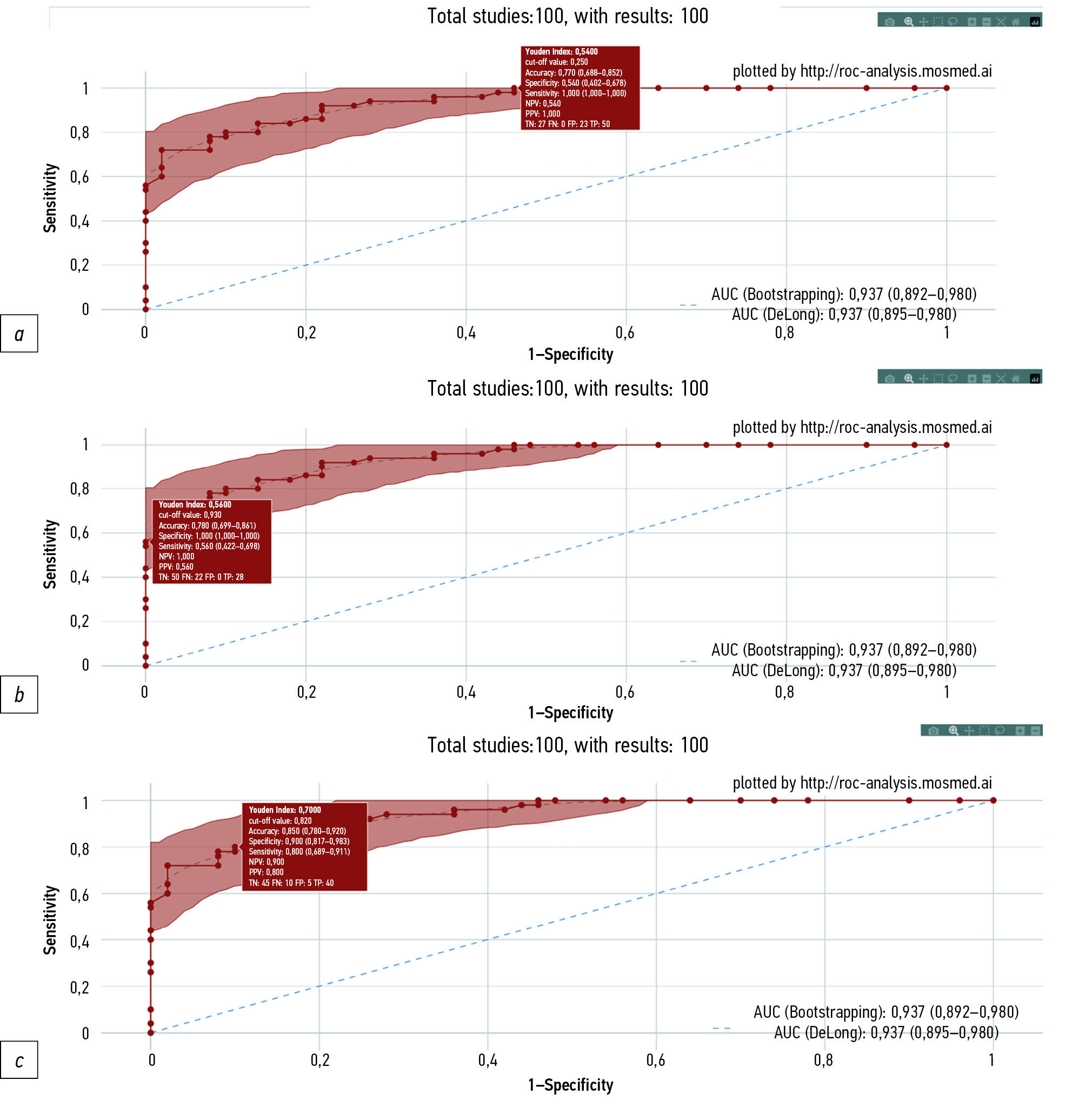
MATERIALS AND METHODS: A dataset of 100 digital mammography studies (50 — “absence of target pathology” and 50 ― “presence of target pathology,” with signs of malignant neoplasms) was processed by software based on artificial intelligence technology that was registered as a medical device in the Russian Federation. Receiver operating characteristic analysis was performed. Limitations of the study include the values of diagnostic accuracy metrics obtained for software based on artificial intelligence technology versions, relevant at the end of 2022.
RESULTS: When set to 80.0% sensitivity, artificial intelligence specificity was 90.0% (95% CI, 81.7–98.3), and accuracy was 85.0% (95% CI, 78.0–92.0). When set to 100% specificity, artificial intelligence demonstrated 56.0% sensitivity (95% CI, 42.2–69.8) and 78.0% accuracy (95% CI, 69.9–86.1). When the sensitivity was set to 100%, the artificial intelligence specificity was 54.0% (95% CI, 40.2–67.8), and the accuracy was 77.0% (95% CI, 68.8–85.2). Two approaches have been proposed, providing an autonomous first interpretation of digital mammography using artificial intelligence. The first approach is to evaluate the X-ray image using artificial intelligence with a higher sensitivity than that of the double-reading mammogram by radiologists, with a comparable level of specificity. The second approach implies that artificial intelligence-based software will determine the mammogram category (“absence of target pathology” or “presence of target pathology”), indicating the degree of “confidence” in the obtained result, depending on the corridor into which the predicted value falls.
CONCLUSIONS: Both proposed approaches for using artificial intelligence-based software for the autonomous first interpretation of digital mammograms can provide diagnostic quality comparable to, if not superior to, double-image reading by radiologists. The economic benefit from the practical implementation of this approach nationwide can range from 0.6 to 5.5 billion rubles annually.
Негізгі сөздер
Толық мәтін
##article.viewOnOriginalSite##Авторлар туралы
Yuriy Vasilev
Moscow Center for Diagnostics and Telemedicine
Email: npcmr@zdrav.mos.ru
ORCID iD: 0000-0002-0208-5218
SPIN-код: 4458-5608
MD, Cand. Sci. (Med)
Ресей, MoscowIlya Tyrov
Moscow Health Care Department
Email: npcmr@zdrav.mos.ru
ORCID iD: 0000-0001-9337-624X
SPIN-код: 8625-3458
Ресей, Moscow
Anton Vladzymyrskyy
Moscow Center for Diagnostics and Telemedicine
Email: npcmr@zdrav.mos.ru
ORCID iD: 0000-0002-2990-7736
SPIN-код: 3602-7120
MD, Dr. Sci. (Med), Professor
Ресей, MoscowKirill Arzamasov
Moscow Center for Diagnostics and Telemedicine
Email: npcmr@zdrav.mos.ru
ORCID iD: 0000-0001-7786-0349
SPIN-код: 3160-8062
MD, Cand. Sci. (Med)
Ресей, MoscowIgor Shulkin
Moscow Center for Diagnostics and Telemedicine
Email: npcmr@zdrav.mos.ru
ORCID iD: 0000-0002-7613-5273
SPIN-код: 5266-0618
Ресей, Moscow
Daria Kozhikhina
Moscow Center for Diagnostics and Telemedicine
Email: npcmr@zdrav.mos.ru
ORCID iD: 0000-0001-7690-8427
SPIN-код: 5869-3854
Ресей, Moscow
Lev Pestrenin
Moscow Center for Diagnostics and Telemedicine
Хат алмасуға жауапты Автор.
Email: PestreninLD@zdrav.mos.ru
ORCID iD: 0000-0002-1786-4329
SPIN-код: 7193-7706
Junior Research Associate
Ресей, MoscowӘдебиет тізімі
- Malignant neoplasms in Russia in 2021 (morbidity and mortality). Ed by A.D. Kaprin, V.V. Starinsky, A.O. Shahzadova. Мoscow; 2022. 252 р. (In Russ).
- The state of oncological assistance to the population of Russia in 2021. Ed by A.D. Kaprin, V.V. Starinsky, A.O. Shahzadova. Мoscow; 2022. 239 р. (In Russ).
- Chen Y, James JJ, Michalopoulou E, et al. Performance of radiologists and radiographers in double reading mammograms: The UK national health service breast screening program. Radiology. 2023;306(1):102–109. doi: 10.1148/radiol.212951
- Euler-Chelpin MV, Lillholm M, Napolitano G, et al. Screening mammography: Benefit of double reading by breast density. Breast Cancer Res Treat. 2018;171(3):767–776. doi: 10.1007/s10549-018-4864-1
- Hickman SE, Woitek R, Le EP, et al. Machine learning for workflow applications in screening mammography: Systematic review and meta-analysis. Radiology. 2022;302(1):88–104. doi: 10.1148/radiol.2021210391
- Liu J, Lei J, Ou Y, et al. Mammography diagnosis of breast cancer screening through machine learning: A systematic review and meta-analysis. Clin Exp Med. 2022. doi: 10.1007/s10238-022-00895-0
- Rozhkova NI, Rojtberg PG, Varfolomeeva AA, et al. Neural network-based segmentation model for breast cancer X-ray screening. Sechenov medical journal. 2020;11(3):4–14 (In Russ). doi: 10.47093/2218-7332.2020.11.3.4-14
- Vasilev JA, Vladzimirskyy AV. Computer vision in radiology: The first stage of the Moscow experiment: Monograph. Moscow: Izdatel’skie resheniya; 2022. 388 р. (In Russ).
- Patent RUS № 2022617324/05.04.2022. Byul. № 4. Morozov SP, Andreichenko AE, Chetverikov SF, et al. A web-based tool for performing ROC analysis of diagnostic test results. Available from: https://www.elibrary.ru/item.asp?id=48373757. Accessed: 10.03.2023. (In Russ).
- Morozov SP, Vladzimirsky AV, Klyashtornyy VG, et al. Clinical acceptance of software based on artificial intelligence technologies (radiology). Moscow; 2019. 45 p. (Ser. Best practices in medical imaging).
- Schaffter T, Buist DS, Lee CI, et al. Evaluation of combined artificial intelligence and radiologist assessment to interpret screening mammograms. JAMA Netw Open. 2020;3(3):e200265. doi: 10.1001/jamanetworkopen.2020.0265
- Wan Y, Tong Y, Liu Y, et al. Evaluation of the combination of artificial intelligence and radiologist assessments to interpret malignant architectural distortion on mammography. Front Oncol. 2022;(12):880150. doi: 10.3389/fonc.2022.880150
- Leibig C, Brehmer M, Bunk S, et al. Combining the strengths of radiologists and AI for breast cancer screening: A retrospective analysis. Lancet Digit Health. 2022;4(7):e507–e519. doi: 10.1016/S2589-7500(22)00070-X
Қосымша файлдар


