

")


乳头乳晕复合体的病理和异常的诊断: 一系列临床病例
- 作者: Karanadze E.N.1, Sinitsyn V.E.2, Karanadze M.A.3
-
隶属关系:
- Clinical Diagnostic Center MEDSI on Krasnaya Presnya
- Lomonosov Moscow State University, Medical Scientific and Educational Center
- The Russian National Research Medical University named after N.I. Pirogov
- 期: 卷 4, 编号 1 (2023)
- 页面: 51-60
- 栏目: 临床病例及临床病例的系列
- URL: https://journals.rcsi.science/DD/article/view/146875
- DOI: https://doi.org/10.17816/DD112093
- ID: 146875
如何引用文章

详细
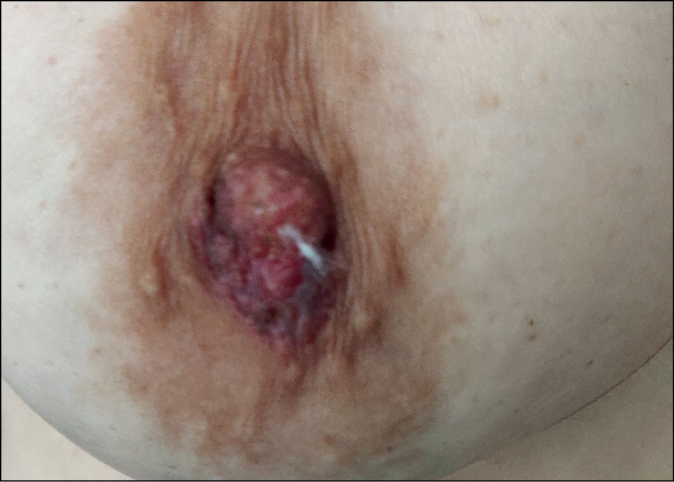
乳头乳晕复合体是一种特殊的解剖学和组织学结构。正常结构的变异性、病理过程的广泛性和诊断成像的复杂性给放射科医生和临床医生带来困难。乳房磁共振成像是检测结构特征、诊断涉及乳头乳晕复合体的良性和恶性疾病的最灵敏方法。在乳腺钼靶和超声检查结果不明确的情况下,磁共振成像作为一种额外的诊断工具非常有用。磁共振成像允许看到乳腺后区,适合诊断乳头瘤、腺瘤、佩吉特氏病、导管原位癌和浸润性癌。
我们在这篇文章中描述了乳头乳晕复合体的病理和异常的临床病例,这可能会对放射科医生、妇科医生和临床住院医师有用。
作者简介
Elena N. Karanadze
Clinical Diagnostic Center MEDSI on Krasnaya Presnya
编辑信件的主要联系方式.
Email: ekaranadze@mail.ru
ORCID iD: 0000-0001-6745-1672
MD, Cand. Sci. (Med.)
俄罗斯联邦, MoscowValentin E. Sinitsyn
Lomonosov Moscow State University, Medical Scientific and Educational Center
Email: info@npcmr.ru
ORCID iD: 0000-0002-5649-2193
SPIN 代码: 8449-6590
MD, Dr. Sci. (Med), Professor
俄罗斯联邦, MoscowMariia A. Karanadze
The Russian National Research Medical University named after N.I. Pirogov
Email: ekaranadze@mail.ru
ORCID iD: 0009-0008-1723-6796
俄罗斯联邦, Moscow
参考
- Stone K, Wheeler A. A Review of anatomy, physiology, and benign pathology of the nipple. Ann Surg Oncol. 2015;22(10):3236–3240. doi: 10.1245/s10434-015-4760-4
- Reisenbichler E, Hanley KZ. Seminars in diagnostic pathology developmental disorders and malformations of the breast. Semin Diagn Pathol. 2019;36(1):11–15. doi: 10.1053/j.semdp.2018.11.007
- Liao CY, Wu YT, Wu WP, et al. Role of breast magnetic resonance imaging in predicting malignant invasion of the nipple-areolar complex: Potential predictors and reliability between inter-observers. Medicine (Baltimore). 2017;96(28):e7170. doi: 10.1097/MD.0000000000007170
- Milon A, Wahab CA, Kermarrec E, et al. Breast MRI: Is faster better? AJR Am J Roentgenol. 2020;214(2):282–95. doi: 10.2214/AJR.19.21924
- Acrpractice parameterfor the performance of contrast-enhanced magnetic resonance imaging (MRI) of the breast. AcoR; 2018. Available from: https://www.acr.org/-/media/ACR/Files/Practice-Parameters/MR-Contrast-Breast.pdf. Accessed: 15.01.2023.
- Lee SJ, Trikha S, Moy L, et al. ACR appropriateness criteria evaluation of nipple discharge. J Am Coll Radiol. 2017;14(5s):S138–53. doi: 10.1016/j.jacr.2017.01.030
- Ferris-James DM, Iuanow E, Mehta TS, et al. Imaging approaches to diagnosis and management of common ductal abnormalities. Radiographics. 2012;32(4):1009–1030. doi: 10.1148/rg.324115150
- Del Riego J, Pitarch M, Codina C, et al. Multimodality approach to the nipple-areolar complex: A pictorial review and diagnostic algorithm. Insights Imaging. 2020 5;11(1):89. doi: 10.1186/s13244-020-00896-1
- Yoon JH, Yoon H, Kim EK, et al. Ultrasonographic evaluation of women with pathologic nipple discharge. Ultrasonography. 2017;36(4):310–320. doi: 10.14366/usg.17013
- Horvat JV, Keating DM, Rodrigues-Duarte H, et al. Calcifications at digital breast tomosynthesis: Imaging features and biopsy techniques. Radiographics. 2019;39(2):307–318. doi: 10.1148/rg.2019180124
- Huppe AI, Overman KL, Gatewood JB, et al. Mammography positioning standards in the digital era: Is the status quo acceptable? AJR Am J Roentgenol. 2017;209(6):1419–1425. doi: 10.2214/AJR.16.17522
- Gao Y, Brachtel EF, Hernandez O, Heller SL. An analysis of nipple enhancement at breast MRI with radiologic-pathologic correlation. Radiographics. 2019;39(1):10–27. doi: 10.1148/rg.2019180039
- Lim HS, Jeong SJ, Lee JS, et al. Paget disease of the breast: Mammographic, US, and MR Imaging findings with pathologic correlation. Radiographics. 2011;31(7):1973–1987. doi: 10.1148/rg.317115070
- Geffroy D, Doutriaux-Dumoulins I. Clinical abnormalities of the nipple-areola complex: The role of imaging. Diagn Interv Imaging. 2015;96(10):1033–1044. doi: 10.1016/j.diii.2015.07.001
- Moon JY, Chang YW, Lee EH, Seo DY. Malignant invasion of the nipple-areolar complex of the breast: Usefulness of breast MRI. AJR Am J Roentgenol. 2013;201(2):448–455. doi: 10.2214/AJR.12.9186
- Maksimov DA, Sergeev AN, Morozov AM, et al. About modern types of surgical treatment for breast cancer (literature review). Journal of new medical technologies, eEdition. 2021;(1):7–13. (In Russ). doi: 10.24412/2075-4094-2021-1-1-1
- Zikiryakhodzhaev AD, Volchenko NN, Saribekyan EK, Rasskazova EA. Lesion of the nipple-areola complex in patients with breast cancer. Problems in oncology. 2017;63(4):593–597. (In Russ).
- Levchuk AL, Khodyrev SA, Shabaev RM. Current state of breast reconstructive surgery. Bulletin of Pirogov National Medical & Surgical Center. 2021;16(2):122–127. (In Russ). doi: 10.25881/20728255-2021-16-2-122
- Berger N, Luparia A, Di Leo G, et al. Diagnostic performance of MRI versus galactography in women with pathologic nipple discharge: A systematic review and meta-analysis. AJR Am J Roentgenol. 2017;209(2):465–471. doi: 10.2214/AJR.16.16682
- Sripathi S, Ayachit A, Kadavigere R, et al. Spectrum of imaging findings in Paget’s disease of the breast: A pictorial review. Insights Imaging. 2015;6(4):419–429. doi: 10.1007/s13244-015-0415-z
- Da Costa D, Taddese A, Cure ML, et al. Common and unusual diseases of the nipple-areolar complex. Radiographics. 2007;27(Suppl. 1):S65–S77. doi: 10.1148/rg.27si075512
- Alhayo ST, Edirimanne S. Clinically challenging case of nipple adenoma. Breast J. 2018;24(6):1084–1085. doi: 10.1111/tbj.13089
补充文件


