

")


肌肉减少症:解决诊断问题的现代方法
- 作者: Smorchkova A.K.1, Petraikin A.V.1, Semenov D.S.1, Sharova D.E.1
-
隶属关系:
- Research and Practical Clinical Center for Diagnostics and Telemedicine Technologies
- 期: 卷 3, 编号 3 (2022)
- 页面: 196-211
- 栏目: 科学评论
- URL: https://journals.rcsi.science/DD/article/view/110721
- DOI: https://doi.org/10.17816/DD110721
- ID: 110721
如何引用文章

详细
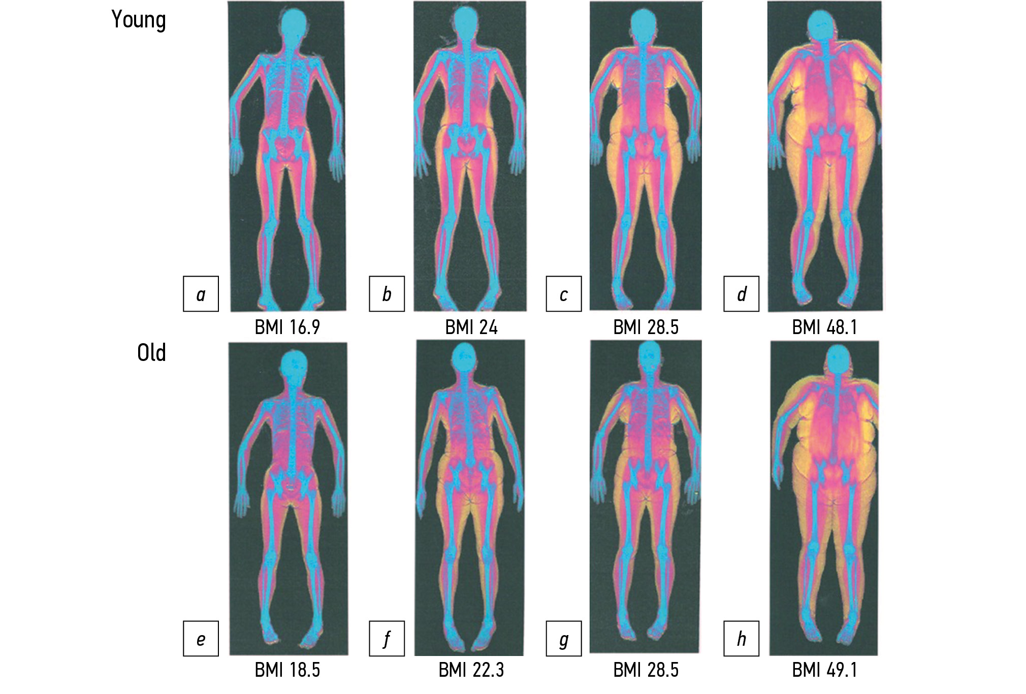
肌肉减少症是医学统计和医疗保健系统的一个相对较新的诊断。然而,由于大量可能的不良后果,例如跌倒风险增加、残疾、住院时间延长和死亡率增加,它对医疗体系造成了社会和经济负担。虽然肌肉减少症没有高度专业化的药物疗法,但预防和及时的非药物治疗可以降低潜在不良反应的风险。诊断»肌肉减少症»不仅需要确认肌力下降,还需要确认肌肉质量下降。仪器诊断包括双能X光吸收测量 (DXA) 和生物阻抗测定法 (BIA)等方法。 这些方法可以辅以人工智能 (AI) 算法,用于在计算机断层扫描和磁共振图像上自动分割肌肉和脂肪组织,然后计算L3椎骨水平的肌肉骨骼指数。 此类软件在莫斯科市统一医疗信息和分析系统 (ERIS EMIAS)统一放射信息服务等系统中使用时,为机会性筛查提供了机会。然而,尽管欧洲老年人肌肉减少症工作组将CT和MRI技术认定为“金标准”,但仍然没有公认的用于诊断肌肉减少症的CT 和MR定量L3介质值。除此之外,还有统一术语“肌肉骨骼指数”的问题。如果这些问题通过进一步的人群研究得到解决,将有可能获得一种用于肌肉减少症的仪器诊断的新方法,并随后将其用于筛查这种疾病。
作者简介
Anastasia K. Smorchkova
Research and Practical Clinical Center for Diagnostics and Telemedicine Technologies
Email: a.smorchkova@npcmr.ru
ORCID iD: 0000-0002-9766-3390
SPIN 代码: 4345-8568
Scopus 作者 ID: 57213145638
俄罗斯联邦, Moscow
Alexey V. Petraikin
Research and Practical Clinical Center for Diagnostics and Telemedicine Technologies
Email: alexeypetraikin@gmail.com
ORCID iD: 0000-0003-1694-4682
SPIN 代码: 6193-1656
MD, Cand. Sci. (Med.)
俄罗斯联邦, MoscowDmitry S. Semenov
Research and Practical Clinical Center for Diagnostics and Telemedicine Technologies
Email: d.semenov@npcmr.ru
ORCID iD: 0000-0002-4293-2514
SPIN 代码: 2278-7290
Scopus 作者 ID: 57213154475
Researcher ID: P-5228-2017
俄罗斯联邦, Moscow
Daria E. Sharova
Research and Practical Clinical Center for Diagnostics and Telemedicine Technologies
编辑信件的主要联系方式.
Email: d.sharova@npcmr.ru
ORCID iD: 0000-0001-5792-3912
SPIN 代码: 1811-7595
俄罗斯联邦, Moscow
参考
- Cruz-Jentoft AJ, Bahat G, Bauer J, et al. Sarcopenia: revised European consensus on definition and diagnosis. Age Ageing. 2019;48(1):16−31. doi: 10.1093/ageing/afy169
- Tkacheva ON, Kotovskaya YV, Runikhina NK, et al. Clinical guidelines on Frailty. Russ J Geriatric Med. 2020;(1):11−46. (In Russ). doi: 10.37586/2686-8636-1-2020-11-46
- Bischoff-Ferrari HA, Orav JE, Kanis JA, et al. Comparative performance of current definitions of sarcopenia against the prospective incidence of falls among community-dwelling seniors age 65 and older. Osteoporos Int. 2015;26(12):2793–2802 doi: 10.1007/s00198-015-3194-y
- da Silva Alexandre T, de Oliveira Duarte YA, Ferreira Santos JL, et al. Sarcopenia according to the European working group on sarcopenia in older people (EWGSOP) versus Dynapenia as a risk factor for disability in the elderly. J Nutr Health Aging. 2014;18(5):547−553. doi: 10.1007/s12603-014-0465-9
- Sousa AS, Guerra RS, Fonseca I, et al. Sarcopenia and length of hospital stay. Eur J Clin Nutr. 2016;70(5):595−601. doi: 10.1038/ejcn.2015.207
- Faulkner JA, Larkin LM, Claflin DR, Brooks SV. Age-related changes in the structure and function of skeletal muscles. Clin Exp Pharmacol Physiol. 2007;34(11):1091−1096. doi: 10.1111/j.1440-1681.2007.04752.x
- Shafiee G, Keshtkar A, Soltani A, et al. Prevalence of sarcopenia in the world: a systematic review and meta-analysis of general population studies. J Diabetes Metab Disord. 2017;16:21. doi: 10.1186/s40200-017-0302-x
- Petermann-Rocha F, Balntzi V, Gray SR, et al. Global prevalence of sarcopenia and severe sarcopenia: a systematic review and meta-analysis. J Cachexia Sarcopenia Muscle. 2022;13(1):86−99. doi: 10.1002/jcsm.12783
- Safonova YA, Zotkin EG. Sarcopenia in older patients with osteoarthritis of large joints. Sci Pract Rheumatol. 2019;57(2):154−159. (In Russ). doi: 10.14412/1995-4484-2019-154-159
- Tsekoura M, Kastrinis A, Katsoulaki M, et al. Sarcopenia and its impact on quality of life. Adv Exp Med Biol. 2017;987:213−218. doi: 10.1007/978-3-319-57379-3_19
- Sepúlveda-Loyola W, Osadnik C, Phu S, et al. Diagnosis, prevalence, and clinical impact of sarcopenia in COPD: a systematic review and meta-analysis. J Cachexia Sarcopenia Muscle. 2020;11(5):1164−1176. doi: 10.1002/jcsm.12600
- Nipp RD, Fuchs G, El-Jawahri A, et al. Sarcopenia is associated with quality of life and depression in patients with advanced cancer. Oncologist. 2018;23(1):97−104. doi: 10.1634/theoncologist.2017-0255
- Beaudart C, Biver E, Reginster JY, et al. Development of a self-administrated quality of life questionnaire for sarcopenia in elderly subjects: the SarQoL. Age Ageing. 2015;44(6):960−966. doi: 10.1093/ageing/afv133
- Geerinck A, Bruyère O, Locquet M, et al. Evaluation of the responsiveness of the SarQoL questionnaire, a patient-reported outcome measure specific to sarcopenia. Adv Ther. 2018;35(11):1842−1858. doi: 10.1007/s12325-018-0820-z
- Geerinck A, Locquet M, Bruyère O, et al. Evaluating quality of life in frailty: applicability and clinimetric properties of the SarQoL questionnaire. J Cachexia Sarcopenia Muscle. 2021;12(2):319−330. doi: 10.1002/jcsm.12687
- Witham MD, Heslop P, Dodds RM, et al. Performance of the SarQoL quality of life tool in a UK population of older people with probable sarcopenia and implications for use in clinical trials: findings from the SarcNet registry. BMC Geriatr. 2022;22(1):368. doi: 10.1186/s12877-022-03077-5
- Russian translation and validation of SarQoL ― quality of life questionnaire for patients with sarcopenia. Sci Pract Rheumatol. 2019;57(1):38−45. (In Russ). doi: 10.14412/1995-4484-2019-38-45
- Gani F, Buettner S, Margonis GA, et al. Sarcopenia predicts costs among patients undergoing major abdominal operations. Surgery. 2016;160(5):1162−1171. doi: 10.1016/j.surg.2016.05.002
- Bruyère O, Beaudart C, Ethgen O, et al. The health economics burden of sarcopenia: a systematic review. Maturitas. 2019;119:61−69. doi: 10.1016/j.maturitas.2018.11.003
- Peterson MD, Rhea MR, Sen A, Gordon PM. Resistance exercise for muscular strength in older adults: a meta-analysis. Ageing Res Rev. 2010;9(3):226−237. doi: 10.1016/j.arr.2010.03.004
- McKendry J, Currier BS, Lim C, et al. Nutritional supplements to support resistance exercise in countering the sarcopenia of aging. Nutrients. 2020;12(7):2057. doi: 10.3390/nu12072057
- Robinson SM, Reginster JY, Rizzoli R, et al. Does nutrition play a role in the prevention and management of sarcopenia? Clin Nutr. 2018;37(4):1121−1132. doi: 10.1016/j.clnu.2017.08.016
- Lozano-Montoya I, Correa-Pérez A, Abraha I, et al. Nonpharmacological interventions to treat physical frailty and sarcopenia in older patients: a systematic overview ― the SENATOR Project ONTOP Series. Clin Interv Aging. 2017;12:721−740. doi: 10.2147/CIA.S132496
- Lappe JM, Binkley N. Vitamin D and sarcopenia/falls. J Clin Densitom. 2015;18(4):478−482. doi: 10.1016/j.jocd.2015.04.015
- Rooks D, Roubenoff R. Development of pharmacotherapies for the treatment of sarcopenia. J Frailty Aging. 2019;8(3):120−130. doi: 10.14283/jfa.2019.11
- Morley JE, Abbatecola AM, Argiles JM, et al. Sarcopenia with limited mobility: an international consensus. J Am Med Dir Assoc. 2011;12(6):403−409. doi: 10.1016/j.jamda.2011.04.014
- Malmstrom TK, Miller DK, Simonsick EM, et al. SARC-F: a symptom score to predict persons with sarcopenia at risk for poor functional outcomes. J Cachexia Sarcopenia Muscle. 2016;7(1):28−36. doi: 10.1002/jcsm.12048
- Bahat G, Yilmaz O, Kiliç C, et al. Performance of SARC-F in regard to sarcopenia definitions, muscle mass and functional measures. J Nutr Health Aging. 2018;22(8):898−903. doi: 10.1007/s12603-018-1067-8
- Porto JM, Nakaishi AP, Cangussu-Oliveira LM, et al. Relationship between grip strength and global muscle strength in community-dwelling older people. Arch Gerontol Geriatr. 2019;82:273−278. doi: 10.1016/j.archger.2019.03.005
- Maggio M, Ceda GP, Ticinesi A, et al. Instrumental and non-instrumental evaluation of 4-meter walking speed in older individuals. PLoS One. 2016;11(4):e0153583. doi: 10.1371/journal.pone.0153583
- Podsiadlo D, Richardson S. The timed “Up & go”: a test of basic functional mobility for frail elderly persons. J Am Geriatr Soc. 1991;39(2):142−148. doi: 10.1111/j.1532-5415.1991.tb01616.x
- Beaudart C, McCloskey E, Bruyère O, et al. Sarcopenia in daily practice: assessment and management. BMC Geriatr. 2016;16(1):170. doi: 10.1186/s12877-016-0349-4
- Stringer HJ, Wilson D. The role of ultrasound as a diagnostic tool for sarcopenia. J Frailty Aging. 2018;7(4):258−261. doi: 10.14283/jfa.2018.24
- Petraikin AV, Smoliarchuk MY, Petryaykin FA, et al. Assessment the accuracy of densitometry measurements using DMA PP2 Phantom. Traumatol Orthopedics Russ. 2019;25(3):124−134. (In Russ). doi: 10.21823/2311-2905-2019-25-3-124-134
- Shen W, Punyanitya M, Wang Z, et al. Total body skeletal muscle and adipose tissue volumes: estimation from a single abdominal cross-sectional image. J Appl Physiol (1985). 2004;97(6):2333−2338. doi: 10.1152/japplphysiol.00744.2004
- Mourtzakis M, Prado CM, Lieffers JR, et al. A practical and precise approach to quantification of body composition in cancer patients using computed tomography images acquired during routine care. Appl Physiol Nutr Metab. 2008;33(5):997−1006. doi: 10.1139/H08-075
- Kim EY, Kim YS, Park I, et al. Prognostic significance of CT-determined sarcopenia in patients with small-cell lung cancer. J Thorac Oncol. 2015;10(12):1795−1799. doi: 10.1097/JTO.0000000000000690
- Baracos V, Kazemi-Bajestani SM. Clinical outcomes related to muscle mass in humans with cancer and catabolic illnesses. Int J Biochem Cell Biol. 2013;45(10):2302−2308. doi: 10.1016/j.biocel.2013.06.016
- Franceschi C, Garagnani P, Morsiani C, et al. The continuum of aging and age-related diseases: common mechanisms but different rates. Front Med (Lausanne). 2018;5:61. doi: 10.3389/fmed.2018.00061
- Ferrucci L, Fabbri E. Inflammageing: chronic inflammation in ageing, cardiovascular disease, and frailty. Nat Rev Cardiol. 2018;15(9):505−522. doi: 10.1038/s41569-018-0064-2
- Zamboni M, Rubele S, Rossi AP. Sarcopenia and obesity. Curr Opin Clin Nutr Metab Care. 2019;22(1):13−19. doi: 10.1097/MCO.0000000000000519
- Batsis JA, Villareal DT. Sarcopenic obesity in older adults: etiology, epidemiology and treatment strategies. Nat Rev Endocrinol. 2018;14(9):513−537. doi: 10.1038/s41574-018-0062-9
- Tomlinson DJ, Erskine RM, Winwood K, et al. Obesity decreases both whole muscle and fascicle strength in young females but only exacerbates the aging-related whole muscle level asthenia Physiol Rep. 2014;2(6):e12030. doi: 10.14814/phy2.12030
- Kim KM, Jang HC, Lim S. Differences among skeletal muscle mass indices derived from height-, weight-, and body mass index-adjusted models in assessing sarcopenia. Korean J Intern Med. 2016;31(4):643−650. doi: 10.3904/kjim.2016.015
- Newman AB, Kupelian V, Visser M, et al. Sarcopenia: alternative definitions and associations with lower extremity function. J Am Geriatr Soc. 2003;51(11):1602−1609. doi: 10.1046/j.1532-5415.2003.51534.x
- Ha J, Park T, Kim HK, et al. Development of a fully automatic deep learning system for L3 selection and body composition assessment on computed tomography. Sci Rep. 2021;11(1):21656. doi: 10.1038/s41598-021-00161-5
- Prado CM, Lieffers JR, McCargar LJ, et al. Prevalence and clinical implications of sarcopenic obesity in patients with solid tumours of the respiratory and gastrointestinal tracts: a population-based study. Lancet Oncol. 2008;9(7):629−635. doi: 10.1016/S1470-2045(08)70153-0
- Martin L, Birdsell L, Macdonald N, et al. Cancer cachexia in the age of obesity: skeletal muscle depletion is a powerful prognostic factor, independent of body mass index. J Clin Oncol. 2013;31(12):1539−1547. doi: 10.1200/JCO.2012.45.2722
- Popuri K, Cobzas D, Esfandiari N, et al. Body composition assessment in axial ct images using fem-based automatic segmentation of skeletal muscle. IEEE Trans Med Imaging. 2016;35(2):512−520. doi: 10.1109/TMI.2015.2479252
- Park J, Gil JR, Shin Y, et al. Reliable and robust method for abdominal muscle mass quantification using CT/MRI: an explorative study in healthy subjects. PLoS One. 2019;14(9):e0222042. doi: 10.1371/journal.pone.0222042
- Fedorov A, Beichel R, Kalpathy-Cramer J, et al. 3D Slicer as an image computing platform for the quantitative imaging network. Magn Reson Imaging. 2012;30(9):1323−1341. doi: 10.1016/j.mri.2012.05.001
- Burns JE, Yao J, Chalhoub D, et al. A machine learning algorithm to estimate sarcopenia on abdominal CT. Acad Radiol. 2020;27(3):311−320. doi: 10.1016/j.acra.2019.03.011
- Blanc-Durand P, Schiratti JB, Schutte K, et al. Abdominal musculature segmentation and surface prediction from CT using deep learning for sarcopenia assessment. Diagn Interv Imaging. 2020;101(12):789−794. doi: 10.1016/j.diii.2020.04.011
- Graffy PM, Liu J, Pickhardt PJ, et al. Deep learning-based muscle segmentation and quantification at abdominal CT: application to a longitudinal adult screening cohort for sarcopenia assessment. Br J Radiol. 2019;92(1100):20190327. doi: 10.1259/bjr.20190327
- Ackermans LL, Volmer L, Wee L, et al. Deep learning automated segmentation for muscle and adipose tissue from abdominal computed tomography in polytrauma patients. Sensors (Basel). 2021;21(6):2083. doi: 10.3390/s21062083
- Ronneberger O, Fischer P, Brox T. U-Net: convolutional networks for biomedical image segmentation. Lecture Notes Computer Sci. 2015:234−241. doi: 10.1007/978-3-319-24574-4_28
- Shelhamer E, Long J, Darrell T. Fully convolutional networks for semantic segmentation. IEEE Trans Pattern Anal Mach Intell. 2017;39(4):640−651. doi: 10.1109/tpami.2016.2572683
- Kanavati F, Islam S, Arain Z, et al. Fully-automated deep learning slice-based muscle estimation from CT images for sarcopenia assessment. Clin Radiol. 2022;77(5):e363−e371. doi: 10.1016/j.crad.2022.01.036
- Kim DW, Kim KW, Ko Y, et al. Assessment of myosteatosis on computed tomography by automatic generation of a muscle quality map using a web-based toolkit: feasibility study. JMIR Med Inform. 2020;8(10):e23049. doi: 10.2196/23049
- Dong X, Dan X, Yawen A, et al. Identifying sarcopenia in advanced non-small cell lung cancer patients using skeletal muscle CT radiomics and machine learning. Thorac Cancer. 2020;11(9):2650−2659. doi: 10.1111/1759-7714.13598
- Graffy PM, Liu J, Pickhardt PJ, et al. Deep learning-based muscle segmentation and quantification at abdominal CT: Application to a longitudinal adult screening cohort for sarcopenia assessment. Br J Radiol. 2019;92(1100):20190327. doi: 10.1259/bjr.20190327
- Petraikin AV, Artyukova ZR, Nizovtsova LA, et al. Analysis of the effectiveness of implementing screening of osteoporosis. Health Care Manager. 2021;(2):31−39. (In Russ). doi: 10.21045/1811-0185-2021-2-31-39
- Morozov SP, Vladzymyrsky AV, Ledikhova NV, et al. Moscow experiment on Computer Vision in radiology: Involvement and participation of Radiologists. Doctor Inform Tech. 2020;(4):14−23. (In Russ). doi: 10.37690/1811-0193-2020-4-14-23
- Senyukova OV, Pyatkovskiy SA, Petraikin AV, et al. Automatic segmentation of muscle and adipose tissue on CT images for assessing human body composition and diagnosing sarcopenia. In: Conference “Information Technologies for personalized medicine” with a block of the summer school for young scientists, November 4, 2021: collection of abstracts. Moscow; 2021. P. 41. (In Russ). doi: 10.14341/cbaipm-2021-41
补充文件


