

")


Latent course of Crohn’s disease: the role of tomographic imaging in diagnosis
- Autores: Shumskaya Y.F.1,2, Nefedova T.S.1, Akhmedzyanova D.A.1, Blokhin I.A.2, Mnatsakanyan M.G.1
-
Afiliações:
- The First Sechenov Moscow State Medical University (Sechenov University)
- Research and Practical Clinical Center for Diagnostics and Telemedicine Technologies
- Edição: Volume 3, Nº 4 (2022)
- Páginas: 394-402
- Seção: Case reports
- URL: https://journals.rcsi.science/DD/article/view/146868
- DOI: https://doi.org/10.17816/DD110952
- ID: 146868
Citar

Resumo
Crohn’s disease with localization in the upper gastrointestinal tract, terminal ileum, or colon is diagnosed based on visualization of the lesion area using endoscopic methods and histological examination. In cases of damage to the small intestine, when endoscopy methods are not informative enough and the use of videocapsular endoscopy has a number of contraindications, it is advised to use radiation diagnostic methods, such as multispiral computed tomography and/or magnetic resonance enterography, to make a diagnosis.
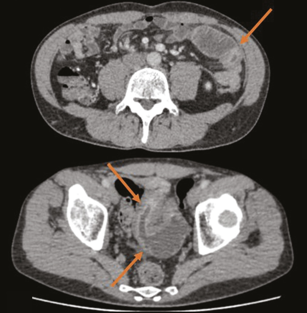
We present a clinical case of ambiguous clinical manifestations of Crohn’s disease with small intestine and rectal involvement. Tomographic imaging was used to confirm the diagnosis. A 44-year-old patient presented with complaints of non-pronounced abdominal pain, dyspepsia. The lab panel showed indirect signs of malabsorption, an increase in fecal calprotectin. An endoscopic examination with histological verification revealed a picture of proctitis. After performing computed tomography and/or magnetic resonance enterography multiple lesions of the small intestine were revealed. This clinical case demonstrates an atypical clinical picture of Crohn’s disease with jejunal, iliac, and rectal lesions.
The patient had no characteristic complaints; the results of endoscopic and morphological studies were not informative. Imaging by means of computed and magnetic resonance tomography has played a crucial role in the diagnosis and successful treatment.
Palavras-chave
Texto integral
##article.viewOnOriginalSite##Sobre autores
Yuliya Shumskaya
The First Sechenov Moscow State Medical University (Sechenov University); Research and Practical Clinical Center for Diagnostics and Telemedicine Technologies
Email: yu.shumskaia@npcmr.ru
ORCID ID: 0000-0002-8521-4045
Rússia, Moscow; Moscow
Tamara Nefedova
The First Sechenov Moscow State Medical University (Sechenov University)
Email: prosto.toma.22@gmail.com
ORCID ID: 0000-0002-6718-8701
Código SPIN: 3097-4977
Rússia, Moscow
Dina Akhmedzyanova
The First Sechenov Moscow State Medical University (Sechenov University)
Email: dina_akhm@mail.ru
ORCID ID: 0000-0001-7705-9754
Código SPIN: 6983-5991
Rússia, Moscow
Ivan Blokhin
Research and Practical Clinical Center for Diagnostics and Telemedicine Technologies
Email: i.blokhin@npcmr.ru
ORCID ID: 0000-0002-2681-9378
Código SPIN: 3306-1387
Rússia, Moscow
Marina Mnatsakanyan
The First Sechenov Moscow State Medical University (Sechenov University)
Autor responsável pela correspondência
Email: mnatsakanyan08@mail.ru
ORCID ID: 0000-0001-9337-7453
Código SPIN: 2015-1822
MD, Dr. Sci. (Med.)
Rússia, MoscowBibliografia
- Ainouche A, Durot C, Soyer P, et al. Unusual intestinal and extra intestinal findings in Crohn’s disease seen on abdominal computed tomography and magnetic resonance enterography. Clin Imaging. 2020;59(1):30–38. doi: 10.1016/j.clinimag.2019.04.010
- Jumani L, Kataria D, Ahmed MU, et al. The Spectrum of extra-intestinal manifestation of Crohn’s disease. Cureus. 2020;12(2):e6928. doi: 10.7759/cureus.6928
- Amado C, Ferreira PG. Pleuroparenchymal fibroelastosis associated with Crohn’s disease: a new aetiology? Eur J Case Rep Intern Med. 2020;7(12):002017. doi: 10.12890/2020_002017
- Dodd EM, Howard JR, Dulaney ED, et al. Pyodermatitis-pyostomatitis vegetans associated with asymptomatic inflammatory bowel disease. Int J Dermatol. 2017;56(12):1457–1459. doi: 10.1111/ijd.13640
- Urbanek M, Neill SM, McKee PH. Vulval Crohn’s disease: difficulties in diagnosis. Clin Exp Dermatol. 1996;21(3):211–214. doi: 10.1111/j.1365-2230.1996.tb00065.x
- Dore M, Junco TP, Galán SA, et al. Pitfalls in diagnosis of early-onset inflammatory bowel disease. Eur J Pediatr Surg. 2018;28(1):39–43. doi: 10.1055/s-0037-1604428
- Cave DR, Hakimian S, Patel K. Current controversies concerning capsule endoscopy. Dig Dis Sci. 2019;64(11):3040–3047. doi: 10.1007/s10620-019-05791-4
- Minordi LM, Larosa L, Papa A, et al. Assessment of Crohn’s disease activity: magnetic resonance enterography in comparison with clinical and endoscopic evaluations. J Gastrointestin Liver Dis. 2019;28:213–224. doi: 10.15403/jgld-183
- Hokama A, Kinjo T, Fujita J. Growing role of magnetic resonance enterography in the management of Crohn disease. Pol Arch Intern Med. 2020;130(9):724–725. doi: 10.20452/pamw.15628
- Clinical guidelines for the diagnosis and treatment of Crohn’s disease in adults (project). Koloproktologiya. 2020;19(2):8–38. (In Russ). doi: 10.33878/2073-7556-2020-19-2-8-38
- Bruining DH, Zimmermann EM, Loftus EV, et al; Society of Abdominal Radiology Crohn’s Disease-Focused Panel. Consensus recommendations for evaluation, interpretation, and utilization of computed tomography and magnetic resonance enterography in patients with small bowel Crohn’s disease. Radiology. 2018;286(3):776–799. doi: 10.1148/radiol.2018171737
- Chavoshi M, Mirshahvalad SA, Kasaeian A, et al. Diagnostic accuracy of magnetic resonance enterography in the evaluation of colonic abnormalities in Crohn’s disease: a systematic review and meta-analysis. Acad Radiol. 2021;28(Suppl 1):S192–S202. doi: 10.1016/j.acra.2021.02.022
- Park EK, Han NY, Park BJ, et al. Value of computerized tomography enterography in predicting Crohn’s disease activity: correlation with Crohn’s disease activity index and C-reactive protein. Iran J Radiol. 2016;13(4):e34301. doi: 10.5812/iranjradiol.34301
- Dubrova SE, Stashuk GA. Possibilities of radiation methods in the diagnosis of inflammatory bowel diseases. Al’manakh klinicheskoi meditsiny. 2016;44(6):757–769. (In Russ). doi: 10.18786/2072-0505-2016-44-6-757-769
- Kurilo DP, Savchenko MI, Soloviev IA, et al. Possibilities of computed tomography angiography in the diagnosis of complicated forms of Crohn’s disease. Bulletin of the Russian military medical academy. 2020;(1):60–65. (In Russ).
Arquivos suplementares

