

")


Том 5, № 2 (2024)

- Жылы: 2024
- Мақалалар: 20
- URL: https://journals.rcsi.science/DD/issue/view/17003
- DOI: https://doi.org/10.17816/DD.52

Original Study Articles
Liver function assessment based on hepatobiliary contrast agent-enhanced magnetic resonance imaging
Аннотация
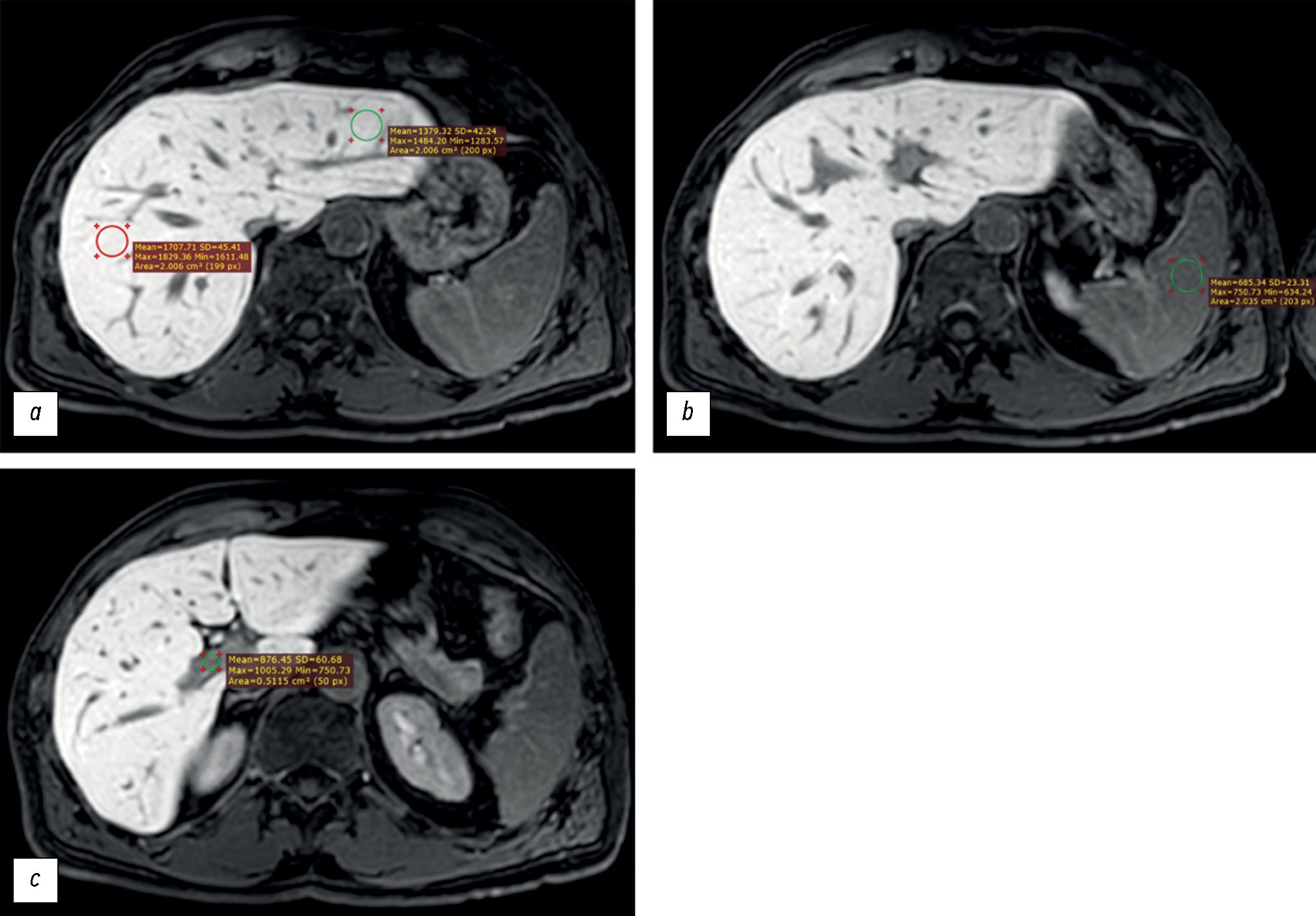
BACKGROUND: Liver function assessment is very important in clinical practice. The use of magnetic resonance imaging for the anatomical and functional evaluation of the liver is possible in actual clinical practice.
AIM: To examine the possibility of using hepatobiliary contrast-enhanced magnetic resonance imaging for the evaluation of liver function.
MATERIALS AND METHODS: Datasets of patients who underwent gadoxetic acid-enhanced magnetic resonance imaging were retrospectively reviewed. Patients were divided into two groups: group 1 included patients with impaired liver function, and group 2 included those with normal liver function. Based on magnetic resonance imaging in the hepatobiliary phase, the liver parenchyma signal intensity and its ratio to spleen signal intensity and portal vein signal intensity were estimated. Differences among these parameters were compared between groups. The correlation between liver parenchyma signal intensity and laboratory blood tests reflecting liver function (total bilirubin, albumen, aspartate aminotransferase, alanine aminotransferase, alkaline phosphatase, gamma glutamyl transpeptidase, and prothrombin time) were analyzed.
RESULTS: Datasets of 53 patients (25 men and 28 women, aged 24–84 years) were analyzed. Group 1 included 19 patients, whereas group 2 included 34. The median liver parenchyma signal intensity was 919.05 [669.65; 1258.35] in group 1 and 1525.13 [1460.5; 1631.4] in group 2 (p=0.0000001). The median ratio of liver parenchyma signal intensity to spleen signal intensity was 1.2 [1.04;1.7] in group 1 and 1.7 [1.46; 1.96] in group 2 (p=0.00076). The median ratio of liver parenchyma signal intensity to portal vein signal intensity was 1.44 [1.29; 1.83] in group 1 and 1.6 [1.43; 1.83] in group 2 (p=0.1). The estimated correlation values between liver parenchyma signal intensity and blood tests parameters were as follows: total bilirubin (r=–0.61; p=0.000001), albumen (r=0.13; p=0.61), aspartate aminotransferase (r=–0.57; p=0.000009), alanine aminotransferase (r=–0.44; p=0.001), alkaline phosphatase (r=–0.45; p=0.0007), gamma glutamyl transpeptidase (r=–0.5; p=0.0003), prothrombin time (r=–0.34; p=0.04).
CONCLUSION: The study reflects the ability to assess liver function using indices (liver parenchyma signal intensity and its ratio to spleen signal intensity) derived from gadoxetic acid-enhanced magnetic resonance imaging. However, this study did not confirm the assumed effectiveness of using the liver parenchyma signal intensity to portal vein signal intensity ratio as an index of liver function. A significant inverse correlation was identified between liver parenchyma signal intensity and blood test parameters in reflecting liver function, except for albumin. The results indicate the possibility of using magnetic resonance imaging to assess liver function.
 137-148


Possibilities and limitations of magnetic resonance imaging in the diagnostics of endocervical adenocarcinomas

Аннотация
BACKGROUND: In recent decades, the incidence of cervical adenocarcinomas has increased from 5% to 20%. Endocervical adenocarcinomas are characterized by a more aggressive course and early metastasis. Owing to the difficulties in the cytological diagnosis of cervical adenocarcinoma, early radiation diagnostics and staging subsequently play a key role. Very few studies have examined the use of magnetic resonance imaging in diagnosing cervical adenocarcinomas.
AIM: To determine the diagnostic informativeness of magnetic resonance imaging in the staging of cervical adenocarcinomas according to the T-criterion and assessing the depth of tumor invasion into the stroma of the cervix and clarify the semiotic signs of adenocarcinoma and features of tumor growth in the uterus.
MATERIALS AND METHODS: In total, 123 patients diagnosed with cervical cancer (C53), who underwent diagnosis and treatment between 2020 and 2023, were examined. The examination results of 22 (18%) patients with cervical adenocarcinoma were analyzed. The average patient age was 56 years. A multiparametric magnetic resonance examination of the pelvic organs was performed on 22 patients using tomographs with a magnetic field strength of 1.5 T. Moreover, 14 (64%) patients underwent surgery including extirpation of the uterus and appendages with pelvic lymphadenectomy. The information value of magnetic resonance imaging was evaluated in 11 patients, whose first stage was surgical treatment.
RESULTS: In this study, cervical adenocarcinoma was detected in 18% among all cases of cervical cancer. The information value of magnetic resonance imaging in assessing the local prevalence of endocervical adenocarcinoma according to the T-criterion was as follows (main value with the corresponding 95% confidence interval): sensitivity, 77.78% (39.99%–97.19%); specificity, 50.00% (1.26%–98.74%); positive predictive value, 87.50% (62.64%–96.69%); negative predictive value, 33.33% (7.30%–76.04%); and accuracy, 72.73% (39.03%–93.98%). The information value of magnetic resonance imaging in assessing the depth of tumor invasion into the cervical stroma was as follows: odds ratio, 3.500 (0.145%–84.694%); sensitivity, 85.7% (0.757%–0.993%); specificity, 33.3% (0.018%–0.0648%); positive predictive value, 75% (0.673%–0.883%); negative predictive value, 50% (0.027%–0.972%).
CONCLUSIONS: The results of this study showed that magnetic resonance imaging is a good tool with high diagnostic informativeness in detecting endocervical cervical adenocarcinoma. The four macrostructures of tumor growth in endocervical adenocarcinoma identified during magnetic resonance imaging data analysis indicate locally aggressive tumor growth and a high frequency of endometrial dropouts. This finding will allow radiologists to structure a descriptive picture, including the verified cervical adenocarcinoma, to enhance methods of developing a treatment plan for the patient.
149-166
Magnetic resonance imaging in the evaluation of pectus excavatum
Аннотация
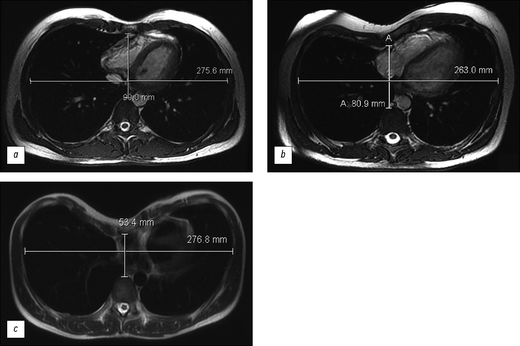
BACKGROUND: Magnetic resonance imaging is more often used to confirm the presence of pectus excavatum and assess compression changes in the heart at this level.
AIM: To evaluate pectus excavatum preoperatively according to magnetic resonance imaging findings.
MATERIALS AND METHODS: A retrospective evaluation of chest magnetic resonance imaging data of 38 patients (male, n=30; female, n=8) was performed. The average age was 19.9 years (±9 years).Cardiac magnetic resonance imaging was performed on a 1.5-T General Electric Optima MR450w GEM scanner with 2D-FIESTA-C pulse sequences, as well as functional assessment of the left and right ventricles. Parameters for surgical treatment of pectus excavatum were as follows: the Haller index, correction index, and sternum rotation angle. Statistical analysis of the relationship between the Haller index, correction index, and sternum rotation angle and ejection fraction of the right ventricle was conducted. A p-value <0.05 was considered significant.
RESULTS: Moderate and severe pectus excavatum were found in 92% of the cases. No significant Pearson correlation was obtained between the Haller index and right ventricular ejection fraction (inspiratory and expiratory ejection fraction, p=0.777 and 0.798, respectively). The mean right ventricular ejection fraction was 46%. A correlation was noted between the Haller index and the correction index (p <0.05). The rotation angle of the sternum, which required modification of surgical intervention, was detected in 44.7% of patients.
CONCLUSION: Magnetic resonance imaging is an informative diagnostic method for pectus excavatum pectus excavatum without radiation exposure and enables detailed preoperative assessment. A correlation was noted between the Haller index and the correction index (p <0.05). Magnetic resonance imaging revealed a decrease in the ejection fraction of the right ventricle.
167-177
Identification of indicators used to assess needs for telemedicine consultations in various profiles of medical care

Аннотация
BACKGROUND: A unified system for assessing the results and real contributions of telemedicine consultations to improving medical care quality in the healthcare system of the Russian Federation has not yet been developed.
AIM: To develop a system of indicators for differentiated assessment of the needs for telemedicine consultations in the provision of medical care.
MATERIALS AND METHODS: In the first stage, reports on the results of on-site activities of national medical research centers in regions of the Russian Federation and their annual public reports (2020–2022) were analyzed to identify indicators that determine the need for telemedicine consultations. The identified indicators were clarified and validated in an open interview with the representatives of the national medical research centers. In the second stage, the value of each indicator was determined based on the expert survey: 18 experts assessed each indicator on a scale of 1–5. Then, the weight coefficient of each indicator was calculated for their subsequent use in planning the coverage of telemedicine consultations.
RESULTS: Three groups of indicators that determined the need for telemedicine consultations for different medical care profiles were as follows: (1) indicators that affect the planned volumes of telemedicine consultations, (2) indicators that characterize the efficiency and effectiveness of telemedicine consultations, and (3) indicators that characterize the validity of requests for telemedicine consultations. Group 1 included indicators of lethality, disability, hospital mortality, frequency of emergency/urgent consultations, and frequency of consultations of patients requiring intensive care. Group 2 included indicators for assessing the effectiveness and efficiency of telemedicine consultations, both subjective (result satisfaction) and objective (number of positive and negative treatment and hospitalization outcomes for cases that received where telemedicine consultations). Group 3 included indicators that characterize the validity of requests for telemedicine consultations: thoroughness of a patient’s examination before a telemedicine consultation and accuracy of the diagnosis. The weight coefficients of group 1 indicators ranged from 0.05 to 1.61 and varied for different profiles.
CONCLUSION: A system of indicators was proposed for the differentiated assessment of the needs for telemedicine consultations when providing medical care.
178-189
Conventional structural magnetic resonance imaging in differentiating chronic disorders of consciousness

Аннотация
BACKGROUND: Differential diagnosis of chronic disorders of consciousness remains one of the most difficult problems even for experienced clinicians.
AIM: To evaluate the inter-expert consistency and capacity of the researcher-developed structural scale based on magnetic resonance imaging to differentiate chronic disorders of consciousness, named, DOC-MRIDS, on a larger sample of patients.
MATERIALS AND METHODS: Sixty patients with a clinically stable status diagnosed with consciousness disorders (vegetative state, n=32; minimally conscious state, n=28) were enrolled. The revised coma recovery scale (CRS-R) was included in the clinical assessment. All patients underwent structural magnetic resonance imaging with 3.0-T Siemens scanners including T2 and T1 sequences. Structural changes were assessed using the DOC-MRIDS scale and included the following features: diffuse cortical atrophy, ventricular enlargement, gyri dilatation, leukoaraiosis, brainstem and/or thalamic degeneration, corpus callosum degeneration, and focal corpus callosum lesions. A total score was calculated. Magnetic resonance imaging data were analyzed by three neuroradiologists, and inter-observer agreement (Krippendorf’s alpha) was assessed.
RESULTS: A high inter-examiner agreement of the DOC-MRIDS scale score was found, with α=0.806 (95% confidence interval 0.757–0.849). The vegetative state group had a higher DOC-MRIDS score than the minimally conscious state group (p <0.005). A negative correlation was obtained between CRS-R and DOC-MRIDS scale scores (ρ=–0.457, p <0.0001), individual clinical scale domains, and magnetic resonance imaging features.
CONCLUSION: When assessing structural changes in patients with chronic consciousness disorders, the use of the DOC-MRIDS scale helps differentiate the type of such disorders with sufficient specificity, sensitivity, and inter-rater agreement. This scale can be used in clinical practice as an additional differential diagnostic tool.
190-202
Remote monitoring of patients with chronic heart failure: A prospective randomized study
Аннотация
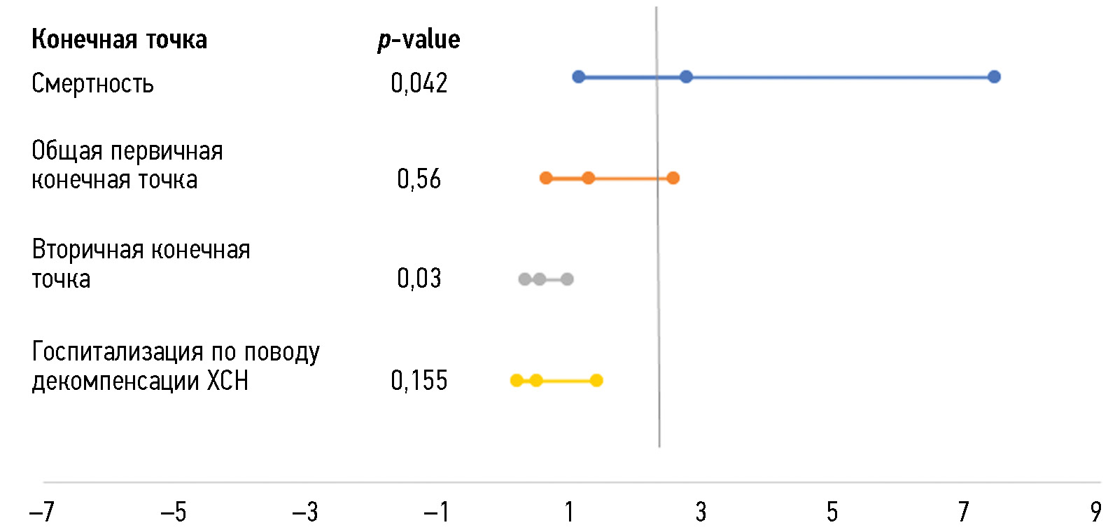
BACKGROUND: Chronic heart failure is one of the key problems of the Russian domestic healthcare system. E-health can be used to improve medical care quality and reduce the of hospitalizations and mortality.
AIM: To examine the effect of telemedicine monitoring on mortality, frequency of hospitalizations, and clinical and functional states of patients with chronic heart failure.
MATERIALS AND METHODS: A prospective, controlled, randomized study was conducted in the Central City Hospital No. 20 in Ekaterinburg (Russia), covering the period from December 2020 to December 2022. Patients with a confirmed diagnosis of chronic heart failure were randomized using the envelope method into three groups: group 1, a telephone control group (n=58); group 2, a remote control group using a Russian medical platform Medsenger (n=52); and group 3, the standard control group (n=103). All patients were examined, including NT-proBNP measurement and echocardiography on the first day of the study and at 3, 6, and 12 months. The occurrence of primary and secondary outcomes was evaluated at these reference points. Stata14 and jamovi software were used for statistical processing.
RESULTS: The study involved 213 participants, and all three groups were comparable in terms of basic demographic and clinical characteristics. The advantage of remote control (groups 1 and 2) over face-to-face observation in reducing cardiovascular mortality was observed after 3 (odds ratio 2.73, 95% confidence interval 1.1–7.39; p=0.042) and 12 (odds ratio 2.1, 95% confidence interval 1.1–3.7; p=0.027) months and that in reducing the occurrence of the combined primary endpoint (odds ratio 2.1, 95% confidence interval 1.1–5.6; p=0.015) after 12 months. The use of the Medsenger platform also demonstrated an advantage over face-to-face observation in the development of the combined secondary endpoint (odds ratio 1.39, 95% confidence interval 0.19–0.81; p=0.011) after 3 months and over telephone control by a nurse after 12 months in reducing cardiovascular mortality (odds ratio 0.177, 95% confidence interval 0.06–0.487; p=0.021) and development of the combined secondary endpoint (odds ratio 0.427, 95% confidence interval 0.189–0.964; p=0.041). When using the Medsenger platform, the ejection fraction increased from 47% initially to 55% after 12 months (p=0.004). The NT-proBNP level decreased from 817 to 582 pg/mL (p <0.001) after 3 months and then to 233 pg/mL after 12 months (p <0.001).
CONCLUSION: Remote monitoring protocols can be a good alternative to the traditional face-to-face monitoring of patients with chronic heart failure, which may improve clinical and functional health indicators.
203-218
Mitral valve calcinosis as an important finding during heart examination

Аннотация
BACKGROUND: Mitral valve calcinosis is a chronic degenerative process in the fibrous structures of the mitral valve. Advanced stages increase the risk of endocarditis and cardiac rhythm disturbances and contribute to cardiovascular mortality. The cause of mitral valve calcinosis is still controversial; however, the contribution of atherosclerosis to its development is currently undisputed. The prevalence of mitral valve calcinosis varies in different age groups and on average is higher in people with cardiovascular disease.
AIM: To assess the prevalence of mitral valve calcinosis in patients undergoing computed tomography angiography and identify the relationship between aortic and mitral valve calcinosis and coronary calcium index and signs of remodeling.
MATERIALS AND METHODS: A retrospective study of 336 patients who underwent computed tomography coronary angiography with intravenous contrast enhancement at the Lomonosov Moscow State University Clinic between November 13, 2020, and May 14, 2022, was conducted.
RESULTS: The prevalence of aortic (16.4%) and mitral (11%) valve calcinosis was high in people undergoing cardiovascular examination, and a relationship was noted between valve calcinosis and coronary calcium index.
CONCLUSION: The detection of mitral valve calcinosis in patients during routine examination is important in predicting further treatment and outcomes because valve calcinosis is an indirect indicator of coronary heart disease risk. Although valve calcinosis is usually an incidental examination finding, it may indicate a high cardiovascular risk and should prompt further evaluation, if clinically necessary.
219-230
Role of teleradiology in the interpretation of ultrasound images acquired in the emergency setting
Аннотация
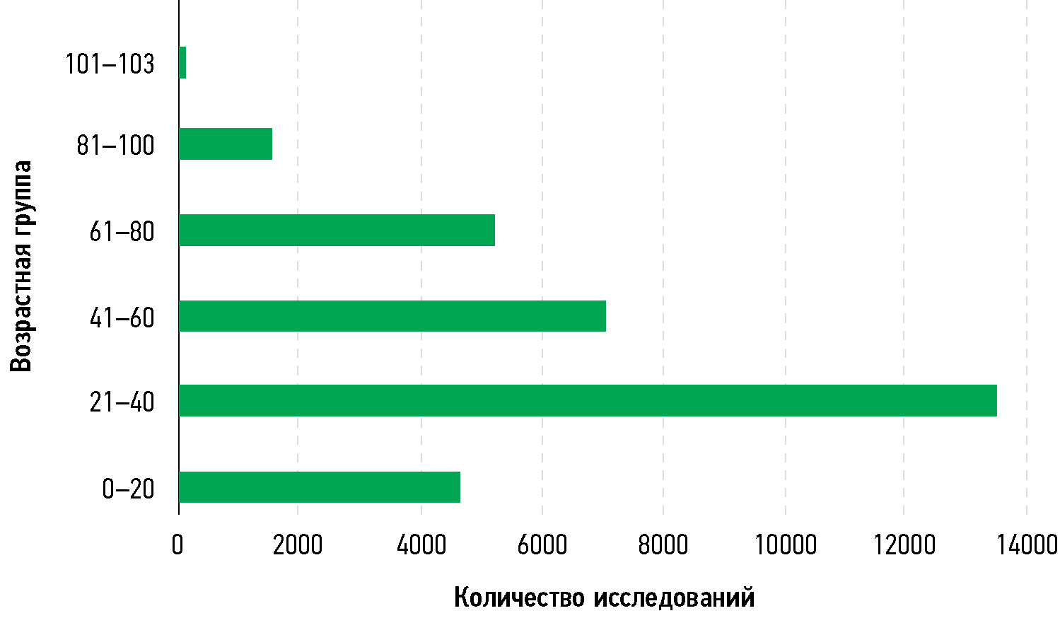
BACKGROUND: Teleradiology has become an important tool in emergency medicine, particularly in the interpretation of emergency ultrasonography. In emergency situations, where time is essential, rapid diagnosis and treatment can mean the difference between life and death. Teleradiology is an innovative alternative to augment the staffing and fill the gaps of onsite radiology personnel in emergency departments or during off-hours.
AIM: To assess the effectiveness/impact of teleradiology in emergency ultrasound interpretation.
MATERIALS AND METHODS: A retrospective study was performed in a cohort of 33,616 patients from 86 hospitals across the USA between January and December 2022. The study involved radiological interpretations of 37,253 ultrasound images acquired in the emergency setting by American Board Certified Radiologists empaneled by a teleradiology service provider, headquartered in Bangalore, India.
RESULTS: The proposed telehealth model provided timely and quality reporting of 37,253 scans of patients with a mean turnaround time of 35.71 min (95% confidence interval 35.50–35.91).
CONCLUSION: This study demonstrates that a structured telesonography program with defined protocols for image capture, transmission, and clinical communication can allow for successful immediate reporting of ultrasound data in the emergency care setting.
231-242
A new artificial intelligence program for the automatic evaluation of scoliosis on frontal spinal radiographs: Accuracy, advantages and limitations

Аннотация
BACKGROUND: Scoliosis is one of the most common spinal deformations that are usually diagnosed on frontal radiographs using Cobb’s method. Automatic measurement methods based on artificial intelligence can overcome many drawbacks of the usual method and can significantly save radiologist’s time.
AIM: To analyze the accuracy, advantages, and disadvantages of a newly developed artificial intelligence program for the automatic diagnosis of scoliosis and measurement of Cobb’s angle on frontal radiographs.
MATERIALS AND METHODS: In total, 114 digital radiographs were used to test the agreement of Cobb’s angle measurements between the new automatic method and the radiologist using the Bland–Altman method on Microsoft Excel. A limited clinical accuracy test was also conducted using 120 radiographs. The accuracy of the system in defining the scoliosis grade was evaluated by sensitivity, specificity, accuracy, and area under the receiver operating characteristic curve.
RESULTS: The agreement of Cobb’s angle measurement between the system and the radiologist’s calculation was found mostly in grade 1 and 2 scoliosis. Only 2.8% of the results showed a clinically significant angle variability of >5°. The diagnostic accuracy metrics of the limited clinical trial in City Mariinsky Hospital (Saint Petersburg, Russia) also proved the reliability of the system, with a sensitivity of 0.97, specificity of 0.88, accuracy (general validity) of 0.93, and area under the receiver operating characteristic curve of 0.93.
CONCLUSION: Overall, the artificial intelligence program can automatically and accurately define the scoliosis grade and measure the angles of spinal curvatures on frontal radiographs.
243-254
Systematic reviews
Priority radiomic parameters for computed tomography of head and neck malignancies: A systematic review

Аннотация
BACKGROUND: Radiomics is the newest and most promising direction in modern radiographic diagnostics. The number of head and neck cancer studies employing radiomics is increasing annually. A systematic review of recent publications (2021–2023) on computed tomography (CT) of head and neck malignancies was performed.
AIM: To present systematized data on parameters for radiomic analysis for head and neck malignancies identified by CT data.
MATERIALS AND METHODS: The literature search was carried out in PubMed. The basic characteristics of the selected articles were extracted, and their quality was assessed using RQS 2.0 and the modified QUADAS-CAD questionnaire. The reproducibility level of radiomic parameters selected for predictive models in different studies was assessed. Eleven articles were selected for the review. In most cases, a high risk of systematic error associated with data imbalance in terms of demographic parameters and level of pathologies was noted.
RESULTS: The range of RQS 2.0 scores for the included articles varied from 19.44% to 50.00% of the maximum possible score. The decreasing research quality was mainly caused by the lack of external result validation (73% of the analyzed articles) and data accessibility and transparency (82%). Inter-study reproducibility of radiomic parameters was low owing to the wide variety of techniques used for image acquisition, image post-processing, extraction, and statistical processing of radiomic parameters.
CONCLUSION: A set of stable radiomic parameters must be successfully introduced into clinical practice. The standardization of radiomics method and creation of an open radiomics database are necessary for this purpose.
255-268
Technical Reports
Optimization of magnetic resonance imaging of the hand

Аннотация
BACKGROUND: Magnetic resonance imaging is one of the leading imaging modalities of the musculoskeletal system. However, when imaging the hand, major problems in magnetic resonance imaging include the lack of specialized coils and reliable fixation devices for the hand, uncomfortable patient posture, motion artifacts, and small anatomical structures in the wrist. These factors inevitably lead to incorrect interpretation.
AIM: To improve the quality of magnetic resonance imaging of the hand by developing an approach to coil selection, scanning protocol, and hand positioning and fixation.
MATERIALS AND METHODS: A positioning device was developed to prevent hand movements. Two types of coils were evaluated. Magnetic resonance images were evaluated comparatively, as well as by a musculoskeletal radiologist.
RESULTS: А head coil is more appropriate when scanning the entire hand, for example, in rheumatic diseases. A knee coil is more appropriate when studying smaller anatomical structures (including the wrist) owing to a smaller field of view and higher resolution. Based on the obtained data, guidelines for the selection of scanning parameters, sequences, and coils for magnetic resonance imaging of the hand were formulated. To prevent motion artifacts, a special fixation device of the patient’s hand was introduced.
CONCLUSION: Certain factors directly affect the qualitative magnetic resonance imaging study of the hand, such as safety protocols, scanning parameters, and hand fixation. The guidelines presented in this study and the use of the developed specialized fixation device may improve the quality of magnetic resonance imaging of the hand.
269-282
Reviews
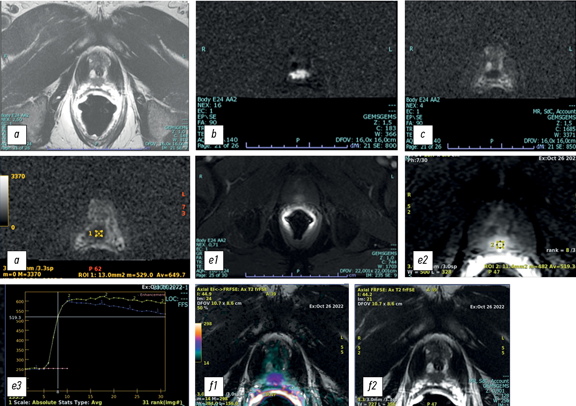
Multiparametric magnetic resonance imaging and magnetic resonance imaging fusion-guided biopsy for the diagnosis of prostate cancer: current status
Аннотация
This review explains the role of multiparametric magnetic resonance imaging, particularly in prostate biopsy, in the detection of prostate cancer. The use of multiparametric magnetic resonance imaging in the diagnosis of prostate cancer has also allowed its use in magnetic resonance imaging-guided biopsies, which according to many studies present high sensitivity and specificity in early diagnosis and staging, in patients with persistently high prostate-specific antigen levels despite previous negative prostate biopsies, and in the follow-up of patients under active surveillance.
To perform a targeted prostate biopsy, three types of magnetic resonance imaging guidance are available: cognitive fusion, direct magnetic resonance imaging-guided biopsy performed within a tomograph (in-bore biopsies), and software coregistration of stored magnetic resonance images with real-time ultrasound using a fusion device, with multiparametric magnetic resonance imaging findings digitally overlaid on real-time transrectal ultrasound images for targeted biopsy.
Each method has its advantages and disadvantages. Magnetic resonance imaging-targeted biopsy improves the quality of histological results compared with other approaches, with approximately 90% correct detection of significant index lesions. Correct staging allows the selection of the best therapeutic options, adequate evaluation of the prognosis, and reduction of the incidence of new biopsies and complications. The current objective is to make magnetic resonance imaging-guided biopsy increasingly available and standardize the technique to minimize inter-operator variability depending on the available system.
283-302
SWOT-analysis: Remote monitoring of blood pressure
Аннотация
Global political and socioeconomic changes have caused a huge strain on the healthcare system. To transition into a new level of medical care, modern technological solutions are needed. Accelerated development of innovations in medicine and the formation of a personalized approach will improve the quality and accessibility of medical services. One of the directions of healthcare development is the use of digital technologies and remote monitoring in assessing the health indicators of citizens. Currently, the federal project of remote monitoring of patients with arterial hypertension, named, “Personal Medical Assistants,” is being implemented in the Russian Federation. Similar to any new technology, remote monitoring has advantages and disadvantages. In this article, a strategic analysis (SWOT-analysis) was performed, considering medical, economic, social, and political aspects that may affect the results of the federal project. For effective implementation of remote monitoring technology in clinical practice, the strengths and weaknesses in the healthcare system and the state as a whole must be emphasized. SWOT-analysis can be used in formulating strategies for the widespread use of new digital technologies in clinical practice.
303-317
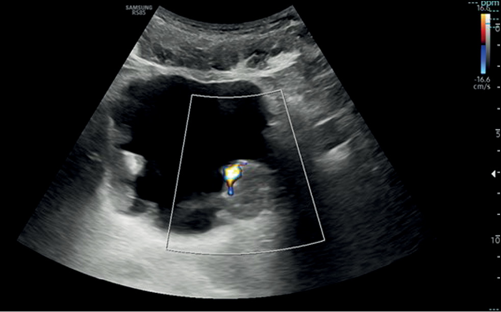
Conventional and innovative imaging modalities in bladder cancer: Techniques and applications
Аннотация
This narrative review describes the current status of imaging in the evaluation of bladder cancer, considering conventional technologies such as ultrasonography, computed tomography urography, and magnetic resonance imaging, as well as novel technologies such as contrast-enhanced ultrasonography and dual-energy computed tomography.
The article is organized by first presenting an introduction on both the anatomy of the bladder (to understand its normal appearance on imaging) and the main features of bladder cancer with reference to epidemiology, clinical picture, classification, and treatment. Subsequently, the role of imaging is discussed, with an explanation of the technique and applications in bladder cancer assessment for each modality.
Imaging plays a critical role in the detection and staging of bladder cancer. In particular, the role of magnetic resonance imaging is expanding because it enables differentiating muscle-invasive bladder cancer from non-muscle-invasive bladder cancer using the Vesical Imaging-Reporting and Data System (VI-RADS), along with conventional technologies, such as computed tomography urography and ultrasonography. Contrast-enhanced ultrasound and dual-energy computed tomography are new imaging modalities that offer special advantages and provide the right approach to patients with oncological conditions. This review ends with the presentation of integrated imaging modalities such as positron emission tomography combined with computed tomography or magnetic resonance imaging, which are promising methods for bladder cancer staging.
318-333
Case reports
Multiple biliary microhamartomas diagnosed in an unsuspecting elderly patient
Аннотация
Multiple biliary hamartomas are a benign incidental finding in the liver. They are not easily detected if one has never seen them, and if appropriate imaging tests are unavailable, and also can be challenging to differentiate from other liver lesions based on imaging alone. Thus, this study aimed to expand the radiologist’s digital image library, enabling a quick and precise differential diagnosis. This paper also highlights the importance of thorough radiological assessment and need for a multidisciplinary approach, involving radiologists, hepatologists, and pathologists, to ensure a precise diagnosis.
The patient presented at the hospital for a computed tomography scan and an abdominal magnetic resonance imaging recommended by his general practitioner to assess the biliary tree (magnetic resonance cholangiopancreatography), owing to persistent abdominal pain. The patient had never undergone an abdominal magnetic resonance imaging previously; hence, the discovery of hepatic lesions was incidental and unexpected.
Magnetic resonance imaging revealed multiple benign lesions in both the hepatic lobes comparable to the Von Meyenburg complex. These lesions are multiple hamartomas and behave differently in all magnetic resonance imaging sequences.
Images acquired with different magnetic resonance imaging sequences were carefully examined. Multiple lesions were found scattered throughout the liver; however, the lesions were benign and consistent with the diagnosis of multiple biliary hamartomas.
Medical practitioners should examine the presence of multiple biliary hamartomas and consider them in the differential diagnosis when patients present with hepatic abnormalities. This can prevent unnecessary interventions and guide appropriate patient management.
334-341
Prospective evaluation of the extensibility of the ascending aorta wall and its vascular prosthesis in a patient with an aneurysm with technically flawless surgical correction and postoperative decrease in functional parameters: A case report

Аннотация
In this clinical case, a patient who had an instrumentally detected aneurysm with the lumen expanding up to 60 mm underwent a surgically flawless prosthetic replacement of the ascending aorta. This treatment led to decreased exercise tolerance, decreased contractile function of the left ventricular myocardium at rest, and enlarged pulmonary artery. The leading factor was a decrease in the volume of systolic expansion of the aorta down to 5 mL (at the initial 13 mL), despite a noticeable increase in the extensibility and a decrease in mechanical stiffness compared with initial indexes of the affected aortic wall. In the literature review, considering mechanical extensibility and elasticity, problems in creating aortic prostheses equivalent to those for healthy biological tissues were discussed.
342-353
Idiopathic enterocolic intussusception: imaging findings in an abdominal emergency

Аннотация
Adult intussusceptions are a rare cause of abdominal obstruction and are usually associated with a neoplastic disease; idiopathic forms are extremely rare. We report a case of enterocolic intussusception in a young woman who experienced symptoms of abdominal obstruction. Imaging findings were reported. On histological examination, no underlying diseases were found. The patient presented at the hospital for computed tomography because of persistent abdominal pain. Computed tomography revealed an enterocolic invagination involving the ileocecal valve and cecum and widespread edematous thickening of the colonic parietal walls.
Idiopathic enterocolic intussusception is an uncommon abdominal urgency in adults. Symptoms can be vague and persistent, delaying an accurate diagnosis. Imaging is crucial in these circumstances to make a diagnosis. Some computed tomography findings, such as a target-like bulk, may be suggestive.
354-360
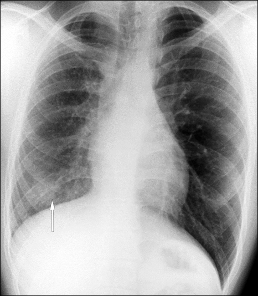
Unilateral pulmonary vein atresia: Difficulties of radiological diagnosis
Аннотация
Pulmonary vein atresia is a rare congenital abnormality that could manifest in isolation or in association with other congenital abnormalities in the cardiovascular system such as pulmonary vein hypoplasia. Pulmonary vein atresia leads to changes in cardiovascular functioning. This abnormality is often diagnosed in children with recurrent pneumonia and hemoptysis. In adulthood, pulmonary vein atresia is much less common, with clinical symptoms such as dyspnea during physical exercises and hemoptysis. However, some patients are asymptomatic. Owing to the nonspecific imaging findings, lung parenchymal changes are often misdiagnosed as another lung disease, including inflammatory genesis disease. In this article, a case of a young man with asymptomatic unilateral pulmonary vein atresia combined with pulmonary artery hypoplasia and interstitial lung changes in a lung with hypoplasia was presented. These pathologies were first identified in a 21-year-old patient by contrast-enhanced computed tomography.
361-369
An unknown situs viscerum inversus totalis, accidentally discovered after computed tomography

Аннотация
Benign situs inversus totalis of the viscerum is often diagnosed accidentally, rarely in adults, and more frequently in children and neonates, affecting both sexes. In this report, a young female patient accidentally discovered a situs inversus totalis after computed tomography for acute abdominal pain. In this uncommon anatomical abnormality, the major visceral organs are reversed in the opposite direction. This report highlights the importance of being aware of and considering situs inversus in clinical practice, particularly when interpreting imaging findings and planning medical procedures. This is critical for differential diagnosis and comorbidities that may affect those patients.
The cause of situs inversus totalis is still unknown; however, this condition is frequently asymptomatic, particularly in infants, and is sometimes associated with other syndromes. The patient arrived at the emergency department with left flank pain, nausea, and fever. In the first ultrasonography, a strange anatomy was suspected; thus, a contrasted computed tomography was performed. The patient had never had a computed tomography scan before. The identification of situs inversus totalis was unexpected and coincidental; the computed tomography images were carefully examined. In patients with chest or abdominal pain, clinicians may consider situs inversus totalis based on computed tomography, particularly if without clinical and imaging history. This knowledge can help in the differential diagnosis, avoiding unneeded interventions. Moreover, comorbidities that affect several systems, particularly cardiovascular and pulmonary systems, affect quite a few patients with situs inversus totalis, who require careful examination and lifelong monitoring.
370-378
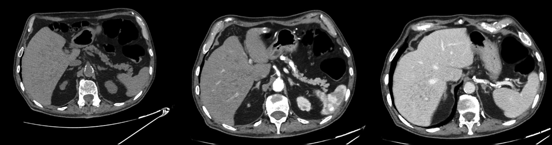
Difficulties in the radiological diagnosis of mature adrenal teratoma mimicking neuroblastoma in a child
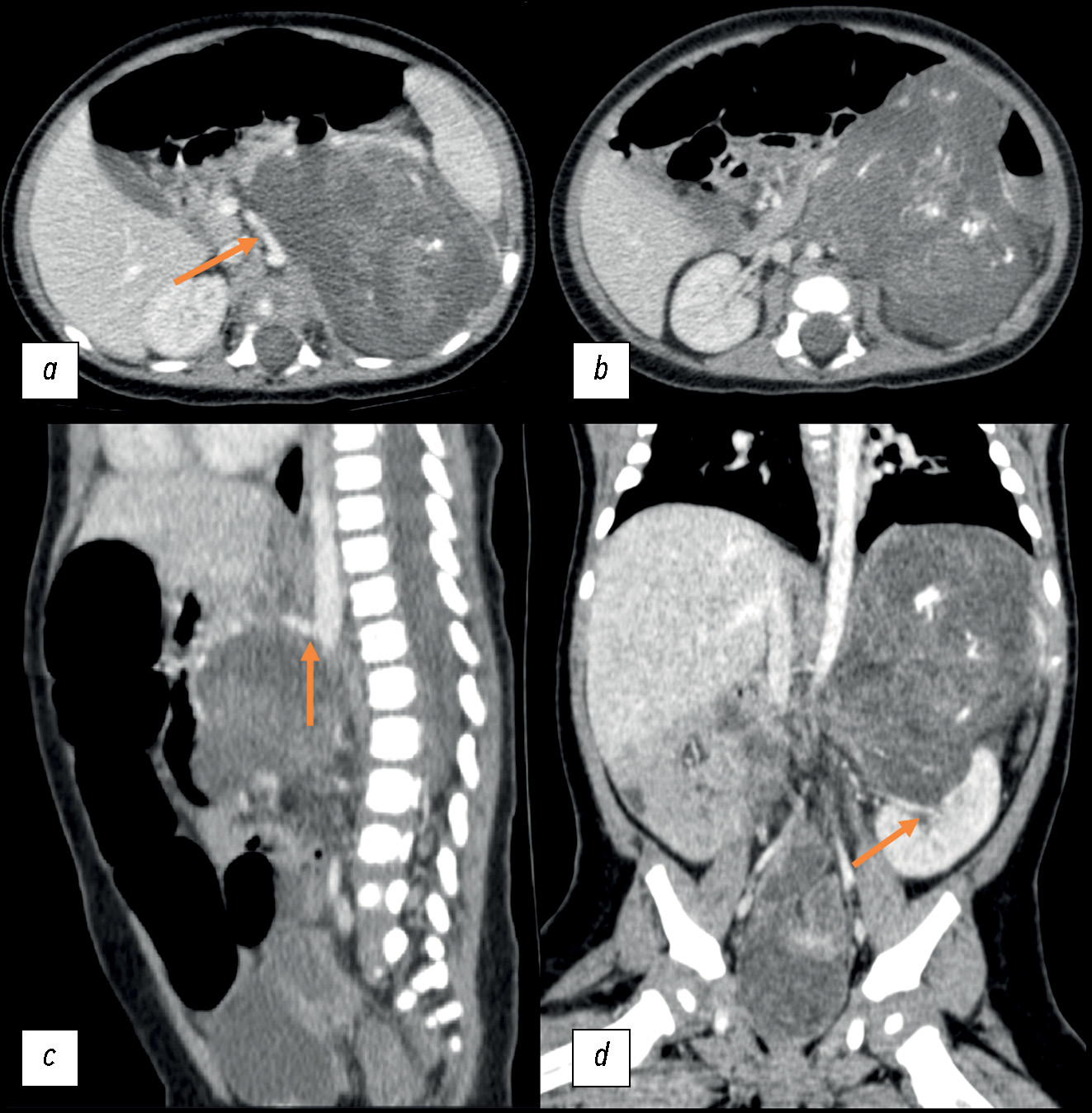
Аннотация
The most common adrenal tumor in young children is neuroblastoma, which can be difficult to differentiate from other conditions such as nephroblastoma, adrenal hemorrhage, angiomyolipoma, myelolipoma, and adenoma. This article describes a case of teratoma, one of the rarest adrenal tumors in children. Initially, despite its large size, it demonstrated all the radiological and histological signs of neuroblastoma. Teratomas are germ cell tumors usually found in the gonads. Adrenal teratomas are extremely rare, accounting for approximately 0.13% of all adrenal tumors. Typically, adrenal teratomas are asymptomatic, as the retroperitoneal space is large enough to accommodate the growth of the tumor without causing symptoms. For the first time in domestic literature, we present a clinical case of adrenal teratoma in a 3-month-old child. The article also presents a detailed description of the diagnostic process and challenges that radiologists and clinicians face when encountering a common tumor in a very rare location for children. This report aimed to help physicians increase awareness of this rare condition and include adrenal teratomas in the potential differential diagnosis of adrenal neoplasms.
379-389