

")


Unilateral pulmonary vein atresia: Difficulties of radiological diagnosis
- Authors: Zharikova V.V.1, Nechaev V.A.1, Kulikova E.A.1, Yudin A.L.2
-
Affiliations:
- Moscow City Oncological Hospital No. 1
- The Russian National Research Medical University named after N.I. Pirogov
- Issue: Vol 5, No 2 (2024)
- Pages: 361-369
- Section: Case reports
- URL: https://journals.rcsi.science/DD/article/view/264845
- DOI: https://doi.org/10.17816/DD619643
- ID: 264845
Cite item

Abstract
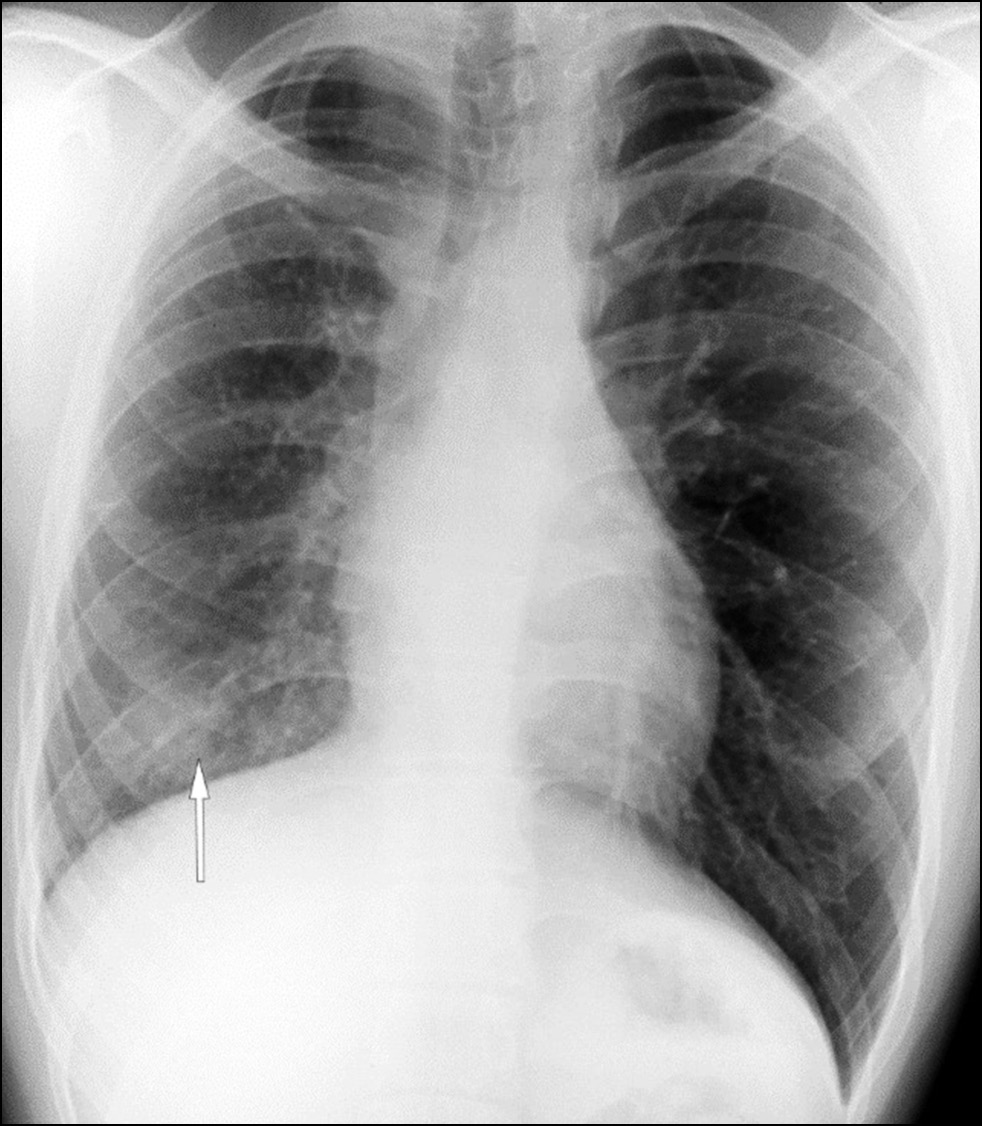
Pulmonary vein atresia is a rare congenital abnormality that could manifest in isolation or in association with other congenital abnormalities in the cardiovascular system such as pulmonary vein hypoplasia. Pulmonary vein atresia leads to changes in cardiovascular functioning. This abnormality is often diagnosed in children with recurrent pneumonia and hemoptysis. In adulthood, pulmonary vein atresia is much less common, with clinical symptoms such as dyspnea during physical exercises and hemoptysis. However, some patients are asymptomatic. Owing to the nonspecific imaging findings, lung parenchymal changes are often misdiagnosed as another lung disease, including inflammatory genesis disease. In this article, a case of a young man with asymptomatic unilateral pulmonary vein atresia combined with pulmonary artery hypoplasia and interstitial lung changes in a lung with hypoplasia was presented. These pathologies were first identified in a 21-year-old patient by contrast-enhanced computed tomography.
Full Text
##article.viewOnOriginalSite##About the authors
Veronika V. Zharikova
Moscow City Oncological Hospital No. 1
Author for correspondence.
Email: ZharikovaVV@zdrav.mos.ru
ORCID iD: 0009-0007-1659-8325
Russian Federation, Moscow
Valentin A. Nechaev
Moscow City Oncological Hospital No. 1
Email: NechaevVA1@zdrav.mos.ru
ORCID iD: 0000-0002-6716-5593
SPIN-code: 2527-0130
MD, Cand. Sci. (Medicine)
Russian Federation, MoscowEvgenia A. Kulikova
Moscow City Oncological Hospital No. 1
Email: kulikovaEA14@zdrav.mos.ru
ORCID iD: 0000-0002-0319-4934
SPIN-code: 2884-4803
Russian Federation, Moscow
Andrey L. Yudin
The Russian National Research Medical University named after N.I. Pirogov
Email: prof_yudin@mail.ru
ORCID iD: 0000-0002-0310-0889
SPIN-code: 6184-8284
MD, Dr. Sci. (Medicine), Professor
Russian Federation, MoscowReferences
- Patil PP. Right pulmonary venous atresia: a rare cause for recurrent unilateral pneumonia. J. Clin. Diagn. Res. 2017;11(9):SD01–SD02. doi: 10.7860/JCDR/2017/25670.10596
- Kim Y, Yoo IR, Ahn MI, Han DH. Asymptomatic adults with isolated, unilateral right pulmonary vein atresia: multidetector CT findings. Br. J. Radiol. 2011;84(1002):109–113. doi: 10.1259/bjr/51344661
- Cohn H-ER, Hicks M, Lacson A, Hicks A. Left hypoplastic lung and hemoptysis — rare familial unilateral pulmonary vein atresia. Clin. Case Rep. 2020;8(9):1698–1703. doi: 10.1002/ccr3.2982
- Reller MD, McDonald RW, Thornburg K, et al. Cardiac embryology: basic review and clinical correlations. J. Am. Soc. Echocardiogr. 1991;4(5):519–532. doi: 10.1016/s0894-7317(14)80388-x
- Heyneman LE, Nolan RL, Harrison JK, McAdams HP. Congenital unilateral pulmonary vein atresia: radiologic findings in three adult patients. Am. J. Roentgenol. 2001;177(3):681–685. doi: 10.2214/ajr.177.3.1770681
- Dixit R, Kumar J, Chowdhury K, et al. Case report: isolated pulmonary vein atresia diagnosted on 128-slice multidetector CT. Indian J. Radiol. Imaging. 2011;21(4):253–256. doi: 10.4103/0971-3026.90681
- Lee SC, Yi JG, Park JH. Cystic lung changes in a thin section CT in an asymptomatic young adult with unilateral pulmonary vein atresia: a case report. J. Korean Soc. Radiol. 2012;67(1):45–48. doi: 10.3348/jksr.2012.67.1.45
- Park S, Cha YK, Kim JS, et al. Isolated Unilateral Pulmonary Artery Hypoplasia with Accompanying Pulmonary Parenchymal Findings on CT: A Case Report. J. Korean Soc. Radiol. 2017;76(5):369–373. doi: 10.3348/jksr.2017.76.5.369
- Cong C-V, Ly T-T, Duc NM. Unilateral pulmonary vein atresia: Literature overview and case report. Radiol. Case Rep. 2022;17(4):1313–1317. doi: 10.1016/j.radcr.2022.01.057
- Pavlenko SM. Pathological physiology. Moscow: Medgiz; 1940. (In Russ).
- Basavarai B, Arun S, Amarinder SM, et al. Unilateral pulmonary vein atresia: diagnostic dilemma unfolded on imaging. BMJ Case Rep. 2018. doi: 10.1136/bcr-2017-224154
- Narayanan R, Shankar B, Paruthikunnan S. Isolated unilateral pulmonary vein atresia. Lung India. 2016;33(5):571–572. doi: 10.4103/0970-2113.188990
Supplementary files


