

")


Диагностическая и экономическая оценка применения комплексного алгоритма искусственного интеллекта, направленного на выявление десяти патологических находок по данным компьютерной томографии органов грудной клетки
- Авторы: Чернина В.Ю.1, Беляев М.Г.1, Силин А.Ю.2, Аветисов И.О.2, Пятницкий И.А.1,3, Петраш Е.А.1,4, Басова М.В.1, Синицын В.Е.5,6, Омельяновский В.В.7,8,9, Гомболевский В.А.1,10
-
Учреждения:
- АЙРА Лабс
- Клинический госпиталь на Яузе
- Техасский университет в Остине
- Национальный медицинский исследовательский центр онкологии имени Н.Н. Блохина
- Московский государственный университет имени М.В. Ломоносова
- Научно-практический клинический центр диагностики и телемедицинских технологий
- Центр экспертизы и контроля качества медицинской помощи
- Российская медицинская академия непрерывного профессионального образования
- Научно-исследовательский финансовый институт
- Институт искусственного интеллекта
- Выпуск: Том 4, № 2 (2023)
- Страницы: 105-132
- Раздел: Оригинальные исследования
- URL: https://journals.rcsi.science/DD/article/view/146880
- DOI: https://doi.org/10.17816/DD321963
- ID: 146880
Цитировать

Аннотация
Обоснование. Технологии искусственного интеллекта призваны помогать в решении проблемы пропуска находок при лучевых исследованиях. Важным вопросом является оценка экономической пользы от внедрения технологий искусственного интеллекта.
Цель ― оценить частоту выявления патологических находок и экономический потенциал применения комплексного искусственного интеллекта для компьютерной томографии органов грудной клетки, валидированного экспертами, по сравнению с рентгенологами без доступа к технологиям в условиях частного медицинского центра.
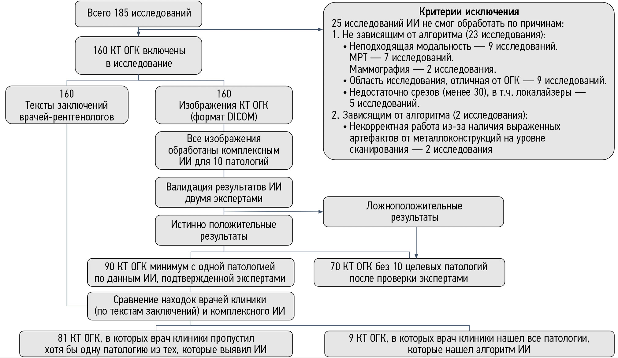
Материалы и методы. Проведено обсервационное одноцентровое ретроспективное исследование. В исследование включались компьютерные томограммы органов грудной клетки без внутривенного контрастирования, выполненные в ООО «Клинический госпиталь на Яузе» (Москва) в период с 01.06.2022 по 31.07.2022. Компьютерные томограммы обработаны комплексным алгоритмом искусственного интеллекта для десяти патологий: инфильтративные изменения в лёгких, характерные для вирусной пневмонии (COVID-19 в условиях пандемии); лёгочные узлы; свободная жидкость в плевральных полостях; эмфизема лёгких; увеличение диаметра грудной аорты; увеличение диаметра ствола лёгочной артерии; коронарный кальциноз; оценка толщины надпочечников; оценка высоты и плотности тел позвонков. Два эксперта анализировали компьютерные томограммы и сравнивали результаты с анализом искусственного интеллекта. Для всех находок, выявленных и не выявленных врачами клиники, определили дальнейшую маршрутизацию в соответствии с клиническими рекомендациями. Для каждого пациента была рассчитана стоимость неоказанных медицинских услуг по прайс-листу клиники.
Результаты. Итоговую группу составили 160 компьютерных томограмм органов грудной клетки с описаниями. С помощью искусственного интеллекта выявлено 90 (56%) исследований с патологиями, из них в 81 (51%) протоколе была пропущена хотя бы одна патология. Общая стоимость неоказанных медицинских услуг «второго этапа» для всех патологий от 81 пациента была оценена в 2 847 760 руб. (37 250,99 долларов или 256 217,95 китайских юаней). Стоимость неоказанных медицинских услуг только для тех патологий, которые пропущены врачами, но выявлены искусственным интеллектом, составила 2 065 360 руб. (27 016,57 долларов или 185 824,05 китайских юаней).
Заключение. Применение искусственного интеллекта для анализа данных компьютерной томографии органов грудной клетки в качестве помощника рентгенолога позволяет существенно уменьшить число случаев пропуска патологий. Использование искусственного интеллекта может принести в 3,6 раза больше стоимости за медицинские услуги по сравнению со стандартной моделью работы рентгенологов без применения таких технологий, и, таким образом, быть рентабельным для применения в условиях частного медицинского центра.
Ключевые слова
Полный текст
Открыть статью на сайте журналаОб авторах
Валерия Юрьевна Чернина
АЙРА Лабс
Email: v.chernina@ira-labs.com
ORCID iD: 0000-0002-0302-293X
SPIN-код: 8896-8051
Scopus Author ID: 57210638679
ResearcherId: AAF-1215-2020
Россия, Москва
Михаил Геннадьевич Беляев
АЙРА Лабс
Email: belyaevmichel@gmail.com
ORCID iD: 0000-0001-9906-6453
SPIN-код: 2406-1772
канд. физ.-мат. наук, профессор
Россия, МоскваАнтон Юрьевич Силин
Клинический госпиталь на Яузе
Email: silin@yamed.ru
ORCID iD: 0000-0003-4952-2347
SPIN-код: 4411-8745
Россия, Москва
Иван Олегович Аветисов
Клинический госпиталь на Яузе
Email: avetisov@yamed.ru
ORCID iD: 0009-0007-3550-7556
Россия, Москва
Илья Аркадьевич Пятницкий
АЙРА Лабс; Техасский университет в Остине
Email: i.pyatnitskiy@ira-labs.com
ORCID iD: 0000-0002-2827-1473
SPIN-код: 6150-4961
Россия, Москва; Остин, США
Екатерина Александровна Петраш
АЙРА Лабс; Национальный медицинский исследовательский центр онкологии имени Н.Н. Блохина
Email: e.a.petrash@gmail.com
ORCID iD: 0000-0001-6572-5369
SPIN-код: 6910-8890
канд. мед. наук
Россия, Москва; МоскваМария Васильевна Басова
АЙРА Лабс
Email: m.basova@ira-labs.com
ORCID iD: 0009-0000-3325-8452
Россия, Москва
Валентин Евгеньевич Синицын
Московский государственный университет имени М.В. Ломоносова; Научно-практический клинический центр диагностики и телемедицинских технологий
Email: vsini@mail.ru
ORCID iD: 0000-0002-5649-2193
SPIN-код: 8449-6590
д-р мед. наук, профессор
Россия, Москва; МоскваВиталий Владимирович Омельяновский
Центр экспертизы и контроля качества медицинской помощи; Российская медицинская академия непрерывного профессионального образования; Научно-исследовательский финансовый институт
Email: vvo@rosmedex.ru
ORCID iD: 0000-0003-1581-0703
SPIN-код: 1776-4270
д-р мед. наук, профессор
Россия, Москва; Москва; МоскваВиктор Александрович Гомболевский
АЙРА Лабс; Институт искусственного интеллекта
Автор, ответственный за переписку.
Email: gombolevskii@gmail.com
ORCID iD: 0000-0003-1816-1315
SPIN-код: 6810-3279
канд. мед. наук
Россия, Москва; МоскваСписок литературы
- Aberle D.R., Adams A.M., Berg C.D., et al.; National Lung Screening Trial Research Team. Reduced lung-cancer mortality with low-dose computed tomographic screening // N Engl J Med. 2011. Vol. 365, N 5. P. 395–409. doi: 10.1056/NEJMoa1102873
- Pastorino U., Silva M., Sestini S., et al. Prolonged lung cancer screening reduced 10-year mortality in the MILD trial: New confirmation of lung cancer screening efficacy // Ann Oncol. 2019. Vol. 30, N 7. P. 1162–1169. doi: 10.1093/annonc/mdz117
- Grover H., King W., Bhattarai N., et al. Systematic review of the cost-effectiveness of screening for lung cancer with low dose computed tomography // Lung Cancer. 2022. N 170. P. 20–33. doi: 10.1016/j.lungcan.2022.05.005
- Николаев А.Е., Гомболевский В.А., Гончар А.П., и др. Случайные находки при скрининге рака легкого методом низкодозной компьютерной томографии // Туберкулез и болезни лёгких. 2018. Т. 96, № 11. С. 60–67. doi: 10.21292/2075-1230-2018-96-11-60-67
- Kilsdonk I.D., de Roos M.P., Bresser P., et al. Frequency and spectrum of incidental findings when using chest CT as a primary triage tool for COVID-19 // Eur J Radiol Open. 2021. N 8. P. 100366. doi: 10.1016/j.ejro.2021.100366
- Бельцевич Д.Г., Мельниченко Г.А., Кузнецов Н.С., и др. Клинические рекомендации Российской ассоциации эндокринологов по дифференциальной диагностике инциденталом надпочечников // Эндокринная хирургия. 2016. Т. 10, № 4. С. 31–42. doi: 10.14341/serg2016431-42
- Эмфизема легких. Клинические рекомендации (утв. Министерством здравоохранения РФ, 2021). Режим доступа: https://www.garant.ru/products/ipo/prime/doc/402775957/. Дата обращения: 17.03.2023.
- Аневризмы грудной и торакоабдоминальной аорты. Клинические рекомендации (утв. Министерством здравоохранения РФ, 2023). Режим доступа: https://www.angiolsurgery.org/library/recommendations/2022/aneurysms_thoracic_thoracoabdominal_aorta/recommendation.pdf. Дата обращения: 17.03.2023.
- Васильев Ю.А., Владзимирский А.В., Арзамасов К.М., и др. Компьютерное зрение в лучевой диагностике: первый этап Московского эксперимента. Монография. 2-е изд., перераб. и доп. / под ред. Ю.А. Васильева, А.В. Владзимирского. Москва: Издательские решения, 2023. 376 с.
- Zakharov A., Pisov M., Bukharaev A., et al. Interpretable vertebral fracture quantification via anchor-free landmarks localization // Med Image Anal. 2023. N 83. P. 102646. doi: 10.1016/j.media.2022.102646
- Goncharov M., Pisov M., Shevtsov A., et al. CT-Based COVID-19 triage: Deep multitask learning improves joint identification and severity quantification // Med Image Anal // 2021. N 71. P. 102054. doi: 10.1016/j.media.2021.102054
- Shirokikh B., Shevtsov A., Dalechina A., et al. Accelerating 3D medical image segmentation by adaptive small-scale target localization // J Imaging. 2021. Vol. 13, N 7. P. 35. doi: 10.3390/jimaging7020035
- Goncharov M., Pisov M., Belyaev M., et al. Quantification of epicardial adipose tissue in low-dose computed tomography images // Lecture Notes in Electrical Engineering. 2022. Vol. 784, N 1. P. 98–107. doi: 10.1007/978-981-16-3880-0_11
- Husereau D., Drummond M., Augustovski F., et al.; CHEERS 2022 ISPOR Good Research Practices Task Force. Consolidated health economic evaluation reporting standards 2022 (cheers 2022) statement: Updated reporting guidance for health economic evaluations // Value Health. 2022. Vol. 25, N 1. P. 3–9. doi: 10.1016/j.jval.2021.11.1351
- Морозов С.П., Владзимирский А.В., Кляшторный В.Г., и др. Клинические испытания программного обеспечения на основе интеллектуальных технологий (лучевая диагностика). Препринт № ЦДТ-2019-1. Серия «Лучшие практики лучевой и инструментальной диагностики». Вып. 23. Москва: ГБУЗ НПКЦ ДиТ ДЗМ, 2019. 33 с.
- Гомболевский В.А., Блохин И.А., Лайпан А.Ш., и др. Методические рекомендации по скринингу рака легкого. Серия «Лучшие практики лучевой и инструментальной диагностики». Вып. 56. Москва: ГБУЗ НПКЦ ДиТ ДЗМ, 2020. 57 с.
- Морозов С.П., Проценко Д.Н., Сметанина С.В., и др. Лучевая диагностика коронавирусной болезни (COVID-19): организация, методология, интерпретация результатов: методические рекомендации. Серия «Лучшие практики лучевой и инструментальной диагностики». Вып. 65. Москва: ГБУЗ НПКЦ ДиТ ДЗМ, 2020. 78 с.
- Временные методические рекомендации. Профилактика, диагностика и лечение новой коронавирусной инфекции (COVID-19). Министерство здравоохранения Российской Федерации. Версия 17 (14.12.2022). Режим доступа: https://static-0.minzdrav.gov.ru/system/attachments/attaches/000/061/254/original/%D0%92%D0%9C%D0%A0_COVID-19_V17.pdf?1671088207. Дата обращения: 17.03.2023.
- Lynch D.A., Austin J.H., Hogg J.C., et al. CT-definable subtypes of chronic obstructive pulmonary disease: A statement of the Fleischner Society // Radiology. 2015. Vol. 277, N 1. P. 192–205. doi: 10.1148/radiol.2015141579
- Hersh C.P., Washko G.R., Estépar R.S., et al. Paired inspiratory-expiratory chest CT scans to assess for small airways disease in COPD // Respir Res. 2013. Vol. 146, N 1. P. 42. doi: 10.1186/1465-9921-14-42
- Walker C., Chung J.H. Muller’s imaging of the chest e-book: Expert radiology series. Elsevier Health Sciences, 2018. 1104 р.
- Erbel R., Aboyans V., Boileau C., et al.; ESC Committee for Practice Guidelines. 2014 ESC Guidelines on the diagnosis and treatment of aortic diseases: Document covering acute and chronic aortic diseases of the thoracic and abdominal aorta of the adult. The Task Force for the Diagnosis and Treatment of Aortic Diseases of the European Society of Cardiology (ESC) // Eur Heart J. 2014. Vol. 35, N 41. P. 2873–2926. doi: 10.1093/eurheartj/ehu281
- Wanhainen A., Verzini F., Van Herzeele I., et al. Clinical practice guidelines on the management of abdominal aorto-iliac artery aneurysmsб // Eur J Vascul Endovascul Sur. 2019. Vol. 57, N 1. P. 8–93. doi: 10.1016/j.ejvs.2018.09.020
- Jurgens P.T., Carr J.J., Terry J.G., et al. Association of abdominal aorta calcium and coronary artery calcium with incident cardiovascular and coronary heart disease events in black and white middle-aged people: The coronary artery risk development in young adults study // J Am Heart Assoc. 2021. Vol. 10, N 24. P. 23–37. doi: 10.1161/JAHA.121.023037
- Galiè N., Humbert M., Vachiery J.L., et al. 2015 ESC/ERS Guidelines for the diagnosis and treatment of pulmonary hypertension: The joint task force for the diagnosis and treatment of pulmonary hypertension of the European society of cardiology (ESC) and the European respiratory society (ERS): Endorsed by: Association for European paediatric and congenital cardiology (AEPC), international society for heart and lung transplantation (ISHLT) // Eur Respir J. 2015. Vol. 46, N 4. P. 903–975. doi: 10.1183/13993003.01032-2015
- Agatston A.S., Janowitz W.R., Hildner F.J., et al. Quantification of coronary artery calcium using ultrafast computed tomography // J Am Coll Cardiol. 1990. Vol. 15, N 4. P. 827–832. doi: 10.1016/0735-1097(90)90282-t
- Журавлев К.Н. КТ-коронарография. Серия «Лучшие практики лучевой и инструментальной диагностики». Вып. 65. Москва: ГБУЗ НПК ЦДиТ ДЗМ, 2020. 35 с.
- Mayo-Smith W.W., Song J.H., Boland G.L., et al. Management of incidental adrenal masses: A white paper of the ACR incidental findings committee // J Am Coll Radiol. 2017. Vol. 14, N 8. P. 1038–1044. doi: 10.1016/j.jacr.2017.05.001
- Белая Ж.Е., Белова К.Ю., Бирюкова Е.В., и др. Федеральные клинические рекомендации по диагностике, лечению и профилактике остеопороза // Остеопороз и остеопатии. 2021. Т. 24, № 2. С. 4–47. doi: 10.14341/osteo12930
- Genant H.K., Jergas M. Assessment of prevalent and incident vertebral fractures in osteoporosis research // Osteoporosis Int. 2003. Vol. 14, N 3. P. 43–55. doi: 10.1007/S00198-002-1348-1
- Патологические переломы, осложняющие остеопороз. Клинические рекомендации. Москва, 2018. 90 с. Режим доступа: https://neirodoc.ru/wp-content/uploads/2019/10/patologicheskie-perelomy-oslozhnyayuschie-osteoporoz-2018.pdf. Дата обращения: 17.03.2023.
- Колбин А.С., Гомон Ю.М., Балыкина Ю.Е., и др. Социально-экономическое и глобальное бремя COVID-19 // Качественная клиническая практика. 2021. № 1. С. 24–34. doi: 10.37489/2588-0519-2021-1-24-34
- Pickhardt P.J., Correale L., Hassan C. AI-based opportunistic CT screening of incidental cardiovascular disease, osteoporosis, and sarcopenia: Cost-effectiveness analysis // Abdom Radiol (NY). 2023. N 3. P. 1181–1198. doi: 10.1007/s00261-023-03800-9
- Behr C.M., Koffijberg H., Degeling K., et al. Can we increase efficiency of CT lung cancer screening by combining with CVD and COPD screening? Results of an early economic evaluation // Eur Radiol. 2022. Vol. 32, N 5. P. 3067–3075. doi: 10.1007/s00330-021-08422-7
- Chintanapakdee W., Mendoza D.P., Zhang E.W., et al. Detection of extrapulmonary malignancy during lung cancer screening: 5-year analysis at a tertiary hospital // J Am Coll Radiol. 2020. Vol. 17, N 12. P. 1609–1620. doi: 10.1016/j.jacr.2020.09.032
- Girling A., Lilford R., Cole A., et al. Headroom approach to device development: Current and future directions // Int J Technol Assess Health Care. 2015. Vol. 31, N 5. P. 331–338. doi: 10.1017/S0266462315000501
- Ijzerman M.J., Steuten L.M. Early assessment of medical technologies to inform product development and market access: A review of methods and applications // Appl Health Econ Health Policy. 2011. Vol. 9, N 5. P. 331–347. doi: 10.2165/11593380-000000000-00000
- Izerman M.J., Koffijberg H., Fenwick E., et al. Emerging use of early health technology assessment in medical product development: A scoping review of the literature // Pharmacoeconomics. 2017. Vol. 35, N 7. P. 727–740. doi: 10.1007/s40273-017-0509-1
- Sahiner B., Pezeshk A., Hadjiiski L.M., et al. Deep learning in medical imaging and radiation therapy // Med Phys. 2019. Vol. 46, N 1. P. 1–36. doi: 10.1002/mp.13264
- Allen B.J., Seltzer S.E., Langlotz C.P., et al. A road map for translational research on artificial intelligence in medical imaging: From the 2018 national institutes of health/RSNA/ACR/The academy workshop // J Am Coll Radiol. 2019. Vol. 16, N 9. P. 1179–1189. doi: 10.1016/j.jacr.2019.04.014
- Xia C., Rook M., Pelgrim G.J., et al. Early imaging biomarkers of lung cancer, COPD and coronary artery disease in the general population: Rationale and design of the ImaLife (Imaging in Lifelines) Study // Eur J Epidemiol. 2020. Vol. 35, N 1. P. 75–86. doi: 10.1007/s10654-019-00519-0
- Ziegelmayer S., Graf M., Makowski M., et al. Cost-effectiveness of artificial intelligence support in computed tomography-based lung cancer screening // Cancers (Basel). 2022. Vol. 14, N 7. P. 1729. doi: 10.3390/cancers14071729
- McDonald R.J., Schwartz K.M., Eckel L.J., et al. The effects of changes in utilization and technological advancements of cross-sectional imaging on radiologist workload // Acad Radiol. 2015. Vol. 22, N 9. P. 1191–1198. doi: 10.1016/j.acra.2015.05.007
- Sokolovskaya E., Shinde T., Ruchman R.B., et al. The effect of faster reporting speed for imaging studies on the number of misses and interpretation errors: A pilot study // J Am Coll Radiol. 2015. Vol. 12, N 7. P. 683–688. doi: 10.1016/j.jacr.2015.03.040
- Geijer H., Geijer M. Added value of double reading in diagnostic radiology, a systematic review // Insights Imaging. 2018. Vol. 9, N 3. P. 287–301. doi: 10.1007/s13244-018-0599-0
- Lauritzen P.M., Hurlen P., Sandbæk G., et al. Double reading rates and quality assurance practices in Norwegian hospital radiology departments: Two parallel national surveys // Acta Radiol. 2015. Vol. 56, N 1. P. 78–86. doi: 10.1177/0284185113519988
- Морозов С.П., Чернина В.Ю., Андрейченко А.Е., и др. Как искусственный интеллект влияет на оценку поражения лёгких при COVID-19 по данным КТ грудной клетки? // Digital Diagnostics. 2021. Т. 2, № 1. C. 27–38. doi: 10.17816/DD60040
Дополнительные файлы


