

")


Unilateral isolated fracture of the pterygoid plate: a case report
- Авторлар: Balzano R.F.1, Testini V.2, Cammarota A.3, Guglielmi G.1,2,4
-
Мекемелер:
- Radiology Unit, Barletta University Campus UNIFG, “Dimiccoli” Hospital
- Department of Clinical and Experimental Medicine, Foggia University School of Medicine
- Radiation Oncology Unit, IRCCS CROB, Rionero in Vulture
- Radiology Unit, Hospital “Casa Sollievo Della Sofferenza”, San Giovanni Rotondo
- Шығарылым: Том 3, № 1 (2022)
- Беттер: 71-77
- Бөлім: Case reports
- URL: https://journals.rcsi.science/DD/article/view/90282
- DOI: https://doi.org/10.17816/DD90282
- ID: 90282
Дәйексөз келтіру

Аннотация
Pterygoid plate fractures are often associated with Le Fort fractures and accompanied by other facial fractures such as frontal sinus and naso-orbital-ethmoid fractures; isolated pterygoid plate fractures are extremely rare.
Le Fort fractures must be surgically treated with fixation of unstable fracture segments to re-establish bone form and function, and the pterygoid process must be surgically stabilized; however, surgical treatment is unnecessary in isolated pterygoid plate fractures.
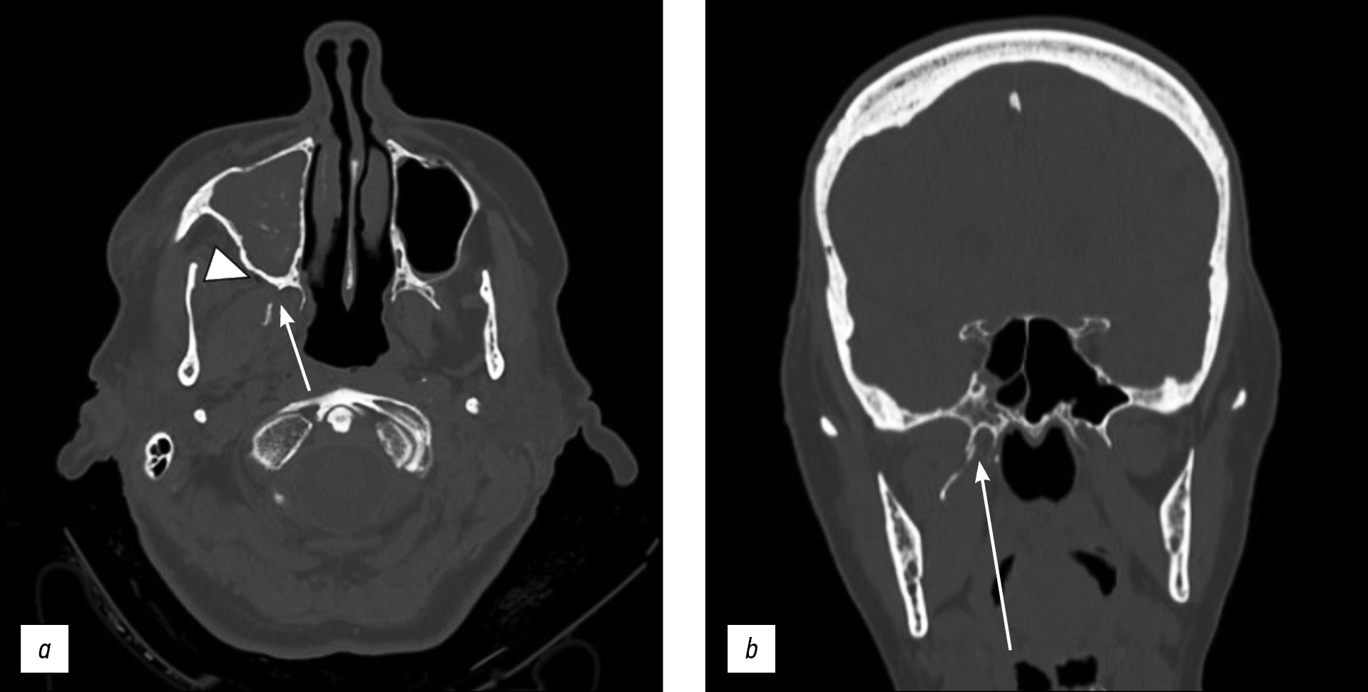
Here, we report a rare case of isolated unilateral fracture of the pterygoid process in a 71-year-old female patient who had a syncopal episode with secondary head injury and a hematoma at the base of the right orbit.
A computed tomography scan showed unilateral right pterygoid plate fracture with signs of emphysema in the ipsilateral masticatory space. The patient also had a fracture of the medial wall of the right maxillary sinus with hemosine, but no fractures of the skull base or theca. She was treated conservatively.
Негізгі сөздер
Толық мәтін
##article.viewOnOriginalSite##Авторлар туралы
Rosario Francesco Balzano
Radiology Unit, Barletta University Campus UNIFG, “Dimiccoli” Hospital
Email: ro.balzano@gmail.com
Италия, Foggia
Valentina Testini
Department of Clinical and Experimental Medicine, Foggia University School of MedicineИталия, Foggia
Aldo Cammarota
Radiation Oncology Unit, IRCCS CROB, Rionero in Vulture
Email: aldo.cammarota@crob.it
Италия, Potenza
Giuseppe Guglielmi
Radiology Unit, Barletta University Campus UNIFG, “Dimiccoli” Hospital; Department of Clinical and Experimental Medicine, Foggia University School of Medicine; Radiology Unit, Hospital “Casa Sollievo Della Sofferenza”, San Giovanni Rotondo
Хат алмасуға жауапты Автор.
Email: giuseppe.guglielmi@unifg.it
ORCID iD: 0000-0002-4325-8330
Medical Doctor, Full Professor of Radiology, Department of Clinical and Experimental Medicine.
Италия, Foggia; Foggia; FoggiaӘдебиет тізімі
- Boffano P, Roccia F, Zavattero E, et al. European Maxillofacial Trauma (EURMAT) project: a multicentre and prospective study. J Craniomaxillofac Surg. 2015;43(1):62–70. doi: 10.1016/j.jcms.2014.10.011
- Winegar BA, Murillo H, Tantiwongkosi B. Spectrum of critical imaging findings in complex facial skeletal trauma. Radiographics. 2013;33(1):3–19. doi: 10.1148/rg.331125080
- Le Fort R: Etude experimentale sur les fractures de la machoire superieure. Rev Chir. 1901;23:208–507.
- Patel BC, Wright T, Waseem M. Le Fort Fractures. In: StatPearls. Treasure Island (FL): StatPearls Publishing; 2021.
- Choi JW, Kim MJ. Treatment of panfacial fractures and three-dimensional outcome analysis: the occlusion first approach. J Craniofac Surg. 2019;30(4):1255–1258. doi: 10.1097/SCS.0000000000005528
- Surya M, Soni P, Bharti R, Jamwal I. Isolated fracture of lateral pterygoid plate by penetrating foreign body ― a rarity indeed. Pol J Radiol. 2017;82:137–140. doi: 10.12659/PJR.900407
- Garg RK, Alsheik NH, Afifi AM, Gentry LR. Pterygoid plate fractures: not limited to Le Fort Fractures. J Craniofac Surg. 2015;26(6):1823–1825. doi: 10.1097/SCS.0000000000001901
- Truong AQ, O’Brien DC, Strong EB, Dublin A. Lateral pterygoid plate fractures associated with mandible fractures. JAMA Facial Plast Surg. 2014;16(6):437–439. doi: 10.1001/jamafacial.2014.645
- Unger JM, Gentry LR, Grossman JE. Sphenoid fractures: prevalence, sites, and significance. Radiology. 1990;175(1):175–180. doi: 10.1148/radiology.175.1.2315477
- Murray GM, Phanachet I, Uchida S, et al. The human lateral pterygoidmuscle: a review of some experimental aspects and possible clinical relevance. Aust Dent J. 2004;49(1):2–8. doi: 10.1111/j.1834-7819.2004.tb00042.x
- Phang SY, Whitehouse K, Lee L, et al. Management of CSF leak in base of skull fractures in adults. Br J Neurosurg. 2016;30(6):596–604. doi: 10.1080/02688697.2016.1229746
- Choi NR, Shin SH, Kim SS, et al. Healing pattern of intentional pterygoid plate fracture after posterior movement of maxilla through Le Fort I osteotomy. J Craniomaxillofac Surg. 2018;46(10):1828–1833. doi: 10.1016/j.jcms.2018.08.003
- Kaeppler G, Cornelius CP, Ehrenfeld M, Mast G. Diagnostic efficacy of cone-beam computed tomography for mandibular fractures. Oral Surg Oral Med Oral Pathol Oral Radiol. 2013;116 (l):98–104. doi: 10.1016/j.oooo.2013.04.004
- De Oliveira DM, Vasconcellos RJ, Filho JR, Cypriano RV. Fracture of the coronoid and pterygoid processes by firearms: case report. Braz Dent J. 2007;18(2):168–170. doi: 10.1590/s0103-64402007000200016
Қосымша файлдар

