

")


Optimization of left ventricular lead implantation based on combined myocardial perfusion scintigraphy and computed tomography data
- Authors: Mishkina A.I.1, Atabekov T.A.1, Sazonova S.I.1, Batalov R.E.1, Popov S.V.1, Zavadovsky K.V.1
-
Affiliations:
- Tomsk National Research Medical Centre, Russian Academy of Sciences
- Issue: Vol 6, No 2 (2025)
- Pages: 229-238
- Section: Original Study Articles
- URL: https://journals.rcsi.science/DD/article/view/310212
- DOI: https://doi.org/10.17816/DD635333
- EDN: https://elibrary.ru/JPGZLU
- ID: 310212
Cite item
Full Text

Abstract
BACKGROUND: Successful cardiac resynchronization therapy in patients with chronic heart failure critically depends on the selection of the optimal implantation site for the left ventricular lead. A hybrid imaging approach combining cardiac venous computed tomography and myocardial perfusion scintigraphy may assist in identifying the target vein and improve procedural efficacy.
AIM: The work aimed to evaluate the feasibility of a multimodal imaging approach for optimizing left ventricular lead implantation in cardiac resynchronization therapy.
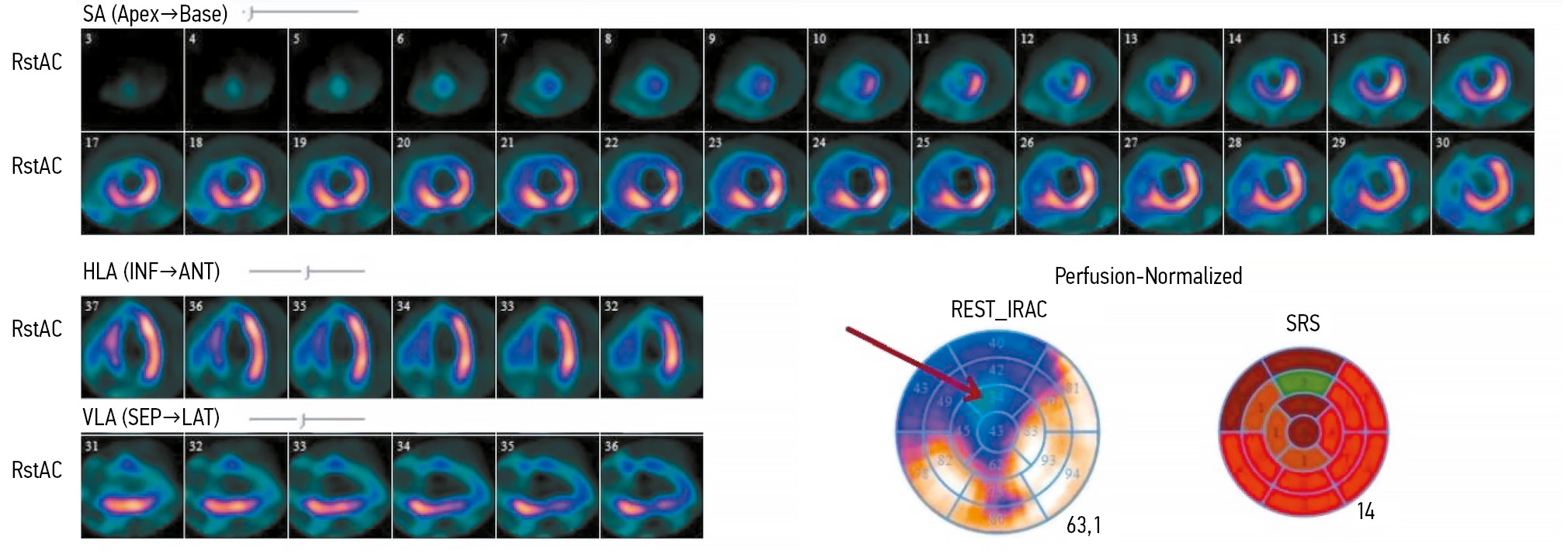
METHODS: It was a prospective, observational, single-center, non-randomized controlled study. Patients with chronic heart failure and indications for cardiac resynchronization therapy in accordance with current guidelines were enrolled. Prior to the procedure, the patients underwent computed tomography of the cardiac veins to visualize venous anatomy and myocardial perfusion scintigraphy to assess the extent of left ventricular perfusion impairment. The optimal site for left ventricular lead placement was identified using a three-dimensional reconstruction of the coronary sinus fused with myocardial perfusion scintigraphy data. To assess the effectiveness of the hybrid approach, a reference group was formed, in which cardiac resynchronization implantation was performed using the standard method, without preprocedural evaluation of coronary venous anatomy or myocardial scarring. Six months after cardiac resynchronization therapy, all patients underwent echocardiography to evaluate treatment effectiveness. Echocardiographic response was defined as a reduction in left ventricular end-systolic volume by ≥15% and/or an increase in ejection fraction by ≥5%.
RESULTS: The imaging group consisted of 40 patients with chronic heart failure, whereas the reference group included 30 patients with the same diagnosis. Six months after cardiac resynchronization therapy, a positive treatment response was observed in 33 patients (82%) in the imaging group, significantly higher than in the reference group (17 patients, 57%), p = 0.031. In the imaging group, the reduction in left ventricular end-systolic volume was statistically significant compared with the reference group and amounted to −52 [−71; −22.5] mL versus −21 [−64; −1] mL, respectively (p = 0.039). The increase in left ventricular ejection fraction was 7.5 [4.5; 15]% in the imaging group and 4.5 [0; 13]% in the reference group, with no statistically significant difference (p = 0.082).
CONCLUSION: The use of cardiovascular imaging methods, including cardiac venous computed tomography and myocardial perfusion scintigraphy, was associated with an increased proportion of responders to cardiac resynchronization therapy.
Full Text
##article.viewOnOriginalSite##About the authors
Anna I. Mishkina
Tomsk National Research Medical Centre, Russian Academy of Sciences
Author for correspondence.
Email: anna123.2013@gmail.com
ORCID iD: 0000-0001-9453-1635
SPIN-code: 9792-6033
MD, Cand. Sci. (Medicine), Cardiology Research Institute
Russian Federation, TomskTariel A. Atabekov
Tomsk National Research Medical Centre, Russian Academy of Sciences
Email: kgma1011@mail.ru
ORCID iD: 0000-0003-2645-4142
SPIN-code: 3274-6898
MD, Cand. Sci. (Medicine), Cardiology Research Institute
Russian Federation, TomskSvetlana I. Sazonova
Tomsk National Research Medical Centre, Russian Academy of Sciences
Email: sazonova_si@mail.ru
ORCID iD: 0000-0003-2799-3260
SPIN-code: 3787-2774
MD, Dr. Sci. (Medicine), Cardiology Research Institute
Russian Federation, TomskRoman E. Batalov
Tomsk National Research Medical Centre, Russian Academy of Sciences
Email: romancer@cardio-tomsk.ru
ORCID iD: 0000-0003-1415-3932
SPIN-code: 1371-4429
MD, Dr. Sci. (Medicine), Cardiology Research Institute
Russian Federation, TomskSergey V. Popov
Tomsk National Research Medical Centre, Russian Academy of Sciences
Email: svp@cardio-tomsk.ru
ORCID iD: 0000-0002-9050-4493
SPIN-code: 6853-7180
MD, Dr. Sci. (Medicine), Professor, academician of the Russian Academy of Science, Cardiology Research Institute
Russian Federation, TomskKonstantin V. Zavadovsky
Tomsk National Research Medical Centre, Russian Academy of Sciences
Email: konstzav@gmail.com
ORCID iD: 0000-0002-1513-8614
SPIN-code: 5081-3495
MD, Dr. Sci. (Medicine), Cardiology Research Institute
Russian Federation, TomskReferences
- Abdin A, Anker SD, Butler J, et al. “Time is prognosis” in heart failure: time-to-treatment initiation as a modifiable risk factor. ESC Heart Failure. 2021;8(6):4444–4453. doi: 10.1002/ehf2.13646 EDN: FYILWM
- Garganeeva AA, Bauer VA, Borel KN. The pandemic of the xxi century: chronic heart failure is the burden of the modern society. Epidemiology (literature review). The Siberian Medical Journal. 2014;29(3):8–12. EDN: TWKRHJ
- Sze E, Samad Z, Dunning A, et al. Impaired recovery of left ventricular function in patients with cardiomyopathy and left bundle branch block. Journal of the American College of Cardiology. 2018;71(3):306–317. doi: 10.1016/j.jacc.2017.11.020 EDN: YHSJTF
- ESC guidelines on cardiac pacing and cardiac resynchronization therapy. Russian Journal of Cardiology. 2022;27(7):289–370. doi: 10.15829/1560-4071-2022-5159 EDN: UTOLNY
- Tokavanich N, Mongkonsritragoon W, Sattawatthamrong S, et al. Outcomes of cardiac resynchronization therapy in congenital heart disease: A meta-analysis and systematic review. Journal of Cardiovascular Electrophysiology. 2023;35(2):249–257. doi: 10.1111/jce.16144 EDN: TNDOUD
- Daubert C, Behar N, Martins RP, et al. Avoiding non-responders to cardiac resynchronization therapy: a practical guide. European Heart Journal. 2017;38(19):1463–1472. doi: 10.1093/eurheartj/ehw270 EDN: YDAKAX
- Dhesi S, Lockwood E, Sandhu RK. Troubleshooting Cardiac Resynchronization Therapy in Nonresponders. Canadian Journal of Cardiology. 2017;33(8):1060–1065. doi: 10.1016/j.cjca.2017.04.007
- Zou J, Hua W, Su Y, et al. SPECT-Guided LV lead placement for incremental CRT efficacy. JACC: Cardiovascular Imaging. 2019;12(12):2580–2583. doi: 10.1016/j.jcmg.2019.07.009
- Hu X, Xu H, Hassea SRA, et al. Comparative efficacy of image-guided techniques in cardiac resynchronization therapy: a meta-analysis. BMC Cardiovascular Disorders. 2021;21(1):255. doi: 10.1186/s12872-021-02061-y EDN: IZWCJW
- Borgquist R, Carlsson M, Markstad H, et al. Cardiac resynchronization therapy guided by echocardiography, MRI, and CT Imaging. JACC: Clinical Electrophysiology. 2020;6(10):1300–1309. doi: 10.1016/j.jacep.2020.05.011 EDN: DOYWDC
- Zavadovskij KV, Saushkin VV, Varlamova YV, et al. Mechanical dyssynchrony for prediction of the cardiac resynchronization therapy response in patients with dilated cardiomyopathy. Kardiologiia. 2021;61(7):14–21. doi: 10.18087/cardio.2021.7.n1420 EDN: JWDUSY
- Viveiros Monteiro A, Martins Oliveira M, Silva Cunha P, et al. Time to left ventricular reverse remodeling after cardiac resynchronization therapy: Better late than never. Revista Portuguesa de Cardiologia. 2016;35(3):161–167. doi: 10.1016/j.repc.2015.11.008
- Mareev VYu, Fomin IV, Ageev FT, et al. Russian Heart Failure Society, Russian Society of Cardiology. Russian Scientific Medical Society of Internal Medicine Guidelines for Heart failure: chronic (CHF) and acute decompensated (ADHF). Diagnosis, prevention and treatment. Kardiologiia. 2018;17(S6):8–158. doi: 10.18087/cardio.2475 EDN: XUAREL
- Butter C, Georgi C, Stockburger M. Optimal CRT implantation—where and how to place the left-ventricular lead? Current Heart Failure Reports. 2021;18(5):329–344. doi: 10.1007/s11897-021-00528-9 EDN: PMWWDH
- Boonyasirinant T, Halliburton SS, Schoenhagen P, et al. Absence of coronary sinus tributaries in ischemic cardiomyopathy: An insight from multidetector computed tomography cardiac venographic study. Journal of Cardiovascular Computed Tomography. 2016;10(2):156–161. doi: 10.1016/j.jcct.2016.01.015
- Stiles MK, Fauchier L, Morillo CA, Wilkoff BL. 2019 HRS/EHRA/APHRS/LAHRS focused update to 2015 expert consensus statement on optimal implantable cardioverter-defibrillator programming and testing. Heart Rhythm. 2020;17(1):e220–e228. doi: 10.1016/j.hrthm.2019.02.034 EDN: RFAGXG
- Wang J, Wang Y, Yang M, et al. Mechanical contraction to guide CRT left-ventricular lead placement instead of electrical activation in myocardial infarction with left ventricular dysfunction: An experimental study based on non-invasive gated myocardial perfusion imaging and invasive electroanatomic mapping. Journal of Nuclear Cardiology. 2020;27(2):419–430. doi: 10.1007/s12350-019-01710-2 EDN: DVOUZZ
- ESC guidelines for the diagnosis and treatment of acute and chronic heart failure. Russian Journal of Cardiology. 2023;28(1):117–224.
- Mansour N, Nekolla SG, Reyes E, et al. Multi-center study of inter-rater reproducibility, image quality, and diagnostic accuracy of CZT versus conventional SPECT myocardial perfusion imaging. Journal of Nuclear Cardiology. 2023;30(2):528–539. doi: 10.1007/s12350-022-03054-w EDN: XJIKCA
- Tada T, Osuda K, Nakata T, et al. A novel approach to the selection of an appropriate pacing position for optimal cardiac resynchronization therapy using CT coronary venography and myocardial perfusion imaging: FIVE STaR method (fusion image using CT coronary venography and perfusion SPECT applied for cardiac resynchronization therapy). Journal of Nuclear Cardiology. 2021;28(4):1438–1445. doi: 10.1007/s12350-019-01856-z
Supplementary files


