

")


Autonomous artificial intelligence for sorting results of preventive radiological examinations of chest organs: medical and economic efficiency
- Authors: Vasilev Y.A.1, Sychev D.A.2, Bazhin A.V.1, Shulkin I.M.1, Vladzymyrskyy A.V.1, Golikova A.Y.1, Arzamasov K.M.1, Mishchenko A.V.2, Bekdzhanyan G.A.2, Goldberg A.S.2, Rodionova L.G.1
-
Affiliations:
- Research and Practical Clinical Center for Diagnostics and Telemedicine Technologies
- Medical Academy of Continuous Professional Education
- Issue: Vol 6, No 1 (2025)
- Pages: 5-22
- Section: Original Study Articles
- URL: https://journals.rcsi.science/DD/article/view/310048
- DOI: https://doi.org/10.17816/DD641703
- ID: 310048
Cite item

Abstract
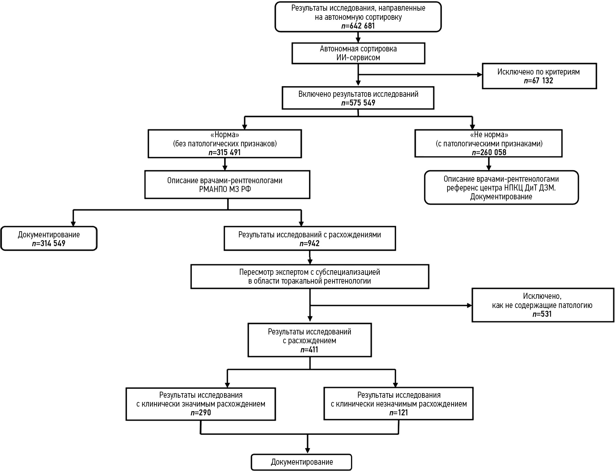
BACKGROUND: This article proposes a model for organizing preventive radiological examinations of chest organs through autonomous sorting of examination results using medical devices based on artificial intelligence technologies, optimized for maximum sensitivity — 1.0 (95% CI: 1.0; 1.0). Sorting involves classifying the results of mass preventive screenings (fluoroscopy and chest X-rays) into two: “not normal” and “normal.” The “not normal” category includes all cases of abnormalities (e.g., pathological conditions, post-disease or post-surgery consequences, and age-related and congenital features), which are sent for interpretation by a radiologist. The “normal” category consists of cases without signs of pathological deviations, which potentially do not require a radiologist’s description.
AIM: To evaluate the feasibility, effectiveness, and efficiency of autonomous sorting of results from preventive radiological examinations of chest organs.
MATERIALS AND METHODS: A prospective multicenter diagnostic study was conducted on the safety and quality of autonomous sorting of results from preventive radiological examinations of chest organs. Analytical and statistical methods of scientific inquiry were used.
RESULTS: The study included results from 575,549 preventive radiological examinations obtained through fluoroscopy and chest X-rays and processed using three medical devices based on artificial intelligence technologies. In autonomous sorting, 54.8% of the preventive radiological examinations of chest organs were classified as “normal,” resulting in a proportional reduction in the radiologist’s workload for interpreting and describing the examination results. Fully correct autonomous sorting was achieved in 99.95% of cases. Clinically significant discrepancies were determined in 0.05% of cases (95% CI: 0.04; 0.06%).
CONCLUSIONS: This study confirmed the medical and economic effectiveness of the model for autonomous sorting of results from preventive radiological examinations of chest organs using medical devices based on artificial intelligence technologies. The next phase should involve updating the regulatory framework and ensuring the legitimacy of the autonomous application of certain medical devices based on artificial intelligence technologies in established conditions and preventive tasks.
Full Text
##article.viewOnOriginalSite##About the authors
Yuriy A. Vasilev
Research and Practical Clinical Center for Diagnostics and Telemedicine Technologies
Email: npcmr@zdrav.mos.ru
ORCID iD: 0000-0002-5283-5961
SPIN-code: 4458-5608
MD, Dr. Sci. (Medicine)
Russian Federation, MoscowDmitry A. Sychev
Medical Academy of Continuous Professional Education
Email: dimasychev@mail.ru
ORCID iD: 0000-0002-4496-3680
SPIN-code: 4525-7556
MD, Dr. Sci. (Medicine), Professor, academician of the Russian Academy of Sciences
Russian Federation, MoscowAlexander V. Bazhin
Research and Practical Clinical Center for Diagnostics and Telemedicine Technologies
Email: BazhinAV@zdrav.mos.ru
ORCID iD: 0000-0003-3198-1334
SPIN-code: 6122-5786
MD, Cand. Sci. (Medicine)
Russian Federation, MoscowIgor M. Shulkin
Research and Practical Clinical Center for Diagnostics and Telemedicine Technologies
Email: ShulkinIM@zdrav.mos.ru
ORCID iD: 0000-0002-7613-5273
SPIN-code: 5266-0618
MD, Cand. Sci. (Medicine)
Russian Federation, MoscowAnton V. Vladzymyrskyy
Research and Practical Clinical Center for Diagnostics and Telemedicine Technologies
Author for correspondence.
Email: vladzimirskijAV@zdrav.mos.ru
ORCID iD: 0000-0002-2990-7736
SPIN-code: 3602-7120
MD, Dr. Sci. (Medicine)
Russian Federation, MoscowAlexandra Yu. Golikova
Research and Practical Clinical Center for Diagnostics and Telemedicine Technologies
Email: GolikovaAY1@zdrav.mos.ru
ORCID iD: 0009-0001-5020-2765
Russian Federation, Moscow
Kirill M. Arzamasov
Research and Practical Clinical Center for Diagnostics and Telemedicine Technologies
Email: ArzamasovKM@zdrav.mos.ru
ORCID iD: 0000-0001-7786-0349
SPIN-code: 3160-8062
MD, Cand. Sci. (Medicine)
Russian Federation, MoscowAndrei V. Mishchenko
Medical Academy of Continuous Professional Education
Email: dr.mishchenko@mail.ru
ORCID iD: 0000-0001-7921-3487
SPIN-code: 8825-4704
MD, Dr. Sci. (Medicine)
Russian Federation, MoscowGevorg A. Bekdzhanyan
Medical Academy of Continuous Professional Education
Email: rmapo@rmapo.ru
ORCID iD: 0009-0007-7150-7166
SPIN-code: 4579-9457
Russian Federation, Moscow
Arcadiy S. Goldberg
Medical Academy of Continuous Professional Education
Email: goldarcadiy@gmail.com
ORCID iD: 0000-0002-2787-4731
SPIN-code: 8854-0469
MD, Cand. Sci. (Medicine)
Russian Federation, MoscowLarisa G. Rodionova
Research and Practical Clinical Center for Diagnostics and Telemedicine Technologies
Email: RodionovaLG@zdrav.mos.ru
ORCID iD: 0009-0008-9862-8205
Russian Federation, Moscow
References
- Boenk EA, Roginko NI, Dzeranova NG, et al. All-Russian medical examination of adult population within the framework of the national project "Healthcare". Vestnik Roszdravnadzora. 2021;(1):21–29. EDN: FIPEZH
- Garifullin TYu, Avdeeva MV, Filatov VN, et al. Improvement of medical check-up process on the basis of lean technologies in outpatient settings. Russian Journal of Preventive Medicine and Public Health. 2023;26(3):30–38. doi: 10.17116/profmed20232603130 EDN: AGSZJF
- Zakharchenko OO, Shikina IB, Terentyeva DS. Results of the medical examination of the adult population over 60 years in the Russian Federation (2016–2021). Preventive And Clinical Medicine. 2023;3(88):103–114. doi: 10.47843/2074-9120_2023_3_103 EDN: YNHXOE
- Ignatyeva VI, Kontsevaya AV, Kalinina AM, et al. Socio-economic effectiveness of early cancer detection during medical checkup. Russian Journal of Preventive Medicine and Public Health. 2024;27(1):36–44. doi: 10.17116/profmed20242701136 EDN: CNVQRC
- Levshin VF, Slepchenko NI, Ryzhova NI, et al. Study of the attitude and participation of the population in the preventive and screening examinations and implementation of these examinations in the health care system. Lechaschi Vrach. 2022;25(10):81–87. doi: 10.51793/OS.2022.25.10.013 EDN: UFZZEB
- Stupina MI, Selezneva PA, Khaptanova VA. Medical screening of patients with coronary heart disease in outpatient settings. Nauchnyy Aspekt. 2024;34(4):4436–4455. (In Russ.) EDN: XOTAYU
- Golubev NA, Ogryzko EV, Tyurina EM, et al. Features of the development of the radiation diagnostics service in the Russian Federation for 2014–2019. Current Problems of Health Care and Medical Statistics. 2021;(2):356–376. doi: 10.24412/2312-2935-2021-2-356-376 EDN: EHSADW
- Ivashikin YM. (2024). Lung imaging screening during preventive medical examinations and medical screening. In: Higher education: scientific research. Proceedings of the Interuniversity International Congress. Moscow: Infinity Publishing House, 2024. P. 139–141. (In Russ.) doi: 10.34660/INF.2024.94.91.106 EDN: BXEOLF
- Trofimova TN, Kozlova OV. Radiology in Saint-Petersburg 2019. Diagnostic radiology and radiotherapy. 2021;4(11):96–99. doi: 10.22328/2079-5343-2020-11-4-96-99 EDN: HTVSUZ
- Tyurin IE. Radiology in the Russian Federation. Journal of Oncology: Diagnostic Radiology and Radiotherapy. 2018;1(4):43–51. EDN: QZSWYK
- Zubova NA. Effectiveness of mass preventive examinations in subjects of the Russian Federation with low morbidity rates of tuberculosis. Social Aspects of Population Health. 2016;4(50):8. doi: 10.21045/2071-5021-2016-50-4-8 EDN: WGIKUN
- Rubis LV. Efficiency of mass preventive examinations of the urban population for the purpose of early diagnosis of tuberculosis in primary health care institutions. Current Problems of Health Care and Medical Statistics. 2021;(3):1–13. doi: 10.24412/2312-2935-2021-3-1-13 EDN: VPLCTZ
- Shelekhov PV. Personnel situation in radiative diagnostics. Current Problems of Health Care and Medical Statistics. 2019;(1):265–275. doi: 10.24411/2312-2935-2019-10018 EDN: ZGZFPV
- Bobrovskaya TM, Vasilev YuA, Nikitin NYu, Arzamasov KM. Approaches to building radiology datasets. Medical Doctor and IT. 2023;(4):14–23. doi: 10.25881/18110193_2023_4_14 EDN: EQHEKE
- Vasiliev YuA, Vlazimirsky AV, Omelyanskaya OV, et al. Methodology for testing and monitoring artificial intelligence-based software for medical diagnostics. Digital Diagnostics. 2023;4(3):252–267. doi: 10.17816/DD321971 EDN: UEDORU
- Vasilev YuA, Arzamasov KM, Kolsanov AV, et al. Experience of application artificial intelligence software on 800 thousand fluorographic studies. Medical Doctor and IT. 2023;(4):54–65. doi: 10.25881/18110193_2023_4_54 EDN: MHCTUB
- Vasiliev YuA, Vladzimirsky AV, Arzamasov KM, et al. The first 10,000 mammography exams performed as part of the “Description and interpretation of mammography data using artificial intelligence” service. Manager Zdravookhranenia. 2023;(8):54–67. doi: 10.21045/1811-0185-2023-8-54-67 EDN: KZHPVW
- Vasilev YuA, Vladzimirsky AV, Arzamasov KM, et al. Computer vision in radiology: stage one of the Moscow experiment. 2nd ed. Moscow: Publishing solutions; 2023. (In Russ.)
- Arzamasov KM, Semenov SS, Kokina DY, et al. Criteria for the applicability of computer vision for preventive studies on the example of chest X-ray and fluorography. Meditsinskaya Fizika. 2022;4(96):56–63. doi: 10.52775/1810-200X-2022-96-4-56-63 EDN: MXKUVL
- Vasilev YuA, Tyrov IA, Vladzymyrskyy AV, et al. A new model of organizing mass screening based on stand-alone artificial intelligence used for fluorography image triage. Public Health and Life Environment. 2023;31(11):23–32. doi: 10.35627/2219-5238/2023-31-11-23-32 EDN: SYIQBX
- Vasilev YuA, Tyrov IA, Vladzymyrskyy AV, et al. Autonomous artificial intelligence for sorting the preventive imaging studies’ results. Russian Journal of Preventive Medicine. 2024;27(7):23–29. doi: 10.17116/profmed20242707123 EDN: ODGHNM
- Morozov SP, Vetsheva NN, Ledikhova NV, et al. Assessing the quality of radiologic studies. Moscow: Moscow Center for Diagnostics and Telemedicine; 2019. (In Russ.)
- Alekseeva TR, Amosov VI, Anikeeva OYu, et al. Chest radiology: national guidelines. Moscow: GEOTAR-Media; 2014. (In Russ.) EDN: VRXFKX
- Vasilev YuA, Vladzymyrskyy AV, Omelyanskaya OV, et al. Assessing the maturity of artificial intelligence technologies for healthcare. Moscow: Moscow Center for Diagnostics and Telemedicine; 2023. (In Russ.)
- Orlov EM, Sokolova ON. Efficiency category in public health services system. Fundamental’nye issledovaniya. 2010;(4):70–75. EDN: MSPQTJ
- Kucherenko VZ, Fleck VO, Putin ME, et al. Evaluation of the effectiveness of medical organizations. Vyalkov AI, editor. Moscow: GEOTAR-Med; 2004.
- Arzamasov KM, Vasilev YuA, Vladzymyrskyy AV, et al. The use of computer vision for the mammography preventive research. Russian Journal of Preventive Medicine and Public Health. 2023;26(6):117–123. doi: 10.17116/profmed202326061117 EDN: YBKHPS
- Arzamasov KM, Vasilev YuA, Vladzymyrskyy AV, et al. An international non-inferiority study for the benchmarking of AI for routine radiology cases: chest X-ray, fluorography and mammography. Healthcare. 2023;11(10):1684. doi: 10.3390/healthcare11121684 EDN: FWVMPQ
- Berlin L. Radiologic errors and malpractice: a burry distinction. American Journal of Roentgenology. 2007;189(3):517–522. doi: 10.2214/AJR.07.2209
- Brady AP. Error and discrepancy in radiology: inevitable or avoidable? Insights into Imaging. 2017;8(1):171–182. doi: 10.1007/s13244-016-0534-1 EDN: FSSDNE
- Bruno MA, Walker EA, Abujudeh HH. Understanding and confronting our mistakes: the epidemiology of error in radiology and strategies for error reduction. RadioGraphics. 2015;35(6):1668–1676. doi: 10.1148/rg.2015150023
- Cascade PN, Kazerooni EA, Gross BH, et al. Evaluation of competence in the interpretation of chest radiographs. Academic Radiology. 2001;8(4):315–321. doi: 10.1016/S1076-6332(03)80500-7
- Morozov S, Guseva E, Ledikhova N, et al. Telemedicine-based system for quality management and peer review in radiology. Insights into Imaging. 2018;9(3):337–341. doi: 10.1007/s13244-018-0629-y EDN: YCIRMT
- Quekel LGBA, Kessels AGH, Goei R, van Engelshoven JMA. Miss rate of lung cancer on the chest radiograph in clinical practice. Chest. 1999;115(3):720–724. doi: 10.1378/chest.115.3.720
- Satia I, Bashagha S, Bibi A, et al. Assessing the accuracy and certainty in interpreting chest X-rays in the medical division. Clinical Medicine. 2013;13(4):349–352. doi: 10.7861/clinmedicine.13-4-349
- Topff L, Steltenpool S, Ranschaert ER, et al. Artificial intelligence-assisted double reading of chest radiographs to detect clinically relevant missed findings: a two-centre evaluation. European Radiology. 2024;34(9):5876–5885. doi: 10.1007/s00330-024-10676-w EDN: RUJICB
- Vasilev YuA, Vladzymyrskyy AV, Omelyanskaya OV, et al. AI-based CXR first reading: current limitations to ensure practical value. Diagnostics. 2023;13(8):1430. doi: 10.3390/diagnostics13081430 EDN: MPQYUP
Supplementary files


