

")


A case report of a mild neurologic deficit with extensive poststroke damage to the subdominant brain hemisphere: analysis of data obtained from magnetic resonance tractography, functional magnetic resonance imaging, and electroencephalography
- Authors: Gumin I.S.1, Gulyaev S.A.1, Beregov M.M.1, Lelyuk V.G.1
-
Affiliations:
- Federal center of brain research and neurotechnologies of the Federal Medical Biological Agency
- Issue: Vol 5, No 3 (2024)
- Pages: 601-612
- Section: Case reports
- URL: https://journals.rcsi.science/DD/article/view/310041
- DOI: https://doi.org/10.17816/DD624967
- ID: 310041
Cite item
Full Text

Abstract
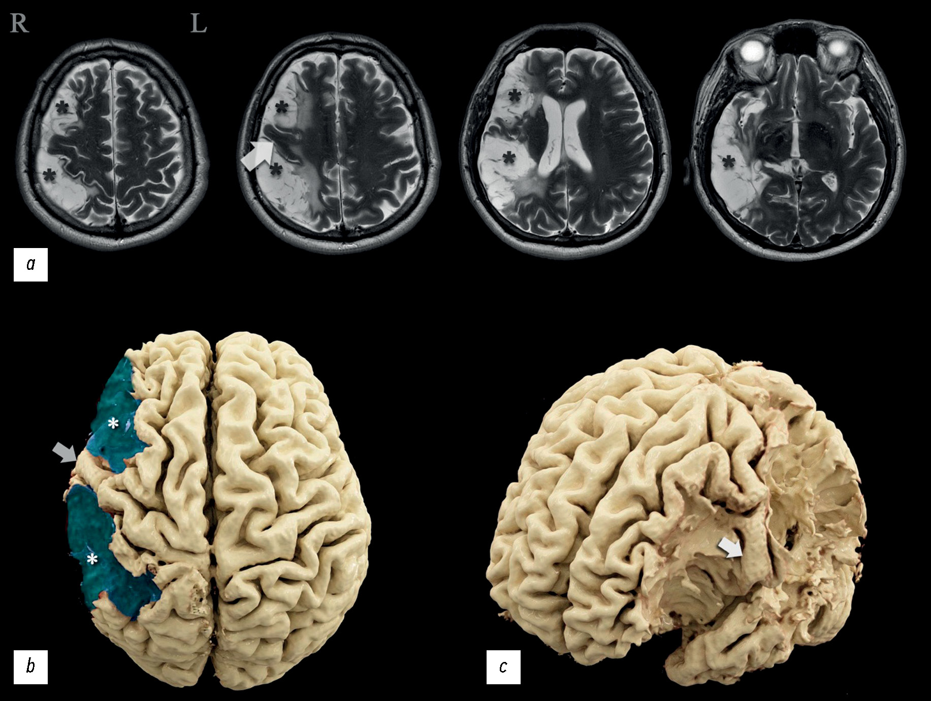
The severity of damage to different brain areas, including the cortex, can vary significantly in the associated neurologic deficit and reduction in the quality of life, often regardless of the lesion volume. The localization of the abnormalities plays a large part. Lesions of the dominant and subdominant hemispheres can differ greatly in both clinical features and effects on the patient’s quality of life. In this case report, a patient admitted for rehabilitation after two ischemic strokes underwent neurological and neuropsychological examination, complex instrumental diagnostics using electroencephalography, magnetic resonance imaging, computed tomography perfusion, magnetic resonance tractography, and functional magnetic resonance imaging. The patient had minimal left-sided hemiparesis, impaired regulation of voluntary activity, mild decrease in neurodynamic indicators, mildly impaired concentration, and a critical view of his condition. Neuroimaging findings demonstrated extensive postinfarction damage to the right subdominant hemisphere of the brain in the middle cerebral artery circulation. A nonconformity between the brain damage volume and the severity of its clinical signs was observed. Based on functional examination data, the dominant hemisphere was determined, and restructuring the functional centers was suggested. This clinical case was compared with similar ones, and their relationship with the data was analyzed. Information that expands the knowledge of the topography of the altered zones involved in motor and speech functions and the ability to perform arithmetic counting was obtained.
Full Text
##article.viewOnOriginalSite##About the authors
Ivan S. Gumin
Federal center of brain research and neurotechnologies of the Federal Medical Biological Agency
Author for correspondence.
Email: ivangumin@mail.ru
ORCID iD: 0000-0003-2360-3261
SPIN-code: 3454-2665
Scopus Author ID: 57223430019
Russian Federation, Moscow
Sergey A. Gulyaev
Federal center of brain research and neurotechnologies of the Federal Medical Biological Agency
Email: gulyaev@fccps.ru
ORCID iD: 0000-0003-0549-0961
MD, Dr. Sci. (Medicine)
Russian Federation, MoscowMikhail M. Beregov
Federal center of brain research and neurotechnologies of the Federal Medical Biological Agency
Email: mikhailberegov@gmail.com
ORCID iD: 0000-0003-1899-8131
SPIN-code: 2559-0307
Russian Federation, Moscow
Vladimir G. Lelyuk
Federal center of brain research and neurotechnologies of the Federal Medical Biological Agency
Email: vglelyuk@fccps.ru
ORCID iD: 0000-0002-9690-8325
MD, Dr. Sci. (Medicine), Professor
Russian Federation, MoscowReferences
- Global Health Estimates 2016: Deaths by cause, age, sex, by country and by region, 2000-2016 [Internet]. Geneva: World Health Organization; 2018 [cited 25.07.2019]. Available from: https://www.who.int/data/gho/data/themes/mortality-and-global-health-estimates
- Benjamin EJ, Muntner P, Alonso A, et al. Heart Disease and Stroke Statistics—2019 Update: A Report From the American Heart Association. Circulation. 2019;139(10):e56–e528. doi: 10.1161/CIR.0000000000000659
- Mijajlović MD, Pavlović A, Brainin M, et al. Post-stroke dementia — a comprehensive review. BMC Medicine. 2017;15(1):11. doi: 10.1186/s12916-017-0779-7
- Pohjasvaara T, Erkinjuntti T, Ylikoski R, et al. Clinical Determinants of Poststroke Dementia. Stroke. 1998;29(1):75–81. doi: 10.1161/01.str.29.1.75
- Barrett AM. Spatial Neglect and Anosognosia After Right Brain Stroke. Continuum (Minneapolis, Minn.). 2021;27(6):1624–1645. doi: 10.1212/CON.0000000000001076
- Broussolle E, Reynolds EH. Anglo-French neurological interactions in the 19th and early 20th centuries: Physicians, places and events. Revue neurologique. 2021;177(8):859–870. doi: 10.1016/j.neurol.2020.10.013
- Chakrabarty M, Pflieger EM, Cardillo E, Chatterjee A. Effects of Chronic Brain Injury on Quality of Life: A Study in Patients With Left- or Right-Sided Lesion. Archives of Rehabilitation Research and Clinical Translation. 2019;2(1):100031. doi: 10.1016/j.arrct.2019.100031
- Howard G, Till JS, Toole JF, et al. Factors Influencing Return to Work Following Cerebral Infarction. JAMA. 1985;253(2):226–232. doi: 10.1001/jama.1985.03350260078030
- Penfield W, Rasmussen T. The cerebral cortex of man. New York: Macmillan Company; 1950.
- Halligan P. Half a brain is enough: the story of Nico. Journal of Neurology Neurosurgery & Psychiatry. 2001;71(4):566. doi: 10.1136/jnnp.71.4.566b
- Gumin IS, Gubskiy IL, Mironov MB, et al. Dyke–Davidoff–Masson syndrome: description of clinical case with diagnostics by EEG, MRI, MR-tractography, fMRI. Neuromuscular Diseases. 2021;11(1):47–57. doi: 10.17650/2222-8721-2021-11-1-47-57
- Agris AR, Almazova AA, Altuhova TA, et al. Narusheniya pis’ma i chteniya u detej: izuchenie i korrekciya. Moscow: “LOGOMAG”; 2018. (In Russ.)
- Bain JS, Yeatman JD, Schurr R, et al. Evaluating arcuate fasciculus laterality measurements across dataset and tractography pipelines. Human Brain Mapping. 2019;40(13):3695–3711. doi: 10.1002/hbm.24626
- Roiha K, Kirveskari E, Kaste M, et al. Reorganization of the primary somatosensory cortex during stroke recovery. Clinical Neurophysiology. 2011;122(2):339–345. doi: 10.1016/j.clinph.2010.06.032
- Sanchez-Panchuelo RM, Francis S, Bowtell R, Schluppeck D. Mapping Human Somatosensory Cortex in Individual Subjects With 7T Functional MRI. Journal of neurophysiology. 2010;103(5):2544–2556. doi: 10.1152/jn.01017.2009
- Alary F, Doyon B, Loubinoux I, et al. Event-Related Potentials Elicited by Passive Movements in Humans: Characterization, Source Analysis, and Comparison to fMRI. Neuroimage. 1998;8(4):377–390. doi: 10.1006/nimg.1998.0377
- Cramer SC, Moore CI, Finklestein SP, Rosen BR. A Pilot Study of Somatotopic Mapping After Cortical Infarct. Stroke. 2000;31(3):668–671. doi: 10.1161/01.str.31.3.668
- Roux FE, Boulanouar K, Ibarrola D, et al. Functional MRI and intraoperative brain mapping to evaluate brain plasticity in patients with brain tumours and hemiparesis. Journal of Neurology Neurosurgery & Psychiatry. 2000;69(4):453–463. doi: 10.1136/jnnp.69.4.453
- Arsalidou M, Taylor MJ. Is 2+2=4? Meta-analyses of brain areas needed for numbers and calculations. Neuroimage. 2011;54(3):2382–2393. doi: 10.1016/j.neuroimage.2010.10.009
- Hawes Z, Sokolowski HM, Ononye CB, Ansari D. Neural underpinnings of numerical and spatial cognition: An fMRI meta-analysis of brain regions associated with symbolic number, arithmetic, and mental rotation. Neuroscience and Biobehavioral Reviews. 2019;103:316–336. doi: 10.1016/j.neubiorev.2019.05.007
- Fedorenko E, Blank IA. Broca’s Area Is Not a Natural Kind. Trends in Cognitive Sciences. 2020;24(4):270–284. doi: 10.1016/j.tics.2020.01.001
- Bach-y-Rita P. Brain plasticity as a basis for recovery of function in humans. Neuropsychologia. 1990;28(6):547–554. doi: 10.1016/0028-3932(90)90033-k
- Wan CY, Schlaug G. Music Making as a Tool for Promoting Brain Plasticity across the Life Span. Neuroscientist. 2010;16(5):566–577. doi: 10.1177/1073858410377805
- Nudo RJ. Remodeling of cortical motor representations after stroke: implications for recovery from brain damage. Molecular Psychiatry. 1997;2(3):188–191. doi: 10.1038/sj.mp.4000188
- Nudo RJ. Postinfarct Cortical Plasticity and Behavioral Recovery. Stroke. 2007;38(2):840–845. doi: 10.1161/01.STR.0000247943.12887.d2
Supplementary files


