Radiological evaluation of a calyceal diverticulum presenting with hematuria

Cover Image

Cite item

Full Text

Abstract

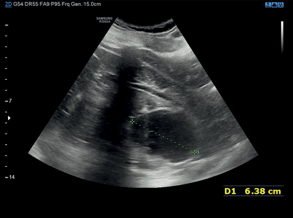
Calyceal diverticula, also known as pyelogenic cysts, are a relatively uncommon condition, which is usually asymptomatic and incidentally diagnosed during routine imaging. In some cases, they may lead to concerning symptoms such as hematuria and flank pain, mimicking a renal tumor. In this case report, the patient suffered from hematuria that was initially suspected as a renal malignancy but was ultimately attributed to a calyceal diverticulum. The presented case allows evaluating one of the rarest and underestimated causes of hematuria and describes the main imaging features of calyceal diverticula. In particular, ultrasonography, computed tomography urography, dual-energy computed tomography, and magnetic resonance urography were performed. Subsequently, this case report also serves an educational purpose.

About the authors

Federica Masino

Foggia University School of Medicine

Email: federicamasino@gmail.com
ORCID iD: 0009-0004-4289-3289

MD

Italy, Foggia

Manuela Montatore

Foggia University School of Medicine

Email: manuela.montatore@unifg.it
ORCID iD: 0009-0002-1526-5047

MD

Italy, Foggia

Laura Eusebi

Carlo Urbani Hospital

Email: lauraeu@virgilio.it
ORCID iD: 0000-0002-4172-5126

MD

Italy, Jesi

Rossella Gifuni

Foggia University School of Medicine

Email: rossella.gifuni@gmial.com
ORCID iD: 0009-0009-9679-3861

MD

Italy, Foggia

Gianmichele Muscatella

Foggia University School of Medicine

Email: muscatella94@gmail.com
ORCID iD: 0009-0004-3535-5802

MD

Italy, Foggia

Marina Balbino

Foggia University School of Medicine

Email: marinabalbino93@gmail.com
ORCID iD: 0009-0009-2808-5708

MD

Italy, Foggia

Giuseppe Sortino

Carlo Urbani Hospital

Email: giuseppesortino@live.it

MD

Italy, Jesi

Lucia Pitoni

Carlo Urbani Hospital

Email: lucia.pitoni22@gmail.com
ORCID iD: 0000-0002-8419-090X

MD

Italy, Jesi

Giuseppe Guglielmi

Foggia University School of Medicine; Dimiccoli Hospital; IRCCS Casa Sollievo della Sofferenza Hospital

Author for correspondence.
Email: giuseppe.guglielmi@unifg.it
ORCID iD: 0000-0002-4325-8330

MD, Professor

Italy, Foggia; Barletta; San Giovanni Rotondo

References

  1. Zhao Y, Zhang R, Yun Y, et al. A case report of renal calyceal diverticulum with hypertension in children and review of literature. BMC pediatrics. 2022;22(1):35. doi: 10.1186/s12887-021-03081-5
  2. Zhang R, Shen W, Li X, et al. A Petal-like Calyceal Diverticulum. Urology. 2015;86(6):e31–e32. doi: 10.1016/j.urology.2015.09.003
  3. Zhang Z, Zhang Y, Wang X, et al. Challenges in the diagnosis of calyceal diverticulum: A report of two cases and review of the literature. Journal of X-ray science and technology. 2020;27(6):1155–1167. doi: 10.3233/XST-190549
  4. Waingankar N, Hayek S, Smith AD, Okeke Z. Calyceal diverticula: a comprehensive review. Reviews in urology. 2014;16(1):29–43.
  5. Kurkov AV, Pominalnaya VM, Nechay VV, et al. A Case Report of Calyceal Diverticulum: Differential Diagnosis for Organ-Preserving Operations. Frontiers in surgery. 2021;8:731796. doi: 10.3389/fsurg.2021.731796
  6. Montatore M, Muscatella G, Eusebi L, et al. Current Status on New Technique and Protocol in Urinary Stone Disease. Current Radiology Reports. 2023;11:161–176. doi: 10.1007/s40134-023-00420-5
  7. Eusebi L, Masino F, Gifuni R, et al. Role of Multiparametric-MRI in Bladder Cancer. Current Radiology Reports. 2023;11:69–80. doi: 10.1007/s40134-023-00412-5

Supplementary files

Supplementary Files
Action
1. JATS XML
2. Fig. 1. Ultrasound examination revealed a large, heterogeneous formation, predominantly anechoic, with rounded, clear boundaries, located in the middle-lower part of the left kidney. The largest diameter was 6.38 cm.

Download (92KB)
3. Fig. 2. Axial sections, results of computed tomographic urography: a — without contrast, during scanning there were no signs of lithiasis in both kidneys, the formation in the left kidney was hypodense with a tomographic density corresponding to fluid (in Hounsfield units); b — in the arterial phase, during scanning a slight increase in the density of the cyst-like cavity was noted; c — in the excretory phase, during scanning there was contrast enhancement of the cavity walls, with gradual filling of the cavity with contrast agent.

Download (172KB)
4. Fig. 3. Processed images obtained by dual-energy computed tomography, coronal plane. In the color-coded iodine distribution maps (a, b), iodine uptake by the excretory system, including the calyceal diverticulum, is represented in bright yellow. In panel b, there is no iodine signal in the cyst area.

Download (304KB)
5. Fig. 4. Magnetic resonance urography, axial sections: a — a hyperintense formation is visualized on the T2-weighted image; b — a hyperintense formation and renal pelvis are visualized on the T1-weighted image in fat-suppression mode after the introduction of a contrast agent in the delayed phase; c — the formation and the excretory system are visualized with the same intensity on the in-phase image; d — diffusion limitation is absent on the diffusion-weighted image with a high b-value.

Download (262KB)
6. Fig. 5. Magnetic resonance urography, coronal sections, T1-weighted images in fat-suppressed mode in the delayed phase after the administration of a contrast agent. Two lesions located in the upper pole of both kidneys are cysts. (In the two subsequent images, the diverticulum was visualized filled with contrast agent, and its funnel-shaped channel communicated with the renal pelvis and calyces).

Download (224KB)

Copyright (c) 2024 Eco-Vector

Creative Commons License
This work is licensed under a Creative Commons Attribution-NonCommercial-NoDerivatives 4.0 International License.

Согласие на обработку персональных данных с помощью сервиса «Яндекс.Метрика»

1. Я (далее – «Пользователь» или «Субъект персональных данных»), осуществляя использование сайта https://journals.rcsi.science/ (далее – «Сайт»), подтверждая свою полную дееспособность даю согласие на обработку персональных данных с использованием средств автоматизации Оператору - федеральному государственному бюджетному учреждению «Российский центр научной информации» (РЦНИ), далее – «Оператор», расположенному по адресу: 119991, г. Москва, Ленинский просп., д.32А, со следующими условиями.

2. Категории обрабатываемых данных: файлы «cookies» (куки-файлы). Файлы «cookie» – это небольшой текстовый файл, который веб-сервер может хранить в браузере Пользователя. Данные файлы веб-сервер загружает на устройство Пользователя при посещении им Сайта. При каждом следующем посещении Пользователем Сайта «cookie» файлы отправляются на Сайт Оператора. Данные файлы позволяют Сайту распознавать устройство Пользователя. Содержимое такого файла может как относиться, так и не относиться к персональным данным, в зависимости от того, содержит ли такой файл персональные данные или содержит обезличенные технические данные.

3. Цель обработки персональных данных: анализ пользовательской активности с помощью сервиса «Яндекс.Метрика».

4. Категории субъектов персональных данных: все Пользователи Сайта, которые дали согласие на обработку файлов «cookie».

5. Способы обработки: сбор, запись, систематизация, накопление, хранение, уточнение (обновление, изменение), извлечение, использование, передача (доступ, предоставление), блокирование, удаление, уничтожение персональных данных.

6. Срок обработки и хранения: до получения от Субъекта персональных данных требования о прекращении обработки/отзыва согласия.

7. Способ отзыва: заявление об отзыве в письменном виде путём его направления на адрес электронной почты Оператора: info@rcsi.science или путем письменного обращения по юридическому адресу: 119991, г. Москва, Ленинский просп., д.32А

8. Субъект персональных данных вправе запретить своему оборудованию прием этих данных или ограничить прием этих данных. При отказе от получения таких данных или при ограничении приема данных некоторые функции Сайта могут работать некорректно. Субъект персональных данных обязуется сам настроить свое оборудование таким способом, чтобы оно обеспечивало адекватный его желаниям режим работы и уровень защиты данных файлов «cookie», Оператор не предоставляет технологических и правовых консультаций на темы подобного характера.

9. Порядок уничтожения персональных данных при достижении цели их обработки или при наступлении иных законных оснований определяется Оператором в соответствии с законодательством Российской Федерации.

10. Я согласен/согласна квалифицировать в качестве своей простой электронной подписи под настоящим Согласием и под Политикой обработки персональных данных выполнение мною следующего действия на сайте: https://journals.rcsi.science/ нажатие мною на интерфейсе с текстом: «Сайт использует сервис «Яндекс.Метрика» (который использует файлы «cookie») на элемент с текстом «Принять и продолжить».