

")


Fundamentals of dual-energy computed tomography and its emerging applications in bladder cancer
- Authors: Masino F.1, Eusebi L.2, Montatore M.1, Muscatella G.1, Gifuni R.1, Ferrara V.2, Marcellini M.3, Guglielmi G.1,4,5
-
Affiliations:
- Foggia University School of Medicine
- “Carlo Urbani” Hospital
- “Senigallia” Hospital
- “Dimiccoli” Hospital
- “IRCCS Casa Sollievo della Sofferenza” Hospital
- Issue: Vol 5, No 3 (2024)
- Pages: 551-566
- Section: Reviews
- URL: https://journals.rcsi.science/DD/article/view/310037
- DOI: https://doi.org/10.17816/DD625405
- ID: 310037
Cite item
Full Text

Abstract
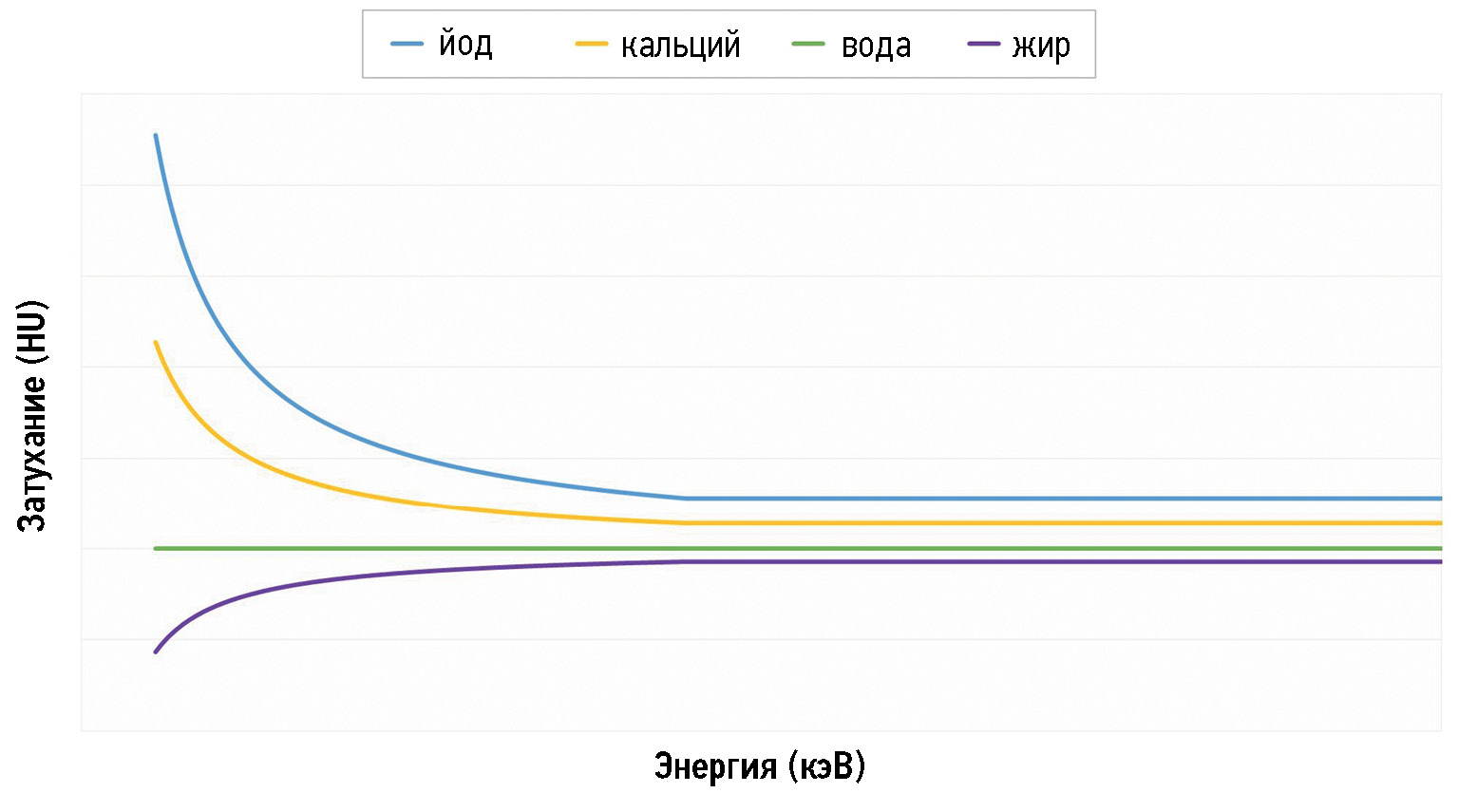
Nowadays, computed tomography urography and multiparametric magnetic resonance are the most often used imaging techniques in patients with bladder cancer; however, dual-energy computed tomography is making its way into the oncological field. This narrative review article aimed to show the fundamentals of dual-energy computed tomography by outlining its physical principles, techniques, protocols, and postprocessing images to help those who used this cutting-edge technology as the first approach to fully understand its emerging applications in the evaluation of bladder cancer, a field yet to be explored. In particular, we discuss the usefulness of dual-energy computed tomography by focusing on the main images obtained, such as the iodine map, comparing them to the images obtained with conventional computed tomography scans.
Dual-energy computed tomography applications may be beneficial for bladder cancer diagnosis, staging, and treatment planning. Nevertheless, its application is limited to its availability in healthcare structures and the training of healthcare personnel who can perform and interpret the scans correctly.
Full Text
##article.viewOnOriginalSite##About the authors
Federica Masino
Foggia University School of Medicine
Email: federicamasino@gmail.com
ORCID iD: 0009-0004-4289-3289
MD
Italy, FoggiaLaura Eusebi
“Carlo Urbani” Hospital
Email: lauraeu@virgilio.it
ORCID iD: 0000-0002-4172-5126
MD
Italy, JesiManuela Montatore
Foggia University School of Medicine
Email: manuela.montatore@unifg.it
ORCID iD: 0009-0002-1526-5047
MD
Italy, FoggiaGianmichele Muscatella
Foggia University School of Medicine
Email: muscatella94@gmail.com
ORCID iD: 0009-0004-3535-5802
MD
Italy, FoggiaRossella Gifuni
Foggia University School of Medicine
Email: rossella.gifuni@unifg.it
ORCID iD: 0009-0009-9679-3861
MD
Italy, FoggiaVincenzo Ferrara
“Carlo Urbani” Hospital
Email: vincenzoferrara4@gmail.com
ORCID iD: 0000-0001-8625-4308
MD
Italy, JesiMassimo Marcellini
“Senigallia” Hospital
Email: massimo.marcellini@sanita.marche.it
ORCID iD: 0000-0002-5281-7819
MD
Italy, SenigalliaGiuseppe Guglielmi
Foggia University School of Medicine; “Dimiccoli” Hospital; “IRCCS Casa Sollievo della Sofferenza” Hospital
Author for correspondence.
Email: giuseppe.guglielmi@unifg.it
ORCID iD: 0000-0002-4325-8330
MD, Professor
Italy, Foggia; Barletta; San Giovanni RotondoReferences
- Sung H, Ferlay J, Siegel RL, et al. Global Cancer Statistics 2020: GLOBOCAN Estimates of Incidence and Mortality Worldwide for 36 Cancers in 185 Countries. CA Cancer J Clin. 2021;71(3):209–249. doi: 10.3322/caac.21660
- Siegel RL, Miller KD, Jemal A. Cancer statistics, 2020. CA Cancer J Clin. 2020;70(1):7–30. doi: 10.3322/caac.21590
- Soria F, Shariat SF, Lerner SP, et al. Epidemiology, diagnosis, preoperative evaluation and prognostic assessment of upper-tract urothelial carcinoma (UTUC). World J Urol. 2017;35(3):379–387. doi: 10.1007/s00345-016-1928-x
- Verma S, Rajesh A, Prasad SR, et al. Urinary bladder cancer: role of MR imaging. Radiographics. 2012;32(2):371–387. doi: 10.1148/rg.322115125
- Rouprêt M, Babjuk M, Burger M, et al. European Association of Urology Guidelines on Upper Urinary Tract Urothelial Carcinoma: 2020 Update. Eur Urol. 2021;79(1):62–79. doi: 10.1016/j.eururo.2020.05.042
- Farling KB. Bladder cancer: Risk factors, diagnosis, and management. Nurse Pract. 2017;42(3):26–33. doi: 10.1097/01.NPR.0000512251.61454.5c
- Eusebi L, Masino F, Gifuni R, et al. Role of multiparametric-MRI in bladder cancer. Current Radiology Reports. 2023;11(5):69–80. doi: 10.1007/s40134-023-00412-5
- Babjuk M, Burger M, Capoun O, et al. European Association of Urology Guidelines on Non-muscle-invasive Bladder Cancer (Ta, T1, and Carcinoma in Situ). Eur Urol. 2022;81(1):75–94. doi: 10.1016/j.eururo.2021.08.010
- Babjuk M, Böhle A, Burger M, et al. EAU Guidelines on Non-Muscle-invasive Urothelial Carcinoma of the Bladder: Update 2016. Eur Urol. 2017;71(3):447–461. doi: 10.1016/j.eururo.2016.05.041
- Kamoun A, de Reyniès A, Allory Y, et al. A consensus molecular classification of muscle-invasive bladder cancer. European Urology. 2020;77(4):420–433. doi: 10.1016/j.eururo.2019.09.006
- Wang Z, Shang Y, Luan T, et al. Evaluation of the value of the VI-rads scoring system in assessing muscle infiltration by bladder cancer. Cancer Imaging. 2020;20(1). doi: 10.1186/s40644-020-00304-3
- Husband JE, Olliff JF, Williams MP, Heron CW, Cherryman GR. Bladder cancer: staging with CT and MR imaging. Radiology. 1989;173(2):435–440. doi: 10.1148/radiology.173.2.2798874
- Wong VK, Ganeshan D, Jensen CT, Devine CE. Imaging and management of Bladder Cancer. Cancers. 2021;13(6):1396. doi: 10.3390/cancers13061396
- Agrawal MD, Pinho DF, Kulkarni NM, et al. Oncologic applications of dual-energy CT in the abdomen. Radiographics. 2014;34(3):589–612. doi: 10.1148/rg.343135041
- Johnson T. Dual-Energy CT: General Principles. AJR Am J Roentgenol. 2012;199(5 Suppl):S3–S8. doi: 10.2214/AJR.12.9116
- Potenta SE, D’Agostino R, Sternberg KM, Tatsumi K, Perusse K. CT Urography for Evaluation of the Ureter. Radiogr Rev Publ Radiol Soc N Am Inc. 2015;35:709–726. doi: 10.1148/rg.2015140209
- Kawashima A, Vrtiska TJ, Leroy AJ, et al. CT Urography. Radiogr Rev Publ Radiol Soc N Am Inc. 2004;24:S35–S54. doi: 10.1148/rg.24si045513
- Caoili EM, Cohan RH. CT urography in evaluation of urothelial tumors of the kidney. Abdom. Radiol. 2016;41:1100–1107. doi: 10.1007/s00261-016-0695-x
- Cheng K, Cassidy F, Aganovic L, Taddonio M, Vahdat N. CT urography: How to optimize the technique. Abdom. Radiol. 2019;44:3786–3799. doi: 10.1007/s00261-019-02111-2
- Silverman SG, Leyendecker JR, Amis ES. What Is the Current Role of CT Urography and MR Urography in the Evaluation of the Urinary Tract? Radiology. 2009;250:309–323. doi: 10.1148/radiol.2502080534
- Shampain KL, Cohan RH, Caoili EM, Davenport MS, Ellis JH. Benign diseases of the urinary tract at CT and CT urography. Abdom. Radiol. 2019;44:3811–3826. doi: 10.1007/s00261-019-02108-x
- Metser U, Goldstein MA, Chawla TP, et al. Detection of urothelial tumors: Comparison of urothelial phase with excretory phase CT urography — a prospective study. Radiology. 2012;264(1):110–118. doi: 10.1148/radiol.12111623
- Kim JK, Park SY, Ahn HJ, Kim CS, Cho KS. Bladder cancer: analysis of multi-detector row helical CT enhancement pattern and accuracy in tumor detection and perivesical staging. Radiology. 2004;231(3):725–731. doi: 10.1148/radiol.2313021253
- Saksena MA, Dahl DM, Harisinghani MG. New imaging modalities in bladder cancer. World J Urol. 2006;24(5):473–480. doi: 10.1007/s00345-006-0118-7
- Parakh A, Lennartz S, An C, et al. Dual-Energy CT Images: Pearls and Pitfalls. Radiographics. 2021;41(1):98–119. doi: 10.1148/rg.2021200102
- Sauter A, Muenzel D, Dangelmaier J, et al. Dual-Layer Spectral Computed Tomography: Virtual Non-Contrast in Comparison to True Non-Contrast Images. Eur J Radiol. 2018;104:108–114. doi: 10.1016/j.ejrad.2018.05.007
- Ananthakrishnan L, Rajiah P, Ahn R, et al. Spectral Detector CT-Derived Virtual Non-Contrast Images: Comparison of Attenuation Values with Unenhanced CT. Abdom Radiol (NY). 2017;42(3):702–709. doi: 10.1007/s00261-016-1036-9
- Fornaro J, Leschka S, Hibbeln D, et al. Dual- and multi-energy CT: approach to functional imaging. Insights Imaging. 2011;2(2):149–159. doi: 10.1007/s13244-010-0057-0
- Silva AC, Morse BG, Hara AK, et al. Dual-energy (spectral) CT: applications in abdominal imaging. Radiographics. 2011;31(4):1031–1050. doi: 10.1148/rg.314105159
- Salameh JP, McInnes MDF, McGrath TA, Salameh G, Schieda N. Diagnostic Accuracy of Dual-Energy CT for Evaluation of Renal Masses: Systematic Review and Meta-Analysis. AJR Am J Roentgenol. 2019;212(4):W100–W105. doi: 10.2214/AJR.18.20527
- Ascenti G, Mazziotti S, Mileto A, et al. Dual-source dual-energy CT evaluation of complex cystic renal masses. AJR Am J Roentgenol. 2012;199(5):1026–1034. doi: 10.2214/AJR.11.7711
- Mileto A, Nelson RC, Samei E, et al. Impact of dual-energy multi-detector row CT with virtual monochromatic imaging on renal cyst pseudoenhancement: in vitro and in vivo study. Radiology. 2014;272(3):767–776. doi: 10.1148/radiol.14132856
- Chandarana H, Megibow AJ, Cohen BA, et al. Iodine quantification with dual-energy CT: phantom study and preliminary experience with renal masses. AJR Am J Roentgenol. 2011;196(6):W693–W700. doi: 10.2214/AJR.10.5541
- Tatsugami F, Higaki T, Nakamura Y, Honda Y, Awai K. Dual-energy CT: minimal essentials for radiologists. Jpn J Radiol. 2022;40(6):547–559. doi: 10.1007/s11604-021-01233-2
- Nolte-Ernsting C, Cowan N. Understanding multislice CT urography techniques: Many roads lead to Rome. Eur Radiol. 2006;16(12):2670–2686. doi: 10.1007/s00330-006-0386-z
- Ge X, Lan ZK, Chen J, Zhu SY. Effectiveness of contrast-enhanced ultrasound for detecting the staging and grading of bladder cancer: a systematic review and meta-analysis. Med Ultrason. 2021;23(1):29–35. doi: 10.11152/mu-2730
- Takahashi N, Vrtiska TJ, Kawashima A, et al. Detectability of urinary stones on virtual nonenhanced images generated at pyelographic-phase dual-energy CT. Radiology. 2010;256(1):184–190. doi: 10.1148/radiol.10091411
- Mangold S, Thomas C, Fenchel M, et al. Virtual nonenhanced dual-energy CT urography with tin-filter technology: determinants of detection of urinary calculi in the renal collecting system. Radiology. 2012;264(1):119–125. doi: 10.1148/radiol.12110851
- Coursey CA, Nelson RC, Boll DT, et al. Dual-energy multidetector CT: how does it work, what can it tell us, and when can we use it in abdominopelvic imaging? Radiographics. 2010;30(4):1037–1055. doi: 10.1148/rg.304095175
- Graser A, Johnson TR, Hecht EM, et al. Dual-energy CT in patients suspected of having renal masses: can virtual nonenhanced images replace true nonenhanced images? Radiology. 2009;252(2):433–440. doi: 10.1148/radiol.2522080557
- Nakagawa M, Naiki T, Naiki-Ito A, et al. Usefulness of advanced monoenergetic reconstruction technique in dual-energy computed tomography for detecting bladder cancer. Jpn J Radiol. 2022;40(2):177–183. doi: 10.1007/s11604-021-01195-5
- Patel BN, Vernuccio F, Meyer M, et al. Dual-Energy CT Material Density Iodine Quantification for Distinguishing Vascular From Nonvascular Renal Lesions: Normalization Reduces Intermanufacturer Threshold Variability. AJR Am J Roentgenol. 2019;212(2):366–376. doi: 10.2214/AJR.18.20115
- De Cecco CN, Darnell A, Rengo M, et al. Dual-energy CT: oncologic applications. AJR Am J Roentgenol. 2012;199(5S):S98–S105. doi: 10.2214/AJR.12.9207
- Vrtiska TJ, Takahashi N, Fletcher JG, et al. Genitourinary applications of dual-energy CT. AJR Am J Roentgenol. 2010;194(6):1434–1442. doi: 10.2214/AJR.10.4404
- Montatore M, Muscatella G, Eusebi L, et al. Current status on new technique and protocol in urinary stone disease. Current Radiology Reports. 2023;11:161–176. doi: 10.1007/s40134-023-00420-5
Supplementary files


