

")


Radiation methods in the diagnosis of primary and recurrent malignant ovarian struma: A case report
- Authors: Nudnov N.V.1, Ivashina S.V.1, Aksenova S.P.1
-
Affiliations:
- Russian Scientific Center of Roentgenoradiology
- Issue: Vol 4, No 2 (2023)
- Pages: 214-225
- Section: Case reports
- URL: https://journals.rcsi.science/DD/article/view/146887
- DOI: https://doi.org/10.17816/DD322846
- ID: 146887
Cite item

Abstract
We provide a rare clinical and diagnostic observation of primary and recurring malignant ovarian struma.
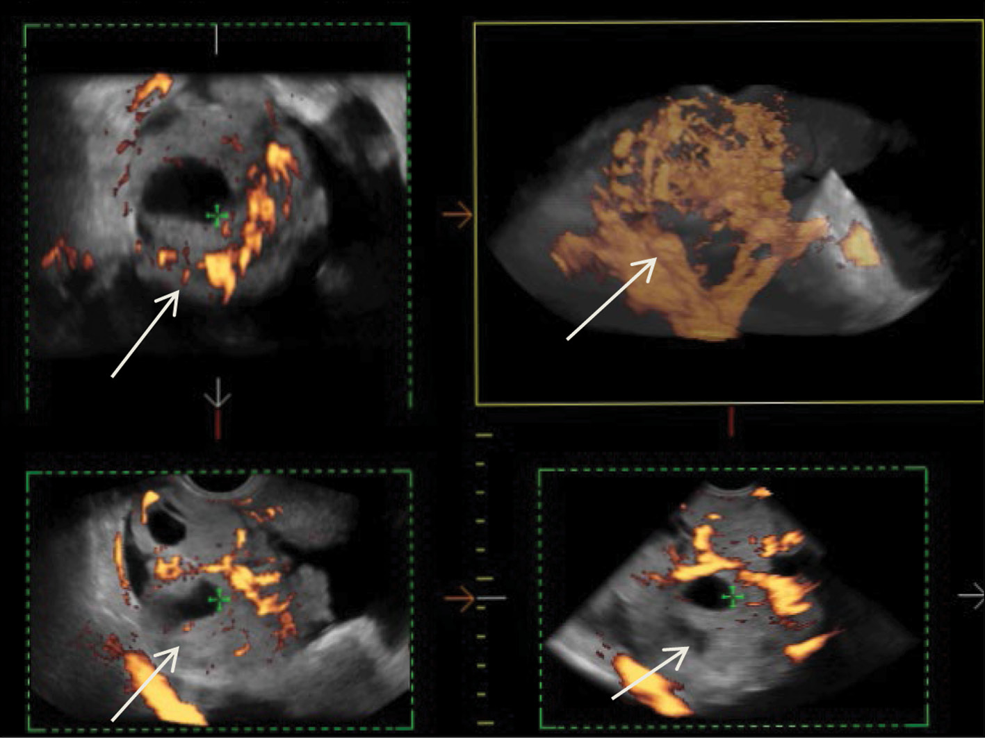
Malignant struma of the right ovary was detected 2 years after surgical treatment of primary benign struma of the left ovary. Six months later, the patient was diagnosed with a disease relapse, visualized exclusively according to radioisotope research methods. In the fourth year of anticancer treatment, ultrasonography revealed recurring foci along the peritoneum. According to the ultrasound data on the pelvic peritoneum and the projection of the removed right ovary, multiple solid nodes with high blood flow were visualized. Peak systolic velocity ranged from 2 to 9 cm/s in minor lesions from 4 to 12 mm, with an RI max of 0.53. For 4 years, the patient underwent radioiodine therapy with 131I with an activity of 6.0 GBq; the patient’s condition during the treatment was satisfactory.
Full Text
##article.viewOnOriginalSite##About the authors
Nikolai V. Nudnov
Russian Scientific Center of Roentgenoradiology
Email: nvnudnov@rncrr.ru
ORCID iD: 0000-0001-5994-0468
SPIN-code: 3018-2527
MD, Dr. Sci. (Med), Professor
Russian Federation, MoscowSvetlana V. Ivashina
Russian Scientific Center of Roentgenoradiology
Email: s.ivashina@bk.ru
ORCID iD: 0000-0002-9287-2636
SPIN-code: 7829-2899
MD, Cand. Sci. (Med), Senior Research Associate
Russian Federation, MoscowSvetlana P. Aksenova
Russian Scientific Center of Roentgenoradiology
Author for correspondence.
Email: fabella@mail.ru
ORCID iD: 0000-0003-2552-5754
SPIN-code: 4858-4627
MD, Cand. Sci. (Med), Research Associate
Russian Federation, MoscowReferences
- Female Genital Tumours. WHO Classification of Tumours, 5th Edition, vol. 4. WHO Classification of Tumours Editorial Board; 2020. Available from: https://publications.iarc.fr/Book-And-Report-Series/Who-Classification-Of-Tumours/Female-Genital-Tumours-2020. Accessed: 15.04.2023.
- Li S, Yang T, Li X. FIGO stage IV and age over 55 years as prognostic predicators in patients with metastatic malignant struma ovarii. Front Oncol. 2020;(10):1983. doi: 10.3389/fonc.2020.584917
- Roth LM, Karseladze AI. Highly differentiated follicular carcinoma arising from struma ovarii: A report of 3 cases, a review of the literature, and a reassessment of so-called peritoneal strumosis. Int J Gynecol Pathol. 2008;27(2):213–222. doi: 10.1097/PGP.0b013e318158e958
- Ayhan S, Kilic F, Ersak B, et al. Malignant struma ovarii: From case to analysis J Obstet Gynaecol Res. 2021;47(9):3339–3351. doi: 10.1111/jog.14902
- Kanasugi M, Nishiyama H, Sanpei M, et al. Ovarian strumal carcinoid: A case report. Fukushima J Med Sci. 2023;69(1):51–55. doi: 10.5387/fms.2022-22
- Smith LP, Brubaker LW, Wolsky RJ. It does exist! Diagnosis and management of thyroid carcinomas originating in struma ovarii. Surg Pathol Clin. 2023;16(1):75–86. doi: 10.1016/j.path.2022.09.008
- Yamauchi S, Kokabu T, Kataoka H, et al. Computed tomography, magnetic resonance imaging, and positron emission tomography/computed tomography findings for the diagnosis of malignant struma ovarii: A case report. J Obstet Gynaecol Res. 2023;49(5):1456–1461. doi: 10.1111/jog.15619
- Yazawa R, Yazawa H, Fukuda K, Ohara M. Struma ovarii with massive ascites mimicking ovarian carcinoma treated with conservative laparoscopic surgery: A case report. Fukushima J Med Sci. 2023;69(1):37–43. doi: 10.5387/fms.2022-30
- Shou L, Lu J, Yang J, et al. Follicular carcinoma originating from struma ovarii: A case report. Medicine (Baltimore). 2023;102(1):e32658. doi: 10.1097/MD.0000000000032658
- Elshafie O, Hussein S, Al Kalbani M, et al. Papillary follicular variant thyroid cancer in a malignant struma ovarii: A report of a rare case. Endocrinol Diabetes Metab Case Rep. 2022;2022:21-0169. doi: 10.1530/EDM-21-0169
- Antonova IB, Fomin DK, Babaeva NA, et al. Malignant ovarian stroma. Literature review and own observation of a rare variant of the tumor. Difficult Patient. 2018;16(8-9):16–18. (In Russ).
- Giovannopoulou E, Saliaris K, Kavoura E, et al. Highly differentiated follicular carcinoma of ovarian origin: A systematic review of the literature. Curr Oncol. 2022;29(12):9105–9116. doi: 10.3390/curroncol29120712
- ResearchGate GmbH [Internet]. Alt C, Bharwani N, Brunesch L, et al.; ESUR Female Pelvis Imaging Working Group. Esur quick guide to female pelvis imaging [cite July 2019]. Available from: https://www.esur.org/fileadmin/content/2019/ESUR_2019_ESUR_Quick_Guide_to_Female_Pelvis_Imaging.pdf. Accessed: 15.04.2023.
- Gil R, Cunha TM, Rolim I. Mature cystic teratoma with high proportion of solid thyroid tissue: A controversial case with unusual imaging findings. J Radiol Case Rep. 2017;11(7):20–30. doi: 10.3941/jrcr.v11i7.2853
- Ozerskaya IA, Chekalova MA, Ivanov VA, Kazaryan GG. Ultrasound signs of ovarian tumors according to a standardized protocol. Medical Imaging. 2023;27(2):110–124. (In Russ). doi: 10.24835/1607-0763-1144
- Fujiwara S, Tsuyoshi H, Nishimura T, et al. Precise preoperative diagnosis of struma ovarii with pseud-Meigs’ syndrome mimicking ovarian cancer with the combination of 131I scintigraphy and 18F-FDG PET: Case report and review of the literature. J Ovarian Res. 2018.11(1):11. doi: 10.1186/s13048-018-0383-2
- Savelli L, Testa AC, Timmerman D, et al. Imaging of gynecologic disease (4): Clinical and ultrasound characteristics of struma ovarii. Ultrasound Obstet Gynecol. 2008;32(2):210–219. doi: 10.1002/uog.5396
- Tamura N, Murakami K, Ozaki R, et al. Current state of management of struma ovarii and preoperative imaging features: A retrospective case series study of 18 patients at a single institution. J Obstet Gynaecol Res. 2023;49(3):1007–1011. doi: 10.1111/jog.15545
- Ranade R, Rachh S, Basu S. Late Manifestation of struma peritonei and widespread functioning lesions in the setting of struma ovarii simulating highly differentiated follicular carcinoma. J Nucl Med Technol. 2015;43(3):231–233. doi: 10.2967/jnmt.114.149294
- Brogsitter C, Wonsak A, Würl K, Kotzerke J. Peritoneal strumosis. Eur J Nucl Med Mol Imaging. 2004;31(7):1057. doi: 10.1007/s00259-004-1548-3
Supplementary files


