

")


Optimized biparametric magnetic resonance imaging protocol for prostate cancer detection
- Authors: Abuladze L.R.1, Semenov D.S.1, Panina O.Y.1,2,3, Vasilev Y.A.1
-
Affiliations:
- Research and Practical Clinical Center for Diagnostics and Telemedicine Technologies
- City Clinical Oncological Hospital No. 1
- Moscow State University of Medicine and Dentistry named after A.I. Evdokimov
- Issue: Vol 3, No 3 (2022)
- Pages: 166-177
- Section: Technical Reports
- URL: https://journals.rcsi.science/DD/article/view/108484
- DOI: https://doi.org/10.17816/DD108484
- ID: 108484
Cite item

Abstract
BACKGROUND: Prostate cancer is one of the most commonly diagnosed cancers in men worldwide. PI-RADS v2.1 contains the requirements for the magnetic resonance imaging protocol, which cannot be fully implemented on a significant component of functioning scanners. Consequently, magnetic resonance imaging approaches vary in different medical organizations and often do not allow for a qualitative interpretation of images and diagnosis of the target pathology.
AIM: To develop a biparametric magnetic resonance imaging protocol optimized for the existing magnetic resonance imaging scanners for the diagnosis of prostate cancer and to allow the screening and detection of neoplasms as early as possible. Simultaneously, the protocol should fulfill the current PI-RADS v2.1 recommendations to the maximum possible extent and meet the requirements of effective workflow in the radiology department.
MATERIALS AND METHODS: Preliminary analysis of prostate magnetic resonance imaging scanning in medical organizations of the Moscow Health Care Department showed the absence of a unified approach. Using the iterative adjustment of scanning parameters, we adjusted the protocol to ensure acceptable quality with maximum available compliance with PI-RADS v2.1.
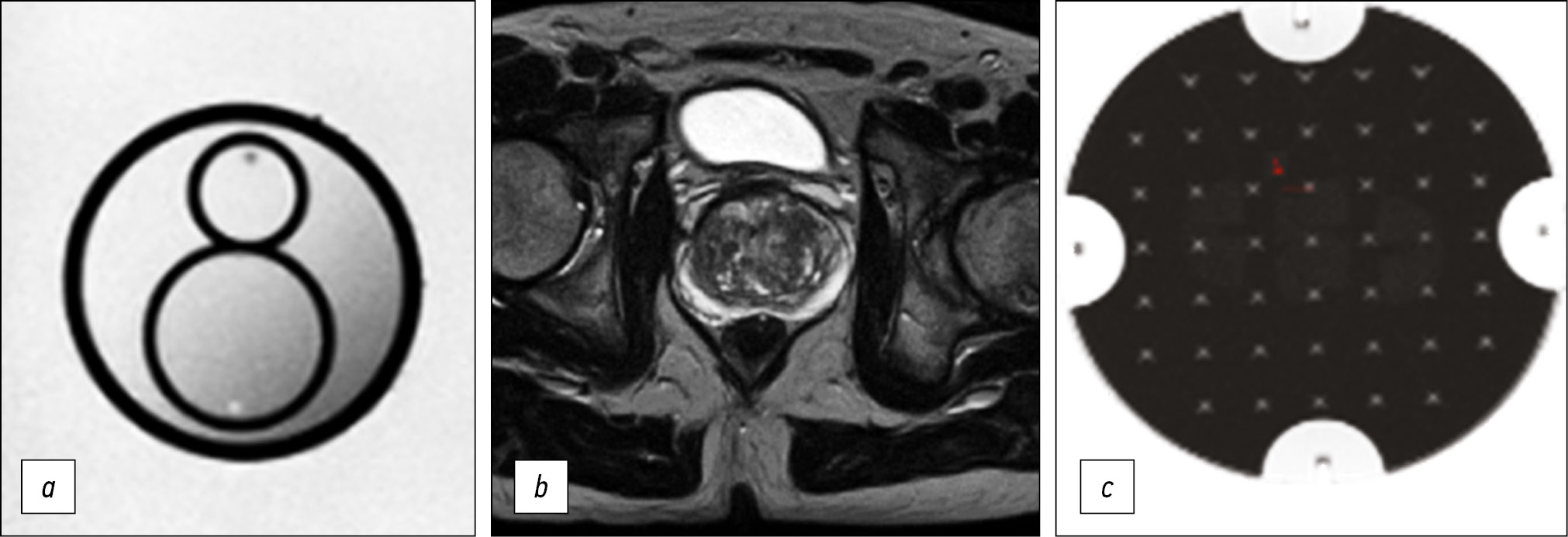
To quantify the quality of the images, we used the magnetic resonance imaging phantom recommended by the American College of Radiology.
RESULTS: The biparametric protocol was developed for Excelart Vantage 1.5 T, including T2-weighted images in three planes and diffusion-weighted images, which took less than 11 min. Moreover, the image quality parameters (intensity inhomogeneity, nonlinearity, resolution, and slice thickness) were within the acceptable ranges recommended by the magnetic resonance imaging manufacturer.
CONCLUSION: The prostate may be effectively evaluated using the proposed magnetic resonance imaging protocol. Introducing it into practice could have a significant impact on the detection of prostate cancer in men. The entire duration of the protocol provides a possibility to supplement it with any sequences, depending on the final purpose of investigation.
Full Text
##article.viewOnOriginalSite##About the authors
Liya R. Abuladze
Research and Practical Clinical Center for Diagnostics and Telemedicine Technologies
Email: l.abuladze@npcmr.ru
ORCID iD: 0000-0001-6745-1672
SPIN-code: 5640-9989
Russian Federation, Moscow
Dmitriy S. Semenov
Research and Practical Clinical Center for Diagnostics and Telemedicine Technologies
Email: d.semenov@npcmr.ru
ORCID iD: 0000-0002-4293-2514
SPIN-code: 2278-7290
Scopus Author ID: 57213154475
ResearcherId: P-5228-2017
Researcher of the Department of Innovative Technologies
Russian Federation, MoscowOlga Y. Panina
Research and Practical Clinical Center for Diagnostics and Telemedicine Technologies; City Clinical Oncological Hospital No. 1; Moscow State University of Medicine and Dentistry named after A.I. Evdokimov
Email: olgayurpanina@gmail.com
ORCID iD: 0000-0002-8684-775X
SPIN-code: 5504-8136
ResearcherId: AAG-6447-2020
Russian Federation, Moscow; Moscow; Moscow
Yuriy A. Vasilev
Research and Practical Clinical Center for Diagnostics and Telemedicine Technologies
Author for correspondence.
Email: dr.vasilev@me.com
ORCID iD: 0000-0002-0208-5218
SPIN-code: 4458-5608
Russian Federation, Moscow
References
- Sung H, Ferlay J, Siegel RL, et al. Global cancer statistics 2020: GLOBOCAN estimates of incidence and mortality worldwide for 36 cancers in 185 countries. CA Cancer J Clin. 2021;71:209–49. doi: 10.3322/caac.21660
- Barentsz JO, Richenberg J, Clements R, et al. ESUR prostate MR guidelines 2012. Eur Radiol. 2012;22(4):746–757. doi: 10.1007/s00330-011-2377-y
- Weinreb JC, Barentsz JO, Choyke PL, et al. PI-RADS prostate imaging ― reporting and data system: 2015, version 2. Eur Urol. 2016;69(1):16–40. doi: 10.1016/j.eururo.2015.08.052
- Park SY, Jung DC, Oh YT, et al. Prostate cancer: PI-RADS version 2 helps preoperatively predict clinically significant cancers. Radiology. 2016;280(1):108–116. doi: 10.1148/radiol.16151133.
- Israël B, van der Leest M, Sedelaar M, et al. Multiparametric magnetic resonance imaging for the detection of clinically significant prostate cancer: what urologists need to know. part 2: interpretation. Eur Urol. 2020;77(4):469–480. doi: 10.1016/j.eururo.2019.10.024
- Tamada T, Kido A, Yamamoto A, et al. Comparison of biparametric and multiparametric mri for clinically significant prostate cancer detection with pi-rads version 2.1. J Magn Reson Imaging. 2021;53(1):283–291. doi: 10.1002/jmri.27283
- Patent RUS 208239 U1. Semenov DS, Petryaykin AV, Vasiliev YuA, et al. Phantom device for configuring protocols of magnetic resonance imaging of the prostate gland in patients with metal structures of the hip joint. (In Russ). Available from: https://www.elibrary.ru/item.asp?id=47429681. Accessed: 15.03.2022.
- Engels RR, Israël B, Padhani AR, et al. Multiparametric magnetic resonance imaging for the detection of clinically significant prostate cancer: what urologists need to know. Part 1: acquisition. Eur Urol. 2020;77(4):457–468. doi: 10.1016/j.eururo.2019.09.021
- Methodology for monitoring the parameters and characteristics of magnetic resonance tomographs under operating conditions Methodological recommendations No. 17 (approved 10.09.2011). (In Russ). Available from: https://docs.cntd.ru/document/456079947. Accessed: 15.03.2022.
- Siegel RL, Miller KD, Fuchs HE, et al. Cancer Statistics, 2021. CA Cancer J Clin. 2021;71(1):7–33. doi: 10.3322/caac.21654
- Ferlay J, Colombet M, Soerjomataram I, et al. Cancer incidence and mortality patterns in Europe: Estimates for 40 countries and 25 major cancers in 2018. Eur J Cancer. 2018;103:356–387. doi: 10.1016/j.ejca.2018.07.005
- Malignant neoplasms in Russia in 2020 (morbidity and mortality). Ed. by A.D. Kaprin, V.V. Starinsky, A.O. Shakhzadova. Moscow; 2021. 252 p. (In Russ).
- Patel AR, Klein EA. Risk factors for prostate cancer. Nat Clin Pract Urol. 2009;6(2):87–95. doi: 10.1038/ncpuro1290
- Sherrer RL, Glaser ZA, Gordetsky JB, et al. Comparison of biparametric MRI to full multiparametric MRI for detection of clinically significant prostate cancer. Prostate Cancer Prostatic Dis. 2019;22(2):331–336. doi: 10.1038/s41391-018-0107-0
- Zawaideh JP, Sala E, Shaida N, et al. Diagnostic accuracy of biparametric versus multiparametric prostate MRI: assessment of contrast benefit in clinical practice. Eur Radiol. 2020;30(7):4039–4049. doi: 10.1007/s00330-020-06782-0
- Van der Leest M, Israël B, Cornel EB, et al. High diagnostic performance of short magnetic resonance imaging protocols for prostate cancer detection in biopsy-naïve men: the next step in magnetic resonance imaging accessibility. Eur Urol. 2019;76(5):574–581. doi: 10.1016/j.eururo.2019.05.029
- Stanzione A, Ponsiglione A, Cuocolo R, et al. Abbreviated protocols versus multiparametric mri for assessment of extraprostatic extension in prostatic carcinoma: A multireader study. Anticancer Res. 2019;39(8):4449–4454. doi: 10.21873/anticanres.13617
- Gelezhe PB, Blokhin IA, Semenov SS, et al. Radiomics of magnetic resonance imaging in prostate cancer: what is currently known? Digital Diagnostics. 2021;2(4):441–452. (In Russ).
Supplementary files


