


Photo gallery: necrobiosis lipoidica and granuloma annulare
- Authors: Olisova O.Y.1, Teplyuk N.P.1, Rogozina V.A.1
-
Affiliations:
- The First Sechenov Moscow State Medical University
- Issue: Vol 28, No 5 (2025)
- Pages: 633-638
- Section: PHOTO GALLERY
- URL: https://journals.rcsi.science/1560-9588/article/view/359059
- DOI: https://doi.org/10.17816/dv692131
- EDN: https://elibrary.ru/ZPHOMJ
- ID: 359059
Cite item

Abstract
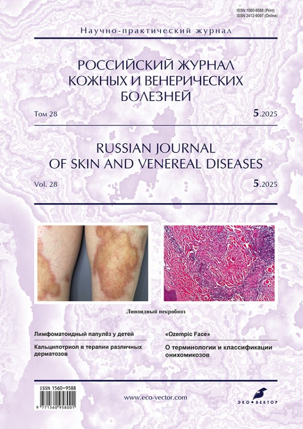
Necrobiosis lipoidica and granuloma annulare are rare chronic, non-infectious (inflammatory) skin diseases.
Necrobiosis lipoidica is characterized by bright-pink papules and plaques that transform into red or yellowish ring-shaped plaques and may be accompanied by telangiectasis, central atrophy, induration, and ulceration. Granuloma annulare is distinguished by isolated ring-shaped plaques or disseminated skin lesions with papules, patches, and bright-pink ring-shaped foci.
Necrobiosis lipoidica and granuloma annulare are common in patients with comorbidities such as type 1 and 2 diabetes mellitus, autosomal dominant maturity-onset diabetes of the young (MODY), endocrinopathy (thyroid and adrenal gland diseases), and chronic upper respiratory tract infections. The high incidence of comorbidities necessitates a multidisciplinary approach to the treatment of these conditions.
Necrobiosis lipoidica and granuloma annulare are treated using topical, systemic, and physical therapy modalities. Topical treatment includes topical glucocorticoids and calcineurin inhibitors. Systemic glucocorticoids are used for systemic therapy. Furthermore, the use of chloroquine, hydroxychloroquine, fumarates, and genetically engineered biological drugs has been reported. Physical therapy modalities include ultraviolet A and B radiation (UVA, UVB), PUVA therapy (UVA + photosensitizer), CO2 lasers, dye lasers, and intense pulsed light (IPL) therapy. However, none of the available therapeutic options show absolute efficacy, and long-term remission is extremely rare.
This photo gallery illustrates the heterogeneity of necrobiosis lipoidica and granuloma annulare symptoms to facilitate timely diagnosis and optimal treatment.
Keywords
Full Text
##article.viewOnOriginalSite##About the authors
Olga Y. Olisova
The First Sechenov Moscow State Medical University
Author for correspondence.
Email: olisovaolga@mail.ru
ORCID iD: 0000-0003-2482-1754
SPIN-code: 2500-7989
MD, Dr. Sci. (Medicine), Professor, Сorresponding Member of the Russian Academy of Sciences
Russian Federation, MoscowNatalia P. Teplyuk
The First Sechenov Moscow State Medical University
Email: Teplyukn@gmail.com
ORCID iD: 0000-0002-5800-4800
SPIN-code: 8013-3256
MD, Dr. Sci. (Medicine), Professor
Russian Federation, MoscowVarvara A. Rogozina
The First Sechenov Moscow State Medical University
Email: varvara.rgzn@gmail.com
ORCID iD: 0000-0002-5471-6130
SPIN-code: 3505-7408
Russian Federation, Moscow
References
Supplementary files


