


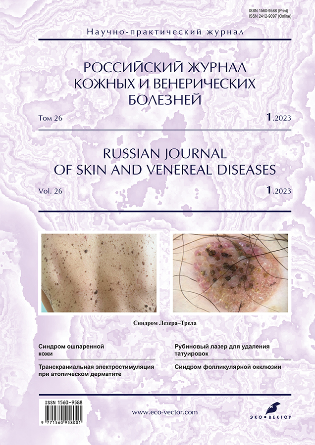
Синдром Лезера–Трела на приёме врача-косметолога
- Авторы: Ромашкина А.С.1, Снарская Е.С.2, Шпак А.М.1
-
Учреждения:
- Клиника дерматологии и косметологии Chistotel
- Первый Московский государственный медицинский университет имени И.М. Сеченова (Сеченовский Университет)
- Выпуск: Том 26, № 1 (2023)
- Страницы: 5-12
- Раздел: ДЕРМАТООНКОЛОГИЯ
- URL: https://journals.rcsi.science/1560-9588/article/view/132590
- DOI: https://doi.org/10.17816/dv119853
- ID: 132590
Цитировать

Аннотация
Себорейные кератомы нередко встречаются в практике врачей дерматологов и косметологов, однако резкое появление, быстрый рост и множественный характер высыпаний может служить маркером злокачественных новообразований.
Синдром Лезера–Трела (эруптивный себорейный кератоз) ― факультативный паранеопластический дерматоз, который характеризуется внезапным появлением на коже себорейных кератом и прогрессирующим увеличением их количества. Заболевание встречается с одинаковой частотой как у мужчин, так и у женщин в возрасте от 40 лет и старше. Этиология и патогенез не изучены подробно: имеются предположения, что развитие синдрома может быть связано со стимуляцией секреции эпидермального фактора роста пулом опухолевых клеток, что приводит к активации кератиноцитов. Манифестация синдрома сочетается с новообразованиями внутренних органов.
Клиническая картина синдрома Лезера–Трела характеризуется внезапным появлением на кожном покрове множества себорейных кератом с типичными клиническими и гистологическими признаками. Наиболее распространённой локализацией множественных очагов себорейного кератоза является кожа спины и груди, конечностей, лица, живота, шеи, подмышечных впадин и паховых складок.
Лечение проводится одновременно с установлением и терапией основного онкологического заболевания и заключается в удалении самых крупных кератом деструктивными методами, в частности путём хирургического иссечения, с помощью радиоволнового метода, криодеструкции, электрокоагуляции. Прогноз благоприятный при раннем обнаружении паранеопластического процесса.
В статье описаны клинические случаи множественного себорейного кератоза, а также тактика обследования данной группы пациентов. Описание случаев преследует цели онконастороженности врачей в отношении данного заболевания и своевременной диагностики злокачественного образования.
Полный текст
Открыть статью на сайте журналаОб авторах
Анастасия Сергеевна Ромашкина
Клиника дерматологии и косметологии Chistotel
Email: RomashkinaAS@mail.ru
ORCID iD: 0000-0002-6775-9797
SPIN-код: 1818-2132
к.м.н.
Россия, 121351, Москва, ул. Партизанская, д. 24Елена Сергеевна Снарская
Первый Московский государственный медицинский университет имени И.М. Сеченова (Сеченовский Университет)
Email: snarskaya-doc@mail.ru
ORCID iD: 0000-0002-7968-7663
SPIN-код: 3785-7859
д.м.н., профессор
Россия, МоскваАнна Михайловна Шпак
Клиника дерматологии и косметологии Chistotel
Автор, ответственный за переписку.
Email: shpak-anya-2011@yandex.ru
ORCID iD: 0000-0002-7395-5305
врач-дерматовенеролог, косметолог
Россия, 121351, Москва, ул. Партизанская, д. 24Список литературы
- Олисова О.Ю., Теплюк Н.П., Грабовская О.В., и др. Кератодермии как паранеопластический синдром // РМЖ. Дерматология. Косметология и пластическая хирургия. 2016. № 10. С. 654–656.
- Теплюк Н.П., Орлова Е.В., Новоселова С.В., и др. Кератодермия как проявление паранеоплазии // Российский журнал кожных и венерических болезней. 2012. № 5. С. 16–19.
- Miyashiro D., Sanches J.A. Paraneoplastic skin disorders: A review // G Ital Dermatol Venereol. 2016. Vol. 151, N 1. Р. 55–76.
- Didona D., Fania L., Didona B., et al. Paraneoplastic dermatoses: A brief general review and an extensive analysis of paraneoplastic pemphigus and paraneoplastic dermatomyositis // Int J Mol Sci. 2020. Vol. 21, N 6. Р. 2178. doi: 10.3390/ijms21062178
- Bernett C.N., Schmieder G.J. Leser trelat sign. In: StatPearls. Treasure Island (FL): StatPearls Publishing, 2022.
- Балуцкий В.В., Виноградов И.А. Синдром Лезера–Трела при раке ободочной кишки: собственное наблюдение // Consilium Medicum. 2020. Т. 22, № 8. С. 90–92. doi: 10.26442/20751753.2020.8.200026
- Гайдина Т.А., Дворников А.С., Скрипкина П.А. Паранеопластический синдром Лезера–Трела: клинические проявления, диагностика, лечение // Архив внутренней медицины. 2022. Т. 12, № 5. С. 325–329. doi: 10.20514/2226-6704-2022-12-5-325-329
- Alsaif F., Alkhayal F.A., Aldahash R., Alhumaidi A. Leser–Trélat sign presenting in a patient with relapsing mycosis fungoides // Case Rep Oncol. 2018. Vol. 11, N 2. Р. 436–441. doi: 10.1159/000490527
- Gori N., Esposito I., Del Regno L., et al. Leser–Trélat sign as a rare manifestation of cutaneous melanoma // Dermatol Reports. 2020. Vol. 12, N 1. Р. 8665. doi: 10.4081/dr.2020.8665
- Al Ghazal P., Körber A., Klode J., Dissemond J. Leser–Trélat sign and breast cancer // Lancet. 2013. Vol. 381, N 9878. Р. 1653. doi: 10.1016/S0140-6736(12)61257-4
- Bölke E., Gerber P.A., Peiper M., et al. Leser–Trélat sign presenting in a patient with ovarian cancer: A case report // J Med Case Rep. 2009. N 3. Р. 8583. doi: 10.4076/1752-1947-3-8583
- Wang N., Yu P.J., Liu Z.L., et al. Malignant acanthosis nigricans with Leser–Trélat sign and tripe palms: A case report // World J Clin Cases. 2020. Vol. 8, N 22. Р. 5632–5638. doi: 10.12998/wjcc.v8.i22.5632
- Khoschbin T., Löser C., Dippel E. [Paraneoplastic skin diseases. (In German).] // Internist (Berl). 2019. Vol. 60, N 8. Р. 775–782. doi: 10.1007/s00108-019-0636-1
- López J.D., García G.E., Ruiz R.J. Leser–Trelat sign, a useful predictor of neoplasms in primary attention // Semergen. 2021. Vol. 47, N 6. Р. e45–e46. doi: 10.1016/j.semerg.2021.02.002
- Wick M.R., Patterson J.W. Cutaneous paraneoplastic syndromes // Semin Diagn Pathol. 2019. Vol. 36, N 4. Р. 211–228. doi: 10.1053/j.semdp.2019.01.001
Дополнительные файлы


