


Leser–Trelat sign in cosmetologist’s practice
- 作者: Romashkina A.S.1, Snarskaya E.S.2, Shpak A.M.1
-
隶属关系:
- Clinic of dermatology and cosmetology Chistotel
- The First Sechenov Moscow State Medical University (Sechenov University)
- 期: 卷 26, 编号 1 (2023)
- 页面: 5-12
- 栏目: DERMATO-ONCOLOGY
- URL: https://journals.rcsi.science/1560-9588/article/view/132590
- DOI: https://doi.org/10.17816/dv119853
- ID: 132590
如何引用文章

详细
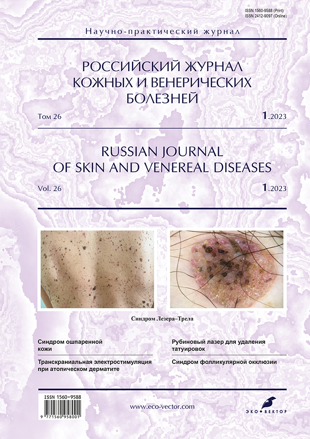
Leser–Trelat syndrome (eruptive seborrheic keratosis) is an unusial paraneoplastic dermatosis characterized by sudden appearance of seborrheic keratomas on the skin and a progressive increase of their number. This condition was first described in 1901 by the German surgeon E. Leser and the French surgeon U. Trelat. It occurs with the same frequency in both men and women aged 40+. The etiology and pathogenesis have not been studied in detail, however, there is evidence that the development of the syndrome may be associated with stimulation of the epidermal growth factor, which leads to stimulation of keratinocytes. The manifestation of the syndrome is usually start at the same time with cancer development, mostly it can be combained with adenocarcinoma of the gastrointestinal tract (47.7%), with intestinal carcinoma (32%), with lymphoproliferative diseases (21%), less often with malignant neoplasms of the lungs, breast, prostate.
The clinical picture of the Leser–Trelat sign is characterized by the sudden appearance оf seborrheic keratomas, which have typical clinical and histological signs. The most typical localization of seborrheic keratosis are back and chest (76%), limbs (18%), face (21%), abdomen (15%), neck (13%), armpits (6%), inguinal folds (3%). Keratosis can appear rapidly, over several months or even weeks. The rapid appearance of multiple seborheic keratomas may precede or develop with the oncological process in the internal organs.
Treatment is carried out at the same time with the establishment and treatment of the underlying disease and consists in removing the largest keratomas by destructive methods (surgical excision, radiowave method, cryodestruction, electrocoagulation).
The prognosis is favorable if paraneoplastic process was early detected.
The article describes clinical cases of multiple seborrheic keratosis and the tactics of examining this group of patients.
作者简介
Anastasia Romashkina
Clinic of dermatology and cosmetology Chistotel
Email: RomashkinaAS@mail.ru
ORCID iD: 0000-0002-6775-9797
SPIN 代码: 1818-2132
MD, Cand. Sci. (Med.)
俄罗斯联邦, 24 Partizanskaya street, 121351 MoscowElena Snarskaya
The First Sechenov Moscow State Medical University (Sechenov University)
Email: snarskaya-doc@mail.ru
ORCID iD: 0000-0002-7968-7663
SPIN 代码: 3785-7859
MD, Dr. Sci. (Med.), Professor
俄罗斯联邦, MoscowAnna Shpak
Clinic of dermatology and cosmetology Chistotel
编辑信件的主要联系方式.
Email: shpak-anya-2011@yandex.ru
ORCID iD: 0000-0002-7395-5305
MD俄罗斯联邦, 24 Partizanskaya street, 121351 Moscow
参考
- Olisova OYu, Teplyuk NP, Grabovskaya OV, et al. Keratodermia as paraneoplastic syndrome. Russkii meditsinskii zhurnal. Dermatologiya. Kosmetologiya i plasticheskaya khirurgiya. 2016;(10):654–656. (In Russ).
- Teplyuk NP, Orlova EV, Novoselova SV, et al. Keratoderma as a manifestation of paraneoplasia. Russ J Skin Veneral Diseases. 2012;(5):16–19. (In Russ).
- Miyashiro D, Sanches JA. Paraneoplastic skin disorders: A review. G Ital Dermatol Venereol. 2016;151(1):55–76.
- Didona D, Fania L, Didona B, et al. Paraneoplastic dermatoses: A brief general review and an extensive analysis of paraneoplastic pemphigus and paraneoplastic dermatomyositis. Int J Mol Sci. 2020;21(6):2178 doi: 10.3390/ijms21062178
- Bernett CN, Schmieder GJ. Leser trelat sign. In: StatPearls. Treasure Island (FL): StatPearls Publishing; 2022.
- Balutsky VV, Vinogradov IA. Leser–Trélat sign in patients with colon cancer: Clinical case. Consilium Medicum. 2020;22(8):90–92. (In Russ). doi: 10.26442/20751753.2020.8.200026
- Gaydina TA, Dvornikov AS, Skripkina PA. Paraneoplastic Leser–Trélat syndrome: Clinical manifestations, diagnosis and treatment. Russ Arch Int Med. 2022;12(5):325–329. (In Russ). doi: 10.20514/2226-6704-2022-12-5-325-329
- Alsaif F, Alkhayal FA, Aldahash R, Alhumaidi A. Leser–Trélat sign presenting in a patient with relapsing mycosis fungoides. Case Rep Oncol. 2018;11(2):436–441. doi: 10.1159/000490527
- Gori N, Esposito I, Del Regno L, et al. Leser–Trélat sign as a rare manifestation of cutaneous melanoma. Dermatol Reports. 2020;12(1):8665. doi: 10.4081/dr.2020.8665
- Al Ghazal P, Körber A, Klode J, Dissemond J. Leser-Trélat sign and breast cancer. Lancet. 2013;381(9878):1653. doi: 10.1016/S0140-6736(12)61257-4
- Bölke E, Gerber PA, Peiper M, et al. Leser–Trélat sign presenting in a patient with ovarian cancer: A case report. J Med Case Rep. 2009;(3):8583. doi: 10.4076/1752-1947-3-8583
- Wang N, Yu PJ, Liu ZL, et al. Malignant acanthosis nigricans with Leser–Trélat sign and tripe palms: A case report. World J Clin Cases. 2020;8(22):5632–5638. doi: 10.12998/wjcc.v8.i22.5632
- Khoschbin T, Löser C, Dippel E. [Paraneoplastic skin diseases. (In German).] Internist (Berl). 2019;60(8):775–782. doi: 10.1007/s00108-019-0636-1
- López JD, García GE, Ruiz RJ. Leser–Trelat sign, a useful predictor of neoplasms in primary attention. Semergen. 2021;47(6):e45–e46. doi: 10.1016/j.semerg.2021.02.002
- Wick MR, Patterson JW. Cutaneous paraneoplastic syndromes. Semin Diagn Pathol. 2019;36(4):211–228. doi: 10.1053/j.semdp.2019.01.001
补充文件


