

")


无对比剂磁共振灌注与相位对比血管成像在脑血流定量评估中的比较:一项前瞻性横断研究
- 作者: Popov V.V.1,2, Stankevich Y.A.1,2, Bogomyakova O.B.1,2, Tulupov A.A.1,2
-
隶属关系:
- International Tomography Institute, Siberian Branch of the Russian Academy of Sciences
- Novosibirsk State University
- 期: 卷 6, 编号 2 (2025)
- 页面: 203-213
- 栏目: 原创性科研成果
- URL: https://journals.rcsi.science/DD/article/view/310210
- DOI: https://doi.org/10.17816/DD636690
- EDN: https://elibrary.ru/MHMUYW
- ID: 310210
如何引用文章
全文:

详细
论证。如何验证通过无对比剂磁共振成像测量的脑血流定量评估结果仍是未解决的问题。最优策略是采用基于另一种生理模型的方法,以提高所得数据的可靠性。
目的:在健康成人中,根据2D相位对比血管成像结果,验证无对比剂磁共振成像对脑组织血流的定量评估结果。
方法。前瞻性纳入18–75岁健康成年人。采用无对比剂磁共振成像评估脑组织灌注,主干血流通过定量2D相位对比血管造影测量,涵盖椎动脉与颈内动脉。脑体积与相对质量由T1加权图像分割获得。主干血流量的数值通过数学转换,并结合脑总质量,换算为脑组织灌注指标。
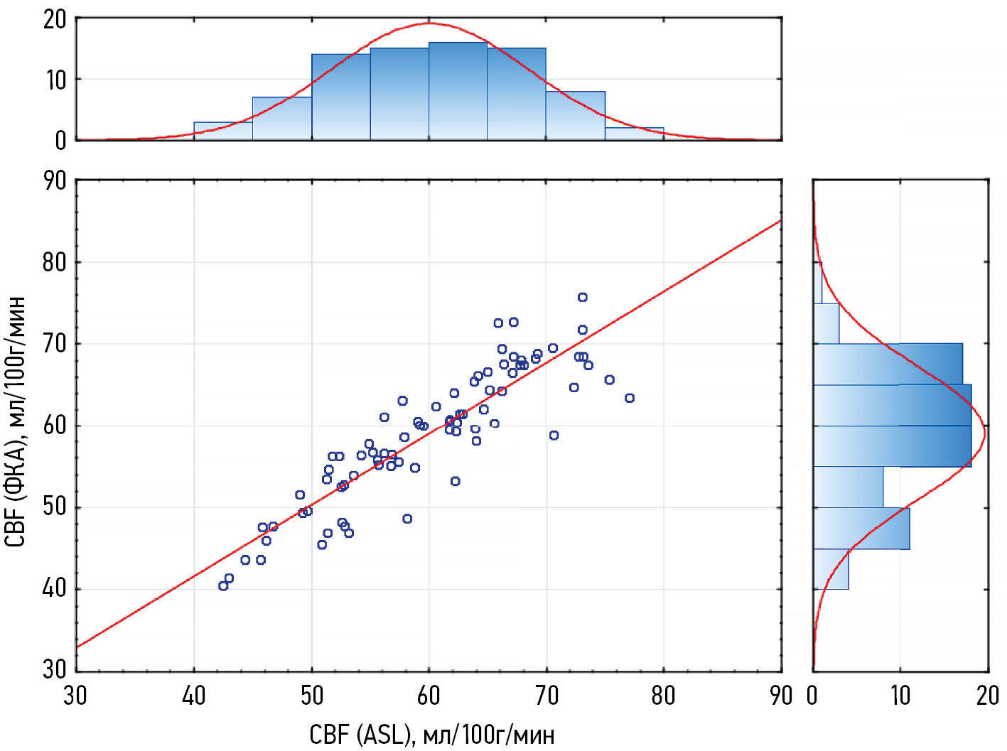
结果。使用两种方法对80名健康成人进行了检查。无对比剂磁共振成像测得白质和灰质平均灌注分别为17.88±2.39和42.06±7.13 ml/100g/min,总脑灌注为59.63±8.56 ml/100g/min。根据相位对比血管成像数据及动脉血流容积速度数值计算得出的总脑灌注为58.96±8.16 ml/s。基于无对比剂磁共振成像与相位对比血管成像所得数据计算的总脑灌注值之间存在显著的强正相关(r=0.892,p<0.001)。
结论。基于不同生理模型的无对比剂磁共振成像与相位对比血管成像所得脑灌注值之间显示出高度正相关。
作者简介
Vladimir V. Popov
International Tomography Institute, Siberian Branch of the Russian Academy of Sciences; Novosibirsk State University
编辑信件的主要联系方式.
Email: popov.v@tomo.nsc.ru
ORCID iD: 0000-0003-3082-2315
SPIN 代码: 5473-0707
MD
俄罗斯联邦, 3a Institutskaya st, unit 1, Novosibirsk, 630090; NovosibirskYuliya A. Stankevich
International Tomography Institute, Siberian Branch of the Russian Academy of Sciences; Novosibirsk State University
Email: stankevich@tomo.nsc.ru
ORCID iD: 0000-0002-7959-5160
SPIN 代码: 6668-5010
MD, Cand. Sci (Medicine)
俄罗斯联邦, 3a Institutskaya st, unit 1, Novosibirsk, 630090; NovosibirskOlga B. Bogomyakova
International Tomography Institute, Siberian Branch of the Russian Academy of Sciences; Novosibirsk State University
Email: bogom_o@tomo.nsc.ru
ORCID iD: 0000-0002-8880-100X
SPIN 代码: 9172-6975
MD, Cand. Sci (Medicine)
俄罗斯联邦, 3a Institutskaya st, unit 1, Novosibirsk, 630090; NovosibirskAndrey A. Tulupov
International Tomography Institute, Siberian Branch of the Russian Academy of Sciences; Novosibirsk State University
Email: taa@tomo.nsc.ru
ORCID iD: 0000-0002-1277-4113
SPIN 代码: 6630-8720
MD, Dr. Sci (Medicine), Professor, Corresponding Member of the Russian Academy of Sciences
俄罗斯联邦, 3a Institutskaya st, unit 1, Novosibirsk, 630090; Novosibirsk参考
- Clement P, Petr J, Dijsselhof MBJ, et al. A Beginner's guide to arterial spin labeling (ASL) image processing. Frontiers in Radiology. 2022;2:929533. doi: 10.3389/fradi.2022.929533 EDN: XFALAS
- Alsop DC, Detre JA, Golay X, et al. Recommended implementation of arterial spin-labeled perfusion MRI for clinical applications: A consensus of the ISMRM perfusion study group and the European consortium for ASL in dementia. Magnetic Resonance in Medicine. 2014;73(1):102–116. doi: 10.1002/mrm.25197
- Taso M, Alsop DC. Arterial spin labeling perfusion imaging. Magnetic Resonance Imaging Clinics of North America. 2024;32(1):63–72. doi: 10.1016/j.mric.2023.08.005EDN: CHWZYK
- Bambach S, Smith M, Morris PP, et al. Arterial spin labeling applications in pediatric and adult neurologic disorders. Journal of Magnetic Resonance Imaging. 2020;55(3):698–719. doi: 10.1002/jmri.27438 EDN: ROGQRS
- Grade M, Hernandez Tamames JA, Pizzini FB, et al. A neuroradiologist’s guide to arterial spin labeling MRI in clinical practice. Neuroradiology. 2015;57(12):1181–1202. doi: 10.1007/s00234-015-1571-z EDN: RHMHQB
- Stankevich YuA, Popov VV, Vasilkiv LM, Tulupov AA. Dynamic assessment of cerebral perfusion blood flow in the early post-stroke period according to non-contrast MRI data. Complex Issues of Cardiovascular Diseases. 2024;13(1):28–35. doi: 10.17802/2306-1278-2024-13-1-28-35 EDN: YAUMMJ
- Yu S, Ma SJ, Liebeskind DS, et al. ASPECTS-based reperfusion status on arterial spin labeling is associated with clinical outcome in acute ischemic stroke patients. Journal of Cerebral Blood Flow & Metabolism. 2017;38(3):382–392. doi: 10.1177/0271678X17697339
- Lyu J, Duan Q, Xiao S, et al. Arterial spin labeling-based MRI estimation of penumbral tissue in acute ischemic stroke. Journal of Magnetic Resonance Imaging. 2022;57(4):1241–1247. doi: 10.1002/jmri.28364 EDN: GRHNQP
- de la Peña MJ, Peña IC, García PG, et al. Early perfusion changes in multiple sclerosis patients as assessed by MRI using arterial spin labeling. Acta Radiol Open. 2019;8(12):2058460119894214. doi: 10.1177/2058460119894214
- Teunissen WHT, Lavrova A, van den Bent M, et al. Arterial spin labelling MRI for brain tumour surveillance: do we really need cerebral blood flow maps? European Radiology. 2023;33(11):8005–8013. doi: 10.1007/s00330-023-10099-z EDN: RCZRAW
- Ngo A, Royer J, Rodriguez-Cruces R, et al. Associations of cerebral blood flow patterns with gray and white matter structure in patients with temporal lobe epilepsy. Neurology. 2024;103(3):e209528. doi: 10.1212/wnl.0000000000209528 EDN: SYGUCB
- Russo A, Silvestro M, Tessitore A, et al. Arterial spin labeling MRI applied to migraine: current insights and future perspectives. The Journal of Headache and Pain. 2023;24(1):71. doi: 10.1186/s10194-023-01597-y EDN: THDPOT
- Xiao Y, Chen S, Zhang Z, et al. Three-dimensional pseudocontinuous arterial spin labeling with dual postlabeling delay for reflecting cerebral blood flow regulation in patients with hydrocephalus: a retrospective cross-sectional study. Quantitative Imaging in Medicine and Surgery. 2024;14(8):5861–5876. doi: 10.21037/qims-24-151 EDN: LISIGE
- Kamphuis ME, Greuter MJW, Slart RHJA, Slump CH. Quantitative imaging: systematic review of perfusion/flow phantoms. European Radiology Experimental. 2020;4(1):15. doi: 10.1186/s41747-019-0133-2 EDN: DTOLUK
- Alisch JSR, Khattar N, Kim RW, et al. Sex and age-related differences in cerebral blood flow investigated using pseudo-continuous arterial spin labeling magnetic resonance imaging. Aging. 2021;13(4):4911–4925. doi: 10.18632/aging.202673 EDN: SGOKGS
- Stankevich Y, Rezakova M, Bogomyakova O, et al. Hemodynamic effects of pathological tortuosity of the internal carotid arteries based on MRI and ultrasound studies. Applied Magnetic Resonance. 2015;46(10):1109–1120. doi: 10.1007/s00723-015-0708-x EDN: UZZHGV
- Stankevich Y, Rezakova M, Olga B, et al. Hemodynamic effects of the carotid abnormalities courses by MRI and ultrasound. Journal of Cardiovascular Magnetic Resonance. 2015;17:P415. doi: 10.1186/1532-429X-17-S1-P415 EDN: UFUKFJ
- Daftari Besheli L, Ahmed A, Hamam O, et al. Arterial spin labeling technique and clinical applications of the intracranial compartment in stroke and stroke mimics - a case-based review. The Neuroradiology Journal. 2022;35(4):437–453. doi: 10.1177/19714009221098806 EDN: KSTCGN
- Azarine A, Garçon P, Stansal A, et al. Four-dimensional flow MRI: principles and cardiovascular applications. RadioGraphics. 2019;39(3):632–648. doi: 10.1148/rg.2019180091
- Boiko AV, Akulov AE, Chupakhin AP, et al. Measurement of viscous flow velocity and flow visualization using two magnetic resonance imagers. Journal of Applied Mechanics and Technical Physics. 2017;58(2):209–213. doi: 10.1134/S0021894417020031EDN: XMXAKZ
- Iutaka T, de Freitas MB, Omar SS, et al. Arterial spin labeling: techniques, clinical applications, and interpretation. Radiographics. 2023;43(1):e220088. doi: 10.1148/rg.220088
- Fantini S, Sassaroli A, Tgavalekos KT, Kornbluth J. Cerebral blood flow and autoregulation: current measurement techniques and prospects for noninvasive optical methods. Neurophotonics. 2016;3(3):031411. doi: 10.1117/1.NPh.3.3.031411
- James JC, Richter D, Tomaske L, et al. Usefulness of computed tomographic perfusion imaging for appropriate diagnosis of acute cerebral vessel occlusion in case of anatomic variations of the circle of Willis. Neurointervention. 2021;16(2):190–193. doi: 10.5469/neuroint.2021.00136 EDN: GDFJES
- Cianfoni A, Colosimo C, Basile M, et al. Brain perfusion CT: principles, technique and clinical applications. La radiologia medica. 2007;112(8):1225–1243. doi: 10.1007/s11547-007-0219-4 EDN: KEWFNT
- Jahng GH, Li KL, Ostergaard L, Calamante F. Perfusion magnetic resonance imaging: a comprehensive update on principles and techniques. Korean Journal of Radiology. 2014;15(5):554. doi: 10.3348/kjr.2014.15.5.554
- Paschoal AM, Woods JG, Pinto J, et al. Reproducibility of arterial spin labeling cerebral blood flow image processing: A report of the ISMRM open science initiative for perfusion imaging (OSIPI) and the ASL MRI challenge. Magnetic Resonance in Medicine. 2024;92(2):836–852. doi: 10.1002/mrm.30081 EDN: QADEOD
- Lee J, Kim HJ. Normal aging induces changes in the brain and neurodegeneration progress: review of the structural, biochemical, metabolic, cellular, and molecular changes. Frontiers in Aging Neuroscience. 2022;14:931536. doi: 10.3389/fnagi.2022.931536 EDN: ZTIDFA
- Hartmann P, Ramseier A, Gudat F, et al. Das Normgewicht des Gehirns beim Erwachsenen in Abhängigkeit von Alter, Geschlecht, Körpergröße und Gewicht. Der Pathologe. 1994;15(3):165–170. doi: 10.1007/s002920050040
- MacDonald ME, Pike GB. MRI of healthy brain aging: A review. NMR in Biomedicine. 2021;34(9):e4564. doi: 10.1002/nbm.4564 EDN: WBJKOS
- Zeinali R, Keshtkar A, Zamani A, Gharehaghaji N. Brain volume estimation enhancement by morphological image processing tools. J Biomed Phys Eng. 2017;7(4):379–388.
- Liu S, Meng T, Russo C, et al. Brain volumetric and fractal analysis of synthetic MRI: A comparative study with conventional 3D T1-weighted images. European Journal of Radiology. 2021;141:109782. doi: 10.1016/j.ejrad.2021.109782 EDN: PFSYZT
- Ota Y, Shah G. Imaging of normal brain aging. Neuroimaging Clinics of North America. 2022;32(3):683–698. doi: 10.1016/j.nic.2022.04.010 EDN: IUITDD
- Nayak KS, Nielsen JF, Bernstein MA, et al. Cardiovascular magnetic resonance phase contrast imaging. Journal of Cardiovascular Magnetic Resonance. 2015;17(1):71. doi: 10.1186/s12968-015-0172-7 EDN: UWNBHL
- Tulupov AA, Korostyshevskaya AM, Savelov AA, et al. Magnetic resonance in the evaluation of circulation and mass transfer in human. Russian Chemical Bulletin. 2021;70(12):2266–2277. doi: 10.1007/s11172-021-3344-7 EDN: SWEVMB
- Wymer DT, Patel KP, Burke WF, Bhatia VK. Phase-contrast MRI: physics, techniques, and clinical applications. RadioGraphics. 2020;40(1):122–140. doi: 10.1148/rg.2020190039 EDN: VAJUKH
- Stankevich YA, Bogomyakova OB, Vasil'kiv LM, Tulupov AA. Features of changes in the hemodynamic characteristics of the main and tissue blood flow in the pathological tortuosity of the internal carotid arteries according to phase-contrast and perfusion magnetic resonance imaging. Clinical Physiology of Circulation. 2019;16(3):217–227. doi: 10.24022/1814-6910-2019-16-3-217-227 EDN: NADGSR
- Han H, Lin Z, Soldan A, et al. Longitudinal Changes in Global Cerebral Blood Flow in Cognitively Normal Older Adults: A Phase-Contrast MRI Study. Journal of Magnetic Resonance Imaging. 2022;56(5):1538–1545. doi: 10.1002/jmri.28133 EDN: DFFVIK
- Taneja K, Liu P, Xu C, et al. Quantitative Cerebrovascular Reactivity in Normal Aging: Comparison Between Phase-Contrast and Arterial Spin Labeling MRI. Front Neurol. 2020; 31(11):758. doi: 10.3389/fneur.2020.00758 EDN: PICWDT
- Chappell M, McConnell F, Golay X, et al. Partial volume correction in arterial spin labeling perfusion MRI: A method to disentangle anatomy from physiology or an analysis step too far? Neuroimage. 2021; 238:118236. doi: 10.1016/j.neuroimage.2021.118236 EDN: SEJXBB
- Zhao M, Mezue M, Segerdahl A, et al. A systematic study of the sensitivity of partial volume correction methods for the quantification of perfusion from pseudo-continuous arterial spin labeling MRI. Neuroimage. 2017;162:384–397. doi: 10.1016/j.neuroimage.2017.08.072
- Muer J, Didier K, Wannebo B, et al. Sex differences in gray matter, white matter, and regional brain perfusion in young, healthy adults. Am J Physiol Heart Circ Physiol. 2024;327(4):847-858. doi: 10.1152/ajpheart.00341.2024.
- Su F, Peng Sh. Range and variability of CBF in young adults: PC-MRI and ASL studies. Int J Imaging Syst Technol. 2024; 34(2):e22986. doi: 10.1002/ima.22986 EDN: APMFSR
- Hu J, Craig M, Knight S, et al. Regional changes in cerebral perfusion with age when accounting for changes in gray-matter volume. Magn Reson Med. 2025;93(4):1807-1820. doi: 10.1002/mrm.30376 EDN: MLWUSN
- Khan MA, Liu J, Tarumi T, et al. Measurement of cerebral blood flow using phase contrast magnetic resonance imaging and duplex ultrasonography. Journal of Cerebral Blood Flow & Metabolism. 2016;37(2):541–549. doi: 10.1177/0271678X16631149
补充文件


