

")


手部磁共振成像:扫描优化
- 作者: Vasilev Y.A.1, Semenov D.S.1, Petraikin A.V.1, Uchevatkin A.A.1, Abuladze L.R.1, Bazhin A.V.1, Sharova D.E.1
-
隶属关系:
- Research and Practical Clinical Center for Diagnostics and Telemedicine Technologies
- 期: 卷 5, 编号 2 (2024)
- 页面: 269-282
- 栏目: 技术说明
- URL: https://journals.rcsi.science/DD/article/view/264838
- DOI: https://doi.org/10.17816/DD568545
- ID: 264838
如何引用文章

详细
论证。磁共振成像是对包括腕关节和手部在内的肌肉骨骼系统病变进行放射诊断的主要方法之一。放射技师和放射科医生在进行手部磁共振成像时遇到的主要问题是缺乏专用线圈和可靠的手部固定装置,以及病人的姿势不舒适。这最终会导致患者在检查过程中过度移动,并降低所获图像的质量。此外,腕关节和手部由许多细小的解剖结构组成,对这些结构的详细观察需要更长的扫描时间。反过来,由于患者的运动活动,这也是造成图像质量差的一个额外风险因素。这就增加了放射科医生对检查做出错误解释的可能性。
目的是通过制定标准化的检查方法,提高手部磁共振成像的图像质量:线圈选择、患者定位、手部固定以及扫描方案和脉冲序列参数的选择。
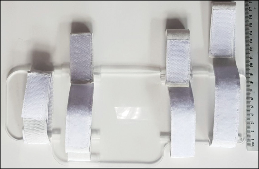
材料和方法。开发了一种防止手部运动的绷带,并使用两种射频线圈进行了检查。一名专门从事肌肉骨骼成像的放射科医生对图像的技术参数进行了比较评估,并根据研究目的对图像质量进行了评估。
结果。当需要扫描整个手部(如风湿病)时,头部线圈更为合适。膝部线圈的视野较小,分辨率较高,可用于对解剖结构(包括腕部)进行更详细的评估。根据获得的数据,我们制定了手部磁共振成像的建议:选择射频线圈、扫描方案和脉冲序列参数。此外,我们还提出了固定患者手部的绷带,以平缓过度运动和防止伪影。
结论。要保证手部磁共振成像的质量,需要考虑以下几个因素:磁共振成像过程中的安全规则、扫描参数的调整以及手部在线圈中的正确固定。遵守本文提出的建议以及使用开发的绷带可以提高手部磁共振成像的质量。
作者简介
Yuriy A. Vasilev
Research and Practical Clinical Center for Diagnostics and Telemedicine Technologies
Email: VasilevYA1@zdrav.mos.ru
ORCID iD: 0000-0002-0208-5218
SPIN 代码: 4458-5608
MD, Cand. Sci. (Medicine)
俄罗斯联邦, MoscowDmitry S. Semenov
Research and Practical Clinical Center for Diagnostics and Telemedicine Technologies
Email: SemenovDS4@zdrav.mos.ru
ORCID iD: 0000-0002-4293-2514
SPIN 代码: 2278-7290
Cand. Sci. (Engineering)
俄罗斯联邦, MoscowAlexey V. Petraikin
Research and Practical Clinical Center for Diagnostics and Telemedicine Technologies
Email: PetryajkinAV@zdrav.mos.ru
ORCID iD: 0000-0003-1694-4682
SPIN 代码: 6193-1656
MD, Dr. Sci. (Medicine)
俄罗斯联邦, MoscowAndrey A. Uchevatkin
Research and Practical Clinical Center for Diagnostics and Telemedicine Technologies
Email: UchevatkinAA@zdrav.mos.ru
ORCID iD: 0000-0001-7284-4737
SPIN 代码: 5575-4511
MD, Cand. Sci. (Medicine)
俄罗斯联邦, MoscowLiya R. Abuladze
Research and Practical Clinical Center for Diagnostics and Telemedicine Technologies
编辑信件的主要联系方式.
Email: drliaabuladze@gmail.com
ORCID iD: 0000-0001-6745-1672
SPIN 代码: 8640-9989
俄罗斯联邦, Moscow
Alexander V. Bazhin
Research and Practical Clinical Center for Diagnostics and Telemedicine Technologies
Email: BazhinAV@zdrav.mos.ru
ORCID iD: 0000-0003-3198-1334
SPIN 代码: 6122-5786
MD, Cand. Sci. (Medicine)
俄罗斯联邦, MoscowDariya E. Sharova
Research and Practical Clinical Center for Diagnostics and Telemedicine Technologies
Email: SharovaDE@zdrav.mos.ru
ORCID iD: 0000-0001-5792-3912
SPIN 代码: 1811-7595
俄罗斯联邦, Moscow
参考
- Ratasvuori MS, Lindfors NC, Sormaala MJ. The clinical significance of magnetic resonance imaging of the hand: an analysis of 318 hand and wrist images referred by hand surgeons. J Plast Surg Hand Surg. 2022;56(2):69–73. doi: 10.1080/2000656X.2021.1933993
- Sergunova KA, Akhmad ES, Petryaikin AV, et al. Safety Fundamentals of Magnetic Resonance Imaging. Moscow: Research and Practical Clinical Center for Diagnostics and Telemedicine Technologies; 2019. (In Russ). EDN: GTOVGS
- Semenov DS, Panina OY, Khoruzhaya AN, et al. All-Russian rating of radiology departments: 2020 competition results. Digital Diagnostics. 2022;3(1):43–54. EDN: SWQWGE doi: 10.17816/DD95661
- Andersson JK, Hansson-Olofsson E, Karlsson J, et al. Cost description of clinical examination and MRI in wrist ligament injuries. J Plast Surg Hand Surg. 2018;52(1):30–36. doi: 10.1080/2000656X.2017.1319845
- Hansford BG. Multimodality Pitfalls of Wrist Imaging With a Focus on Magnetic Resonance Imaging. Top Magn Reson Imaging. 2020;29(5):263–272. doi: 10.1097/RMR.0000000000000254
- Burns JE, Tanaka T, Ueno T, et al. Pitfalls That May Mimic Injuries of the Triangular Fibrocartilage and Proximal Intrinsic Wrist Ligaments at MR Imaging. RadioGraphics. 2011;31(1):63–78. doi: 10.1148/rg.311105114
- Guidelines for MR Imaging of Sports Injuries [Internet]. European Society of Musculoskeletal Radiology. Available from: https://essr.org/content-essr/uploads/2016/10/ESSR-MRI-Protocols-Fingers.pdf
- Sudoł-Szopińska I, Jurik A, Eshed I, et al. Recommendations of the ESSR Arthritis Subcommittee for the Use of Magnetic Resonance Imaging in Musculoskeletal Rheumatic Diseases. Semin Musculoskelet Radiol. 2015;19(04):396–411. doi: 10.1055/s-0035-1564696
- Magnetic resonance imaging of the wrist joint [Internet]. Moscow Standard of Radiologic Diagnostics. [cited 10 Oct 2023] Available from: https://standard.telemedai.ru/issledovanie/magnitno-rezonansnaya-tomografiya-luchezapyastnogo-sustava (In Russ)
- Bazhin AV, Blinov NN, Vasilev YuA, et al. Standards for the performance of magnetic resonance imaging. Moscow: Moscow State Medical and Dental University named after A.I. Evdokimov; 2019. (In Russ).
- Eschweiler J, Li J, Quack V, et al. Anatomy, Biomechanics, and Loads of the Wrist Joint. Life. 2022;12(2):188. doi: 10.3390/life12020188
- Rubin DA, Roberts CC, Bencardino JT, et al. ACR Appropriateness Criteria® Chronic Wrist Pain. J Am Coll Radiol. 2018;15(5):S39–S55. doi: 10.1016/j.jacr.2018.03.021
- Dreckmann SC, von Schroeder HP, Novak CB, et al. Utility of Specialized Imaging for Diagnosis of Chronic Wrist Pain. J Wrist Surg. 2019;08(06):497–502. doi: 10.1055/s-0039-1697022
- El-Deek AMF, Dawood EMAE-HH, Mohammed AAM. Role of ultrasound versus magnetic resonance imaging in evaluation of non-osseous disorders causing wrist pain. Egypt J Radiol Nucl Med. 2019;50(1):8. doi: 10.1186/s43055-019-0008-9
- Götestrand S, Björkman A, Björkman-Burtscher IM, et al. Visualization of wrist anatomy — a comparison between 7T and 3T MRI. Eur Radiol. 2022;32(2):1362–1370. doi: 10.1007/s00330-021-08165-5
- Kocharian A, Adkins MC, Amrami KK, et al. Wrist: Improved MR Imaging with Optimized Transmit-Receive Coil Design. Radiology. 2002;223(3):870–876. doi: 10.1148/radiol.2233010824
- Hand/Wrist 16 [Internet]. Siemens Healthineers. [cited 10 Oct 2023] Available from: https://www.siemens-healthineers.com/magnetic-resonance-imaging/options-and-upgrades/coils/hand-wrist-16
- Hand and Wrist MRI Coil [Internet]. ScanMed: A DirectMed Company. [cited 10 Oct 2023] Available from: https://www.scanmed.com/wrist-mri-coil
- Peterfy CG, Olech E, DiCarlo JC, et al. Monitoring cartilage loss in the hands and wrists in rheumatoid arthritis with magnetic resonance imaging in a multi-center clinical trial: IMPRESS (NCT00425932). Arthritis Res Ther. 2013;15(2):R44. doi: 10.1186/ar4202
- Vassa R, Garg A, Omar IM. Magnetic resonance imaging of the wrist and hand. Polish J Radiol. 2020;85(1):461–488. doi: 10.5114/pjr.2020.99034
补充文件


