

")


Возможности применения магнитно-резонансной томографии с использованием гепатотропных контрастных веществ для функциональной оценки печени
- Авторы: Агеева С.Ф.1, Синицын В.Е.1, Мершина Е.А.1, Ручьева Н.А.2, Петрова Е.И.3
-
Учреждения:
- Московский государственный университет имени М.В. Ломоносова
- Национальный медицинский исследовательский центр трансплантологии и искусственных органов имени академика В.И. Шумакова
- Отраслевой клинико-диагностический центр ПАО «Газпром»
- Выпуск: Том 5, № 2 (2024)
- Страницы: 137-148
- Раздел: Оригинальные исследования
- URL: https://journals.rcsi.science/DD/article/view/264828
- DOI: https://doi.org/10.17816/DD624826
- ID: 264828
Цитировать

Аннотация
Обоснование. Оценка функции печени при различных заболеваниях остаётся важной клинической задачей. Применение магнитно-резонансной томографии с гепатотропным контрастным веществом для оценки функции печени представляет существенный научный и практический интерес.
Цель — изучить возможность функциональной оценки печени на основании показателей, полученных по данным магнитно-резонансной томографии с контрастированием гепатотропным веществом.
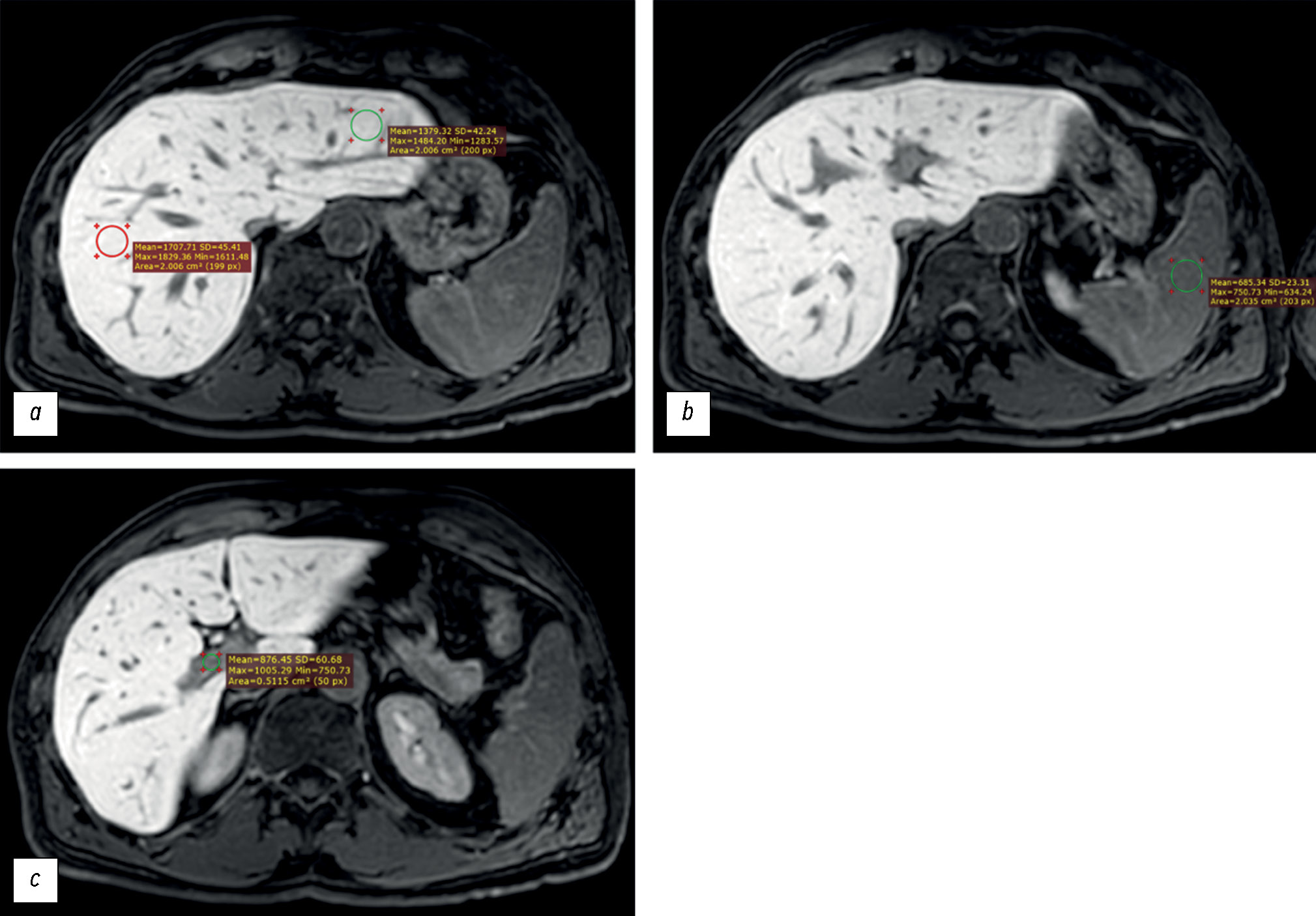
Материалы и методы. Были проанализированы данные пациентов, которым выполнялась магнитно-резонансная томография с внутривенным контрастированием гадоксетовой кислотой. Пациенты были разделены на две группы: с нарушенной (первая группа) и с нормальной (вторая группа) функцией печени. По данным магнитно-резонансных исследований оценивались следующие параметры: интенсивность сигнала печени, её отношение к интенсивности сигнала селезёнки и к интенсивности сигнала в просвете воротной вены. Были оценены показатели лабораторных анализов крови, отражающие функции печени: общий билирубин, альбумин, аланинаминотрансфераза, аспартатаминотрансфераза, γ-глутамилтранспептидаза, щелочная фосфатаза, протромбиновое время. Был проведён анализ статистической значимости различий между группами по параметрам магнитно-резонансной томографии, оценивалось наличие корреляционной связи между значениями интенсивности сигнала печени и данными лабораторных анализов крови.
Результаты. Были проанализированы данные 53 пациентов (25 мужчин и 28 женщин в возрасте от 24 до 84 лет). В первую группу вошло 19 человек, во вторую — 34 человека. Были установлены статистически значимые различия показателей интенсивности сигнала печени и её отношения к интенсивности сигнала селезёнки между исследуемыми группами. В первой группе значение интенсивности сигнала печени составило 919,05 [669,65; 1258,35], во второй — 1525,13 [1460,5; 1631,4] (p=0,0000001). Отношение интенсивности сигнала печени к интенсивности сигнала селезёнки в первой группе составило 1,2 [1,04; 1,7], во второй — 1,7 [1,46; 1,96] (p=0,00076). Отношение интенсивности сигнала печени к интенсивности сигнала в просвете воротной вены составило 1,44 [1,29; 1,83] в первой группе, 1,6 [1,43; 1,83] — во второй (p=0,1). Была оценена корреляция между интенсивностью сигнала печени и общим билирубином (r=–0,61; p=0,000001), альбумином (r=0,13; p=0,61), аспартатаминотрансферазой (r=–0,57; p=0,000009), аланинаминотрансферазой (r=–0,44; p=0,001), щелочной фосфатазой (r=–0,45; p=0,0007), γ-глутамилтранспептидазой (r=–0,5; p=0,0003), протромбиновым временем (r=–0,34; p=0,04). По шкале Чеддока заметная сила корреляционной связи была выявлена между показателем интенсивности сигнала печени и значениями общего билирубина, аспартатаминотрансферазы. Умеренная сила — между показателем интенсивности сигнала печени и значениями аланинаминотрансферазы, щелочной фосфатазы, γ-глутамилтранспептидазы, протромбинового времени.
Заключение. Продемонстрирована эффективность применения параметров магнитно-резонансной томографии (интенсивность сигнала печени и её отношение к интенсивности сигнала селезёнки) в функциональной оценке печени. В исследовании не подтвердилось предположение об эффективности применения такого параметра, как отношение значения интенсивности сигнала печени к интенсивности сигнала в просвете воротной вены. Были установлены статистически значимые обратные связи между значениями интенсивности сигнала печени и показателями лабораторных анализов крови, отражающих функции печени, за исключением альбумина. Результаты свидетельствуют о возможности использования магнитно-резонансной томографии для функциональной оценки печени.
Полный текст
Открыть статью на сайте журналаОб авторах
София Фаильевна Агеева
Московский государственный университет имени М.В. Ломоносова
Автор, ответственный за переписку.
Email: son.ageeva13@gmail.com
ORCID iD: 0009-0003-9563-6756
SPIN-код: 9695-3717
Россия, Москва
Валентин Евгеньевич Синицын
Московский государственный университет имени М.В. Ломоносова
Email: vsini@mail.ru
ORCID iD: 0000-0002-5649-2193
SPIN-код: 8449-6590
д-р мед. наук, профессор
Россия, МоскваЕлена Александровна Мершина
Московский государственный университет имени М.В. Ломоносова
Email: elena_mershina@mail.ru
ORCID iD: 0000-0002-1266-4926
SPIN-код: 6897-9641
канд. мед. наук
Россия, МоскваНаталья Александровна Ручьева
Национальный медицинский исследовательский центр трансплантологии и искусственных органов имени академика В.И. Шумакова
Email: rna1969@yandex.ru
ORCID iD: 0000-0002-8063-4462
SPIN-код: 2196-8300
канд. мед. наук
Россия, МоскваЕкатерина Игоревна Петрова
Отраслевой клинико-диагностический центр ПАО «Газпром»
Email: doc_mri@mail.ru
ORCID iD: 0009-0005-0355-8098
канд. мед. наук
Россия, МоскваСписок литературы
- Peng Y., Qi X., Guo X. Child–Pugh Versus MELD Score for the Assessment of Prognosis in Liver Cirrhosis // Medicine. 2016. Vol. 95, N 8. P. e2877. doi: 10.1097/MD.0000000000002877
- Ликарь Ю.Н., Ахаладзе Д.Г., Румянцев А.Г. Гепатобилиарная сцинтиграфия в предоперационной оценке функции планируемого остатка печени (обзор литературы и собственные примеры) // Российский журнал детской гематологии и онкологии. 2020. Т. 7, № 1. С. 62–69. EDN: VWDZUW doi: 10.21682/2311-1267-2020-7-1-62-69
- Chernyak V., Fowler K.J., Heiken J.P., Sirlin C.B. Use of gadoxetate disodium in patients with chronic liver disease and its implications for liver imaging reporting and data system (LI-RADS) // Journal of Magnetic Resonance Imaging. 2019. Vol. 49, N 5. P. 1236–1252. doi: 10.1002/jmri.26540
- Welle C.L., Guglielmo F.F., Venkatesh S.K. MRI of the liver: choosing the right contrast agent // Abdominal Radiology. 2020. Vol. 45, N 2. P. 384–392. doi: 10.1007/s00261-019-02162-5
- Furlan A., Borhani A.A., Heller M.T., Yu R.K., Tublin M.E. Non-focal liver signal abnormalities on hepatobiliary phase of gadoxetate disodium-enhanced MR imaging: a review and differential diagnosis // Abdominal Radiology. 2016. Vol. 41, N 7. P. 1399–1410. doi: 10.1007/s00261-016-0685-z
- Cho S.H., Kang U.R., Kim J.D., Han Y.S., Choi D.L. The value of gadoxetate disodium-enhanced MR imaging for predicting posthepatectomy liver failure after major hepatic resection: A preliminary study // Eur J Radiol. 2011. Vol. 80, N 2. P. e195–e200. doi: 10.1016/j.ejrad.2011.08.008
- Collettini F., Elkilany A., Seta M.D., et al. MR imaging of hepatocellular carcinoma: prospective intraindividual head-to-head comparison of the contrast agents gadoxetic acid and gadoteric acid // Sci Rep. 2022. Vol. 12, N 1. P. 18583. doi: 10.1038/s41598-022-23397-1
- Galle P.R., Forner A., Llovet J.M., et al. EASL Clinical Practice Guidelines: Management of hepatocellular carcinoma // J Hepatol. 2018. Vol. 69, N 1. P. 182–236. doi: 10.1016/j.jhep.2018.03.019
- Yang M., Zhang Y., Zhao W., et al. Evaluation of liver function using liver parenchyma, spleen and portal vein signal intensities during the hepatobiliary phase in Gd-EOB-DTPA-enhanced MRI // BMC Med Imaging. 2020. Vol. 20, N 1. P. 119. doi: 10.1186/s12880-020-00519-7
- Bastati N., Wibmer A., Tamandl D., et al. Assessment of Orthotopic Liver Transplant Graft Survival on Gadoxetic Acid–Enhanced Magnetic Resonance Imaging Using Qualitative and Quantitative Parameters // Invest Radiol. 2016. Vol. 51, N 11. P. 728–734. doi: 10.1097/RLI.0000000000000286
- Мнацаканян М.К., Рубцова Н.А., Кабанов Д.О., и др. Роль магнитно-резонансной томографии с гадоксетовой кислотой в оценке функционального резерва печени // Российский электронный журнал лучевой диагностики. 2022. Т. 12, № 1. С. 43–55. EDN: GXFGZS doi: 10.21569/2222-7415-2022-12-1-43-55
- Zhang W., Wang X., Miao Y., Hu C., Zhao W. Liver function correlates with liver-to-portal vein contrast ratio during the hepatobiliary phase with Gd-EOB-DTPA-enhanced MR at 3 Tesla // Abdominal Radiology. 2018. Vol. 43, N 9. P. 2262–2269. doi: 10.1007/s00261-018-1462-y
- Lee N.K., Kim S., Kim G.H., et al. Significance of the “Delayed hyperintense portal vein sign” in the hepatobiliary phase MRI obtained with Gd-EOB-DTPA // Journal of Magnetic Resonance Imaging. 2012. Vol. 36, N 3. P. 678–685. doi: 10.1002/jmri.23700
- Vincent J.L., Moreno R., Takala J., et al. The SOFA (Sepsis-related Organ Failure Assessment) score to describe organ dysfunction/ failure // Intensive Care Med. 1996. Vol. 22, N 7. P. 707–710. doi: 10.1007/BF01709751
- Zaccherini G., Weiss E., Moreau R. Acute-on-chronic liver failure: Definitions, pathophysiology and principles of treatment // JHEP Reports. 2021. Vol. 3, N 1. P. 100176. doi: 10.1016/j.jhepr.2020.100176
Дополнительные файлы


